http://pubs.acs.org/journal/acsodf
Article
Impact of Increased Sonication-Induced Dispersion of Sepiolite on
Its Interaction with Biological Macromolecules and Toxicity/
Proliferation in Human Cells
David Adame Brooks, Olivier Piétrement, Elodie Dardillac, Ayesha Jayantha, Manuel A. Lores Guevara,
Fidel Antonio Castro-Smirnov, Pilar Aranda, Eduardo Ruiz-Hitzky, and Bernard S. Lopez*
Downloaded via 200.55.162.196 on June 12, 2023 at 17:02:35 (UTC).
See https://pubs.acs.org/sharingguidelines for options on how to legitimately share published articles.
Cite This: ACS Omega 2023, 8, 1026−1036
ACCESS
Read Online
Metrics & More
Article Recommendations
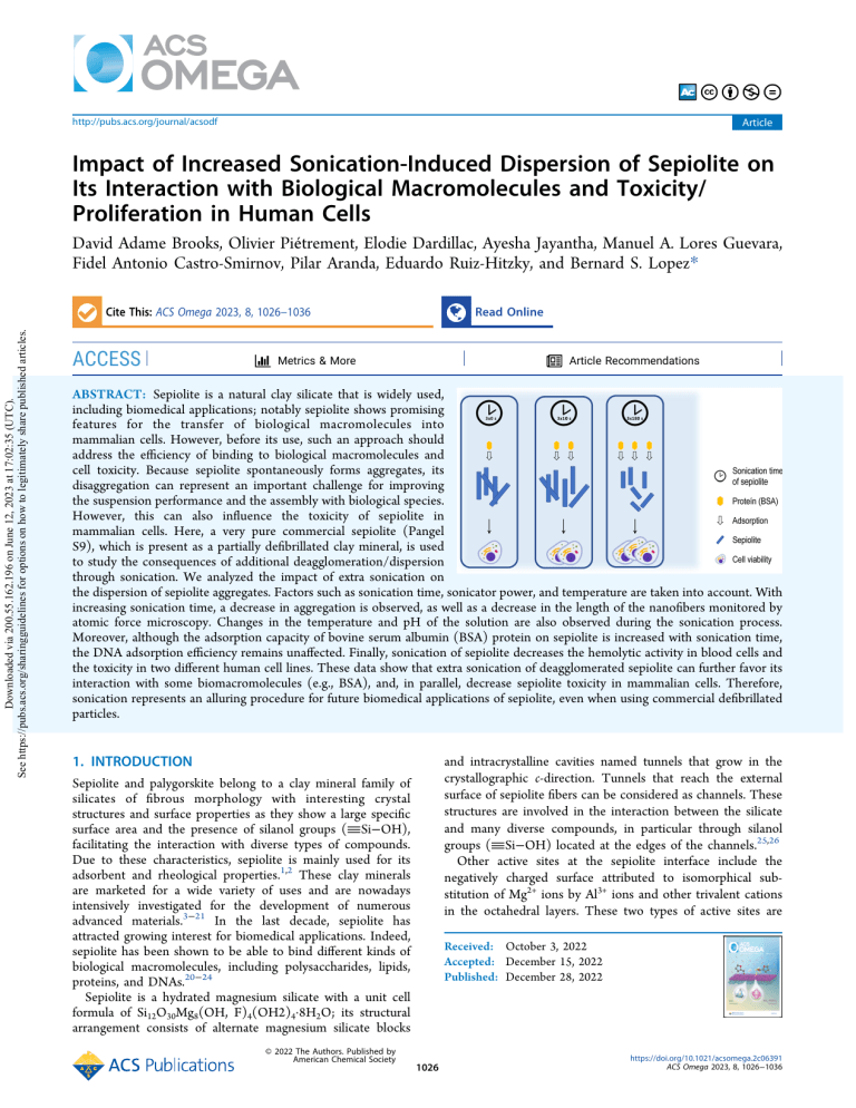
ABSTRACT: Sepiolite is a natural clay silicate that is widely used,
including biomedical applications; notably sepiolite shows promising
features for the transfer of biological macromolecules into
mammalian cells. However, before its use, such an approach should
address the efficiency of binding to biological macromolecules and
cell toxicity. Because sepiolite spontaneously forms aggregates, its
disaggregation can represent an important challenge for improving
the suspension performance and the assembly with biological species.
However, this can also influence the toxicity of sepiolite in
mammalian cells. Here, a very pure commercial sepiolite (Pangel
S9), which is present as a partially defibrillated clay mineral, is used
to study the consequences of additional deagglomeration/dispersion
through sonication. We analyzed the impact of extra sonication on
the dispersion of sepiolite aggregates. Factors such as sonication time, sonicator power, and temperature are taken into account. With
increasing sonication time, a decrease in aggregation is observed, as well as a decrease in the length of the nanofibers monitored by
atomic force microscopy. Changes in the temperature and pH of the solution are also observed during the sonication process.
Moreover, although the adsorption capacity of bovine serum albumin (BSA) protein on sepiolite is increased with sonication time,
the DNA adsorption efficiency remains unaffected. Finally, sonication of sepiolite decreases the hemolytic activity in blood cells and
the toxicity in two different human cell lines. These data show that extra sonication of deagglomerated sepiolite can further favor its
interaction with some biomacromolecules (e.g., BSA), and, in parallel, decrease sepiolite toxicity in mammalian cells. Therefore,
sonication represents an alluring procedure for future biomedical applications of sepiolite, even when using commercial defibrillated
particles.
1. INTRODUCTION
Sepiolite and palygorskite belong to a clay mineral family of
silicates of fibrous morphology with interesting crystal
structures and surface properties as they show a large specific
surface area and the presence of silanol groups (�Si−OH),
facilitating the interaction with diverse types of compounds.
Due to these characteristics, sepiolite is mainly used for its
adsorbent and rheological properties.1,2 These clay minerals
are marketed for a wide variety of uses and are nowadays
intensively investigated for the development of numerous
advanced materials.3−21 In the last decade, sepiolite has
attracted growing interest for biomedical applications. Indeed,
sepiolite has been shown to be able to bind different kinds of
biological macromolecules, including polysaccharides, lipids,
proteins, and DNAs.20−24
Sepiolite is a hydrated magnesium silicate with a unit cell
formula of Si12O30Mg8(OH, F)4(OH2)4·8H2O; its structural
arrangement consists of alternate magnesium silicate blocks
© 2022 The Authors. Published by
American Chemical Society
and intracrystalline cavities named tunnels that grow in the
crystallographic c-direction. Tunnels that reach the external
surface of sepiolite fibers can be considered as channels. These
structures are involved in the interaction between the silicate
and many diverse compounds, in particular through silanol
groups (�Si−OH) located at the edges of the channels.25,26
Other active sites at the sepiolite interface include the
negatively charged surface attributed to isomorphical substitution of Mg2+ ions by Al3+ ions and other trivalent cations
in the octahedral layers. These two types of active sites are
Received: October 3, 2022
Accepted: December 15, 2022
Published: December 28, 2022
1026
https://doi.org/10.1021/acsomega.2c06391
ACS Omega 2023, 8, 1026−1036
ACS Omega
http://pubs.acs.org/journal/acsodf
central to the adsorption mechanisms that occur at the external
surfaces of fibrous clay minerals.26
Natural raw sepiolite appears at the electronic microscope as
bundles resulting from the agglomeration of their silicate
microfibers, which can be disaggregated in concentrated water
dispersions by applying high-speed mechanical shearing or by
ultrasonication. The resulting products at the industrial level
are considered samples of “rheological grade” sepiolite
products, which have been commercialized as thickening or
suspending agents (e.g., Pangel S9).27,28 However, such
materials still contain aggregates, and it has been shown that
their further dispersion can improve efficacy regarding many
different applications. For instance, detangled sepiolite
promotes the transfer of DNA into mammalian cells, but the
efficiency can be optimized through additional sonication of
sepiolite (sSep) before the synthesis of the bionanocomposite
sSep/DNA.20
Therefore, for the use of sepiolite in different applications,
increased disaggregation can be important for wider spreading
and the formation of stable and homogeneous suspensions that
are crucial steps to improve their performance. Some
mechanical irradiation (ultrasounds) has also been proposed
as a procedure to efficiently defibrillate sepiolite to generate
colloidal routes to obtain new heterostructured functional
nanomaterials.26
The individual particles of sepiolite (even those that have
been previously defibrillated) can easily aggregate in water
through hydrogen bonding and van der Waals interactions to
form bundles and aggregates, limiting clay dispersion.29 Of
note, a recent study reports on the colloidal dispersion
behavior of individual sepiolite fibers by analyzing the diffusive
motion of some natural clays. The authors show that sepiolite
nanoclay demonstrates rich Brownian-type rotational dynamics.30 To improve the dispersibility of sepiolite, several
strategies have been evaluated, and the most common
approaches include mechanical treatment, addition of chemical
dispersants to the suspension, and chemical modification of the
mineral surface.28 Chemical treatments can modify the
chemical properties of sepiolite and/or its toxicity in
mammalian cells. Mechanical treatment can consist of
ultrasound or high-speed shear processes, among others,
which are capable of dispersing particle aggregates into smaller
aggregates or simple particles without damaging the crystalline
structure. Sonication has been presented as a relatively simple
approach to disaggregate sepiolite fibers and improve their
properties and characteristics.26 Sonication involves the
process of cavitation generation, which consists of the creation,
growth, and collapse of bubbles formed in the liquid due to
high intensity ultrasound irradiation.31 In recent years, this
technique has received much attention, especially due to the
advent of nanotechnological applications for the assembly of
nanoparticles to generate advanced functional nanoarchitectures, including sepiolite-based materials.5,11 The sonication
process makes it possible to improve the homogeneity of
nanomaterials in suspensions and potentially achieve a smaller
size distribution. In the field of nanomaterials and their
application in biotechnology, the quality of the suspensions is
very important, since this will determine the properties that
can interfere with the nanomaterial interaction(s) with other
molecules. This process can potentially alter the main physical
and chemical properties of nanomaterials, such as size,
morphology, increased contact surface area and surface charge
distribution.32,33 The literature has reported the importance of
Article
controlling the sonication process and the impact that it can
have on the particle parameters.34−38 More specifically,
sonication of commercial sepiolite (Pangel S9) strongly
stimulates the transfer of DNA into mammalian cells.21
Therefore, it is essential to evaluate whether sonication affects
the interaction of sepiolite with biological macromolecules and
its potential detrimental effects on cell viability. In this respect,
it is important to determine how different factors, such as
power, duration, and temperature, can affect the quality of the
dispersion. Although these considerations have been taken into
account by some researchers, work in this area remains limited.
Bihari et al. (2008) studied the stability of dispersions of
different nanomaterials using different ultrasound energies with
different dispersion stabilizers. Hartmann et al. (2015)
highlighted that although work has been carried out to
understand the different factors that affect the dispersion
quality of nanomaterials, there is still no well-defined and
universally accepted sonication procedure.
Here, we studied the process of deagglomeration/dispersion
of sepiolite nanofibers to identify the optimum conditions for
this process to produce detangled sepiolite for future biological
applications. We first focused on the analysis of factors such as
sonication time, sonicator power, temperature, and pH.
Second, as a proof of concept, we analyzed the adsorption
capacity of a protein (bovine serum albumin, BSA) and DNA
on sonicated sepiolite (sSep). Finally, to assess whether
sonication can affect sepiolite toxicity in mammalian cells, we
analyzed the impact of the sonication of sepiolite on hemolytic
activity in blood cells and toxicity/proliferation in human cell
lines.
2. MATERIALS AND METHODS
2.1. Preparation of Sepiolite Suspension Using
Vortex Dispersion. In this study, sepiolite (Sep) obtained
from Vicálvaro-Vallecas deposits, Madrid (Spain), was
generously supplied by TOLSA, S.A. with the trade name of
Pangel S9. A 2 mg/mL sepiolite suspension was prepared in 10
mM Tris−HCl buffer, pH = 7.5, under vigorous vortexing at a
maximum speed for a minimum time of 10 min to properly
disperse the clay.
2.2. Preparation of Sepiolite Suspensions Using
Ultrasound. Four tubes were prepared with sepiolite in 10
mL of a suspension in 10 mM Tris−HCl buffer, pH = 7.5, at a
concentration of 2 mg/mL for ultrasonication. The clay was
dispersed using a Vibra-Cell VC-50-1 (Sonics & Materials)
with a standard Ti probe with a diameter and length of 13 mm
and 138 mm, respectively, and a generator output power of 50
W. The sepiolite suspension was sonicated three times with an
on/off pulse duration of 10 s at different sonication times (10,
20, 60, and 180 s). The procedure was repeated for each
sonication time with amplitude settings of 30, 50, and 100%.
2.3. Effective Acoustic Power. The effective acoustic
power delivered to the sonicated suspension is an important
parameter for obtaining reproducible dispersions. This is
different from the electrical input or output power of the
generator indicated by the manufacturer as this is the actual
power that is delivered to the suspension during sonication.41
Among many methods for the calculation of effective delivered
power, the most commonly used method is calorimetry.41 This
is known to be a simple and efficient way to directly measure
the effective power delivered to a suspension.42 In this method,
the increase in temperature in the liquid at a given ultrasonic
1027
https://doi.org/10.1021/acsomega.2c06391
ACS Omega 2023, 8, 1026−1036
ACS Omega
http://pubs.acs.org/journal/acsodf
different concentrations, 500 μL of sSep was added at a
concentration of 100 μg/mL. The samples were completed
with a 10 mM Tris−HCl, pH = 7.5 solution, until 1 mL was
obtained for each sample. All sSep/protein mixtures were
stirred overnight at 25 °C. Finally, the mixtures were
centrifuged for 5 min at 5000 rpm, and the protein
concentrations in the supernatants were measured using a
NanoDrop ND1000 spectrophotometer.39,40
2.8. sSep− DNA Synthesis. For these samples, a Tris−
HCl/CaCl2 solution consisting of 25 mL of 20 mM Tris−HCl
at pH = 7.5, 5 mL of 100 mM CaCl2, and 20 mL of distilled
water was prepared; 1 mL of sSep dispersion was taken at 2
mg/mL and centrifuged for 5 min at 5000 rpm, the
supernatant was removed, and the precipitate was resuspended
in 10 mL of Tris−HCl/CaCl2 solution. Plasmid DNA was
used, which was obtained by amplifying a bacterial culture and
was purified using the NucleoBond Plasmid Purification
Protocol. For each DNA sample at different concentrations,
500 μL of sSep in Tris−HCL/CaCl2 was added at a
concentration of 100 μg/mL. The samples were completed
with 10 mM Tris−HCl, pH = 7.5 solution, until a volume of 1
mL was obtained for each sample. These samples were
incubated under stirring overnight at 25 °C.
The mixtures were centrifuged for 5 min at 5000 rpm, and
the DNA concentrations in the supernatants were measured
using a NanoDrop ND1000 spectrophotometer.
2.9. Calculation of Adsorption Isotherms. To determine the adsorption isotherms for the different proteins used
in our experiment, the concentration of the adsorbent phase
was calculated using eq 3.46
equipment setting is recorded over time, and the effective
power delivered is calculated using eq 1
i dT y
P = jjj zzz·M · Cp
k dt {
(1)
where P is the delivered acoustic power, T is the temperature, t
is the time, CP is the specific heat of the liquid (4.18 J.g−1. K−1
for water), and M is the mass of the liquid.
2.4. Calorimetric Experiment. The temperature was
measured using a digital thermometer (GESA Termómetros,
S.L). The probe was introduced 4 cm below the surface of the
liquid without touching the walls of the tube. The increase in
temperature was recorded for 5 min at 30 s intervals. The data
obtained allowed the construction of a temperature versus time
graph; from this graph, the linear fit and the slope (which is the
increase in temperature over time) were obtained. These data
enable calculation of the effective acoustic power using eq 1.
The experiment was repeated three times for each of the three
amplitudes used.
2.5. Differential Centrifugation. The centrifugation
study was performed for sSep solutions at different sonication
times (10, 20, 60, and 180 s). Particles of different sizes in a
suspension settle at different rates, and larger particles and
denser sediment settle faster. By increasing the centrifugal
force, the sedimentation rates can be increased. An Eppendorf
541712 centrifuge was used at varying angular velocity values
of 0, 500, 1000, 2000, and 5000 rpm. Aliquots with a volume of
200 μL were taken at the mean height of the liquid in each
sample, and the absorbance at a wavelength of 350 nm was
measured using an Epoch Microplate spectrophotometer
(BioTeK Instruments). The experiment was repeated three
times for each sonication time.
2.6. Experimental Determination of the Sedimentation Velocity and the Aggregation Index. 2.6.1. Sedimentation Velocity. The sedimentation velocity was
determined using the data obtained for sedimentation of
sepiolite suspensions with different sonication times. The
samples were shaken so that they were as uniform as possible
throughout the entire tube. Subsequently, the samples were left
to rest, and the absorbance was measured at 0, 15, 30, and 60
min.
After determining the pairs of absorbance values as a
function of time, the sedimentation curve was obtained. With
this graphical representation, the sedimentation velocity was
calculated as a function of the sonication time. From the
experimental data obtained, the values for the slopes at the
origin, (dAbs/dt), coincide with the sedimentation velocities.
2.6.2. Aggregation Index. The aggregation index (AI) was
calculated using eq 2.43,44
ij
yz
ODMin
zz·100
AI = jjj
j ODMax ODMin zz
k
{
Article
qe =
(C 0
C) × V
m
(3)
where C0 is the initial value of the protein concentrations (μg/
mL), C is the protein concentration (μg/mL) in the
supernatant, V denotes the volume (mL), and m (μg) is the
amount of clay.
The adsorption performance was calculated, as shown in eq
4.46,47
(%) =
(C0 C)
× 100
C0
(4)
2.10. Hemolytic Activity. Blood cells were extracted from
human blood drawn from normal patients. The cells were
washed three times with phosphate-buffered saline (PBS)
solution. A suspension of erythrocytes was prepared by
forming 2 mL of packed erythrocytes in 100 mL of PBS
solution. A 0.1% sodium carbonate (Na2CO3) solution and a
negative control PBS pH 7.4 were used as positive and negative
controls, respectively.
For the evaluation of the compounds, 0.2 mL of erythrocyte
solution was used, to which 10 μL of the sSep dispersion was
added and brought to a volume of 10 mL with PBS. The
positive control was evaluated by taking 0.2 mL of the
erythrocyte solution and completing the 10 mL volume with
0.1% Na2CO3. For the negative control, 0.2 mL of erythrocyte
solution was taken, and the volume was made up to 10 mL
with PBS. Once the compound was added, the samples were
carefully mixed and allowed to stand for 1 h at 37 °C.
The samples were centrifuged at 2000 rpm for 10 min. The
absorbance at 545 nm was measured with a spectrophotometer
(Pharmacia LKB·ULTROSPEC III).
(2)
where ODMax represents the maximum in the absorbance band
of the spectrum, ODMin is the minimum absorbance; sepiolite
shows light absorption predominantly at approximately 300−
350 nm,.45 The values for the max and min wavelengths were
350 and 600 nm, respectively.
2.7. sSep-Protein Synthesis. Protein solutions were
prepared at different concentrations (5, 10, 20, 50, 100, 150,
and 200 μg/mL). The protein used was bovine serum albumin
(BSA): 2 mg/mL stock solutions were prepared in 10 mM
Tris−HCl buffer, pH = 7.5. For each protein sample at
1028
https://doi.org/10.1021/acsomega.2c06391
ACS Omega 2023, 8, 1026−1036
ACS Omega
http://pubs.acs.org/journal/acsodf
Article
Figure 1. Calorimetric analysis with three different powers (%), as a function of the sonication times. Each point corresponds to three independent
experiments (in triplicate for each repeat).
Figure 2. Analysis of pH as a function of sonication time for different input powers. Each point corresponds to three independent experiments (in
triplicate for each repeat).
In each test, three replicates for each sSep dispersion and
each reading were made. The hemolytic activity of sepiolite at
different concentrations was calculated from the average
absorbance for three repetitions and was expressed as a
percentage using eq 5.48
%H =
ODS•ODNC
· 100
ODPC•ODNC
homodimer-1 probe was used to measure the dead cells (FL-2
channel).
2.12. AFM Imaging. Atomic force microscopy (AFM)
imaging of sepiolite was performed using a freshly cleaved mica
surface (V1 quality, EMS) treated with 50 μM spermidine for 1
min. Excess spermidine solution was blotted with a filter paper,
and 3−5 μL of sepiolite dispersion was deposited onto the
mica surface, incubated for 1−2 min, and rinsed with 25 μL of
ultrapure water. The surface was blotted and dried. Imaging
was carried out in the peak force mode with SCANASYST-Air
probes (Bruker) with a Multimode system (Bruker) operating
with a Nanoscope V controller (Bruker). All images were
collected at a scan frequency of 1 Hz and a resolution of 1024
× 1024 pixels. Images were analyzed with Nanoscope V and
ImageJ software. A third-order polynomial function was used
to remove the background.
(5)
where ODS, ODNC, and ODPC are the optical density for the
sample, negative control, and positive control, respectively.
2.11. Human Cell Toxicity Assay. The cell lines U2OS
(human osteosarcoma) and RG37 (SV40-transformed human
fibroblasts) were maintained at 37 °C with 5% CO2 in
modified Eagle’s medium (MEM). One day after seeding of
the cells, sepiolite (10 μg/mL) was added to the culture
medium. Cells were collected 1 day after exposure to sepiolite.
Cell pellets were resuspended in PBS and stained with the
LIVE/DEAD Viability/Cytotoxicity Kit from Invitrogen
(#L3224) according to the manufacturer’s instructions. Flow
cytometry analyses were performed using an Accuri C6 flow
cytometer (BD Biosciences). A Calcein AM probe was used to
measure the live cells (FL-1 channel), and an ethidium
3. RESULTS AND DISCUSSION
3.1. Calorimetric Analysis. Although in principle, the
increase of temperature produced by the treatment should not
induce major transformation of the materials, we performed a
calorimetric study. The calorimetric data reveal an increase in
temperature with increasing sonication time (Figure 1). The
1029
https://doi.org/10.1021/acsomega.2c06391
ACS Omega 2023, 8, 1026−1036
ACS Omega
http://pubs.acs.org/journal/acsodf
Article
Figure 3. Centrifugation velocity of the sSep suspensions. Centrifugation was performed for 5 min for each speed indicated in the figure with
different sonication times (indicated in the figure). Each point corresponds to three independent experiments (in triplicate for each repeat).
Figure 4. AI (a) and images of sepiolite nanofibers taken under a microscope (Leica Microsystems Microscope) (b). A standard transillumination
technique of optical microscopy (bright field) was used, the dimensions of the images were 1024 × 1024, 16 bit per pixel and the objective 100×/
1.45 (100× magnification and 1.45 numerical aperture). Data are taken from the average values for at least three independent experiments carried
out in triplicate. Error bars represent the standard deviations. (b) Images of sepiolite nanofibers taken under a microscope (Leica Microsystems
Fluorescence Microscope). Data are taken from the average values for at least three independent experiments carried out in triplicate. Error bars
represent the standard deviations.
effective acoustic power delivered to the suspension at 30, 50,
and 100% amplitude is 0.60 ± 0.02, 0.87 ± 0.03, and 1.18 ±
0.03 W, respectively.
The vibrational waves generated by ultrasound irradiation
promote the creation of microbubbles, which undergo a
process of expansion and collapse. This indicates that despite
the high-energy source, most of the energy is lost during the
generation of these microbubbles, and only a small fraction is
actually delivered to the fibers in the suspension exposed to the
ultrasound waves.41,49
Some studies have highlighted the importance of controlling
the effective acoustic power compared to the input power of
ultrasonic equipment for better control of dispersion during
sonication.49,50 In addition, the pH also evolves with sonication
time and power. For the two lowest acoustic powers, a slight
pH decrease is observed with increasing sonication time, while
a stronger pH decrease with time is recorded for the highest
power (Figure 2).
The lixiviation of magnesium from the clay can also affect
the pH. One can suggest that with sonication and increasing
sonication time, more magnesium can be lixiviated, resulting in
a pH decrease. This can also affect the charge at the surface of
the clay, which can be important in view of the interactions
with the biomolecule.
As previously observed, there is a relationship between
sonication time and temperature, but this parameter in turn
can generate pH variations. Indeed, the temperature affects the
pH since its variation is affected by the elements composing
the solutions. An increase in temperature stimulates the
dissociation of salts, acids, and bases, increasing the
concentration of ions in the solution. In addition, increasing
the temperature will decrease the viscosity and increase the
mobility of the ions.
Since sepiolite absorbs biomolecules on its surface, it is
important to consider the surface charge. The pH changes can
lead to alterations in the surface charge of the fibers, leading to
modifications in the adsorption of some ions.51−53
3.2. Dispersion of sSep. To estimate the dispersion
efficiency, the sonicated nanoparticle solutions were subjected
to centrifugation that enables separation of particles according
to their mass. The absorbance in the UV−Vis range for the
resulting supernatant is the most widely used method of
detection for analytical centrifugation.54
1030
https://doi.org/10.1021/acsomega.2c06391
ACS Omega 2023, 8, 1026−1036
ACS Omega
http://pubs.acs.org/journal/acsodf
Article
Figure 5. Sedimentation velocity (a) and 24 h of sedimentation of sepiolite sonicated for different times (indicated in the figure). The samples were
left to rest, and the absorbance was measured at 0, 15, 30, and 60 min. The values correspond to the average of at least three independent
experiments, each carried out in triplicate. Error bars represent the standard deviations. (b) Representative images after sedimentation of sSep.
White arrow: sepiolite pellet.
Nonsonicated sepiolite samples were observed to reach
centrifugation equilibrium at a speed lower than 500 rpm. This
suggests that although the samples are defibrillated prior to
sonication, they still contain aggregates and that the sizes of the
aggregates or particles are large. In contrast, the sonicated
samples reach centrifugation equilibrium at speeds higher than
1000 rpm (Figure 3), suggesting that the aggregates are
smaller. No significant differences are observed for centrifugation speeds higher than 1000 rpm (Figure 3).
These data show that sonication leads to smaller aggregates/
particles, even using Pangel S9, that is, corresponding to a
previously detangled sepiolite sample. However, no differences
are observed for varying sonication time. Therefore, to
distinguish possible more subtle differences for different
sonication times, we performed gentler analyses, and we
measured the AI and the spontaneous sedimentation velocity.
Indeed, the size of the particles also affects the sedimentation
velocity, and the larger particles will precipitate at a higher
velocity.
We analyzed the stability of the suspension and the
aggregation by UV−Vis spectroscopy. Figure 4a shows the
AI values obtained from analysis of the UV−Vis spectra and
images obtained from fluorescence microscopy of the
sonicated samples (Figure 4b).
The AI is predominantly affected by the particle size. The
light scattering intensity increases with particle size, leading to
greater AI values. Values below 10 usually represent solutions
with insignificant amounts of soluble aggregates.43 The data
shown in Figure 4 show that nonsonicated sepiolite
(defibrillated) still contains aggregates but that sonication
dissociates these aggregates with an efficiency that increases
with sonication time, with a strong effect obtained as soon as
10 s of sonication.
Then, we analyzed the impact of sonication on the
spontaneous sedimentation of sepiolite fibers (Figure 5).
Figure 5a shows the variation in sedimentation for the different
sonication times. The different degrees of sedimentation
should be noted (Figure 5b). A strong decrease is observed
immediately after 10 s of sonication, which then progressively
decreases with increasing sonication time (Figure 5a). Several
theoretical and/or experimental studies have been carried out
to evaluate the sedimentation process and to determine the
sedimentation velocity of isolated particles and aggregates of
particles settling in blocks. It has been found that there is an
influence of shape, aggregation, and particle size on the
sedimentation velocity of suspensions.55,56 Here, the exposed
sedimentation velocity data are consistent with the analysis of
the AI (see above).
AFM images of sepiolite nanoclays have shown prominent
size distribution and varied morphology.30 The AFM analysis
performed here (Figure 6) confirmed (i) the presence of
aggregates in the nonsonicated initial sample of sepiolite and
the dissociation of sepiolite aggregates upon sonication.
Remarkably, a sonication time of 60 s leads to a shortening
of the nanofiber length compared to that for a sonication time
of 10 s (Figure 6).
62% of the sepiolite nanofibers are found to have a length
ranging between 100 and 400 nm without ultrasonic treatment.
This value does not change for sepiolite sonicated for 10 s
(63%) but decreases to 44% for sepiolite sonicated for 60 s
(Figure 6c).
The maximum lengths of the nonsonicated sepiolite are
found to range from 1900−2000 nm for 0.75% of the
nanofibers. With increasing sonication time, the range of
maximum lengths decreases to 1800−1900 nm for 0.25% of
the fibers, upon 10 s of sonication, and to [1600−1700] for
0.17% of the fibers upon 60 s of sonication. This shows a
decrease in the maximum length with increasing sonication
time. The proportion of nanofibers with lengths less than 100
nm is increased from 7 to 10% and 16% after treatment for 0,
10, and 60 s, respectively.
3.3. Impact of Sonication Time on the Adsorption of
BSA and DNA. To assess whether the investigated sepiolite
dispersion parameters impact the binding of biological
macromolecules, we first measured the adsorption isotherms
for sSep for two different kinds of essential biological
molecules, that is, one model protein (bovine serum albumin,
BSA) or DNA (Figure 7).
Remarkably, the two kinds of biological molecules behave
differently. Indeed, while BSA protein adsorption increases
with the dispersion of nanofibers (higher sonication time),
DNA adsorption does not show significant difference with
increasing sonication time (Figure 7).
1031
https://doi.org/10.1021/acsomega.2c06391
ACS Omega 2023, 8, 1026−1036
ACS Omega
http://pubs.acs.org/journal/acsodf
Article
Figure 6. Sepiolite length distribution. Samples sonicated at (a) 0, (b) 10, and (c) 60; the insets show the AFM images for each distribution of
sepiolite fibers [scan size 8 × 8 μm2 for (a,b), and scan size 4 × 4 μm2 for (c)]. Measurements were made on about 400 molecules for each
experimental condition. The vertical scale bar is 100 nm.
Observations of the Sep/DNA bionanocomposites using
TEM and AFM confirm that the DNA-sepiolite fiber assembly
partially covers the surface of the sepiolite nanoparticles, and
that, additionally, it is possible for more than two nanofibers to
be linked by one DNA plasmid chain.20 This aspect could be
related to the low variation in DNA adsorption in relation to
the sonication time.
3.4. Hemolytic Activity. To estimate the consequences of
sepiolite sonication on human cells, we first monitored
hemolytic (H) activity on erythrocytes, which reflects toxicity.
Indeed, in the case where we would like to use sepiolite as a
carrier for therapeutic molecules in future medical development, its impact on erythrocytes in blood becomes a
paramount parameter, as discussed previously.8,9,57 Interestingly, while nonsonicated sepiolite exhibits H values that range
from approximately 80% (Figure 8), sonication of sepiolite
decreases the H values as a function of sonication time.
Notably, the H value drops to only 20% with sepiolite
sonicated for 180 s (Figure 8). Some authors have reported
changes in the hemolytic activity of sepiolite.58,59 These
changes are associated with the dimensions of the fibers, as
well as the sources of the deposits, since this clay can contain
impurities. Note that in our study, a shortening of the length of
the nanofibers was observed.
These data suggest that sonication of sepiolite significantly
reduces the toxicity of this mineral and that stronger treatment
increases the dispersion of aggregates and reduces the fiber
size, preventing sepiolite toxicity.
3.5. Impact of Sonication on Human Cell Toxicity.
Hemolytic activity can be considered as a toxicity assay.
However, erythrocytes are very specific cells, without a
nucleus, and, thus, show limited metabolism; therefore, we
analyzed the impact of sepiolite sonication on the toxicity of
two different human cell lines: RG37 (a human SV40
transformed fibroblasts) and U2OS cells (from a human
osteosarcoma). After 24 h of exposure, nonsonicated sepiolite
1032
https://doi.org/10.1021/acsomega.2c06391
ACS Omega 2023, 8, 1026−1036
ACS Omega
http://pubs.acs.org/journal/acsodf
Article
Figure 7. Adsorption isotherms for (a) BSA and (b) DNA on sepiolite. Each point has error bars for three different experiments carried out in
triplicate for each condition. Reaction conditions: 10 mM Tris−HCl pH = 7.5 and a sepiolite concentration of 2 mg/mL. 100 μg of sepiolite was
used in each experiment. Adsorption occurred at 25 °C under agitation overnight using a variable speed Thermo Scientific tube revolver/rotator.
data might appear paradoxical because sonication should
increase dispersion, and more cells should be affected.
However, sepiolite fibers can be spontaneously internalized
into cells but can also be spontaneously rejected from the cell,
as we have previously shown.21 The final toxicity depends on
the final balance between these parameters. Indeed, toxicity
depends on the equilibrium internalization/externalization
from the cells; thanks to sonication, smaller aggregates should
be more easily rejected, being thus less toxic, although more
cells should be targeted. In contrast bigger aggregates should
be less easily rejected and the fact that they are stuck in the cell
leads to cell toxicity.
4. CONCLUSIONS
Here, we report that increasing the sepiolite sonication time
enhances the adsorption efficiency for BSA, while this does not
affect DNA binding efficiency. Finally, sonication of sepiolite
diminishes both the hemolytic activity, and the cell
proliferation decreases, suggesting that cell toxicity is decreased
due to sonication of the sepiolite fibers.
The information presented throughout this work confirms
the presence of aggregates in nonsonicated initial commercial
samples of sepiolite but that their disaggregation is possible
through sonication. Notably, a shortening of the nanofibers is
observed for 3 × 60 s of ultrasonic treatment, compared to 3 ×
10 s of treatment in the same experimental conditions.
Increasing sonication time further might increase disaggregation and might shorten further the length of the fibers.
Additionally, increased disaggregation might lead to the release
of short fibers trapped in aggregates. However, it could be
Figure 8. Hemolytic activity (H) of human red blood cells as a
function of sonication time of sepiolite. Data show the average values
taken from at least three independent experiments carried out in
triplicate. Error bars represent the standard deviations.
increases the frequency of dead cells and, in parallel, decreases
the frequency of living cells in both cell lines (Figure 9).
Sonication of sepiolite rescues viability in both cell lines.
Indeed, the frequency of dead cells drops, and in parallel, the
frequency of living cells increases to levels close to that of
untreated cells (Figure 9). However, the sonication time does
not significantly influence the cell viability.
Collectively, the above reported data show that sonication of
sepiolite decreases its toxicity. The fact that sonication time has
no effect on the toxicity suggests that the toxicity
predominantly results from the fiber aggregates rather than
the size of the fibers (compare Figure 9 with Figure 6). These
1033
https://doi.org/10.1021/acsomega.2c06391
ACS Omega 2023, 8, 1026−1036
ACS Omega
http://pubs.acs.org/journal/acsodf
Article
Figure 9. Toxicity of sepiolite in two different human cell lines. (a). U2OS cells. (b). RG37 human SV40-transformed fibroblasts. Red bars:
frequency of dead cells; blue bars: frequency of living cells normalized to the untreated cells. The sonication times are indicated in the figures. The
values shown correspond to the mean values from three independent experiments. Statistics; Mann−Whitney tests; *: p < 0.05; ns; not significant.
GraphPad Prism software was used for statistical analysis.
expected that a plateau level could be reached for long
sonication times. Remarkably, sonication of the sepiolite
samples used here does not impact the efficiency of DNA
binding but improves the efficiency of binding of one model
protein, BSA, and, importantly, decreases the toxicity in
mammalian cells. We cannot exclude that further increase of
sonication time might allow to reach a threshold above which
the DNA binding would be improved. Possibly, the binding of
proteins might be improved, and the cell toxicity further
decreased. However, plateau levels might also be reached for
these parameters. Note that our data suggest that sepiolite
microfibril aggregation could result in cell toxicity. Therefore,
commercially available sepiolite (Pangel S9), supplied as a
partially defibrillated clay mineral, could still be significantly
improved by sonication, especially useful for future biomedical
applications. The fact that ultrasonication improves the
binding of biological macromolecules (such as proteins) and,
in parallel, reduces the cell toxicity, might represent a very
alluring improvement to use sepiolite in some biomedical
applications, such as non-viral vector for cancer treatment.
■
orcid.org/0000-0001-5088-0155;
Email: [email protected]
Authors
David Adame Brooks − Université de Paris Cité, INSERM
U1016, UMR 8104 CNRS, Institut Cochin, Equipe
Labellisée Ligue Contre le Cancer, Paris 75014, France;
Centro de Biofísica Médica, Universidad de Oriente, Santiago
de Cuba CP 90500, Cuba
Olivier Piétrement − Laboratoire Interdisciplinaire Carnot de
Bourgogne, CNRS UMR 6303, Université de BourgogneFranche-Comté, Dijon Cedex 21078, France; orcid.org/
0000-0002-0018-7202
Elodie Dardillac − Université de Paris Cité, INSERM U1016,
UMR 8104 CNRS, Institut Cochin, Equipe Labellisée Ligue
Contre le Cancer, Paris 75014, France
Ayesha Jayantha − Laboratoire Interdisciplinaire Carnot de
Bourgogne, CNRS UMR 6303, Université de BourgogneFranche-Comté, Dijon Cedex 21078, France
Manuel A. Lores Guevara − Centro de Biofísica Médica,
Universidad de Oriente, Santiago de Cuba CP 90500, Cuba
Fidel Antonio Castro-Smirnov − Universidad de las Ciencias
Informáticas, La Habana 19370, Cuba
Pilar Aranda − Instituto de Ciencia de Materiales de Madrid,
CSIC, Madrid 28049, Spain
AUTHOR INFORMATION
Corresponding Author
Bernard S. Lopez − Université de Paris Cité, INSERM
U1016, UMR 8104 CNRS, Institut Cochin, Equipe
Labellisée Ligue Contre le Cancer, Paris 75014, France;
1034
https://doi.org/10.1021/acsomega.2c06391
ACS Omega 2023, 8, 1026−1036
ACS Omega
http://pubs.acs.org/journal/acsodf
Eduardo Ruiz-Hitzky − Instituto de Ciencia de Materiales de
Madrid, CSIC, Madrid 28049, Spain; orcid.org/00000003-4383-7698
(7) Ruiz-Hitzky, E.; Sobral, M. M. C.; Gómez-Avilés, A.; Nunes, C.;
Ruiz-García, C.; Ferreira, P.; Aranda, P. Clay-Graphene Nanoplatelets
Functional Conducting Composites. Adv. Funct. Mater. 2016, 26,
7394−7405.
(8) Piétrement, O.; Castro-Smirnov, F. A.; Le Cam, E.; Aranda, P.;
Ruiz-Hitzky, E.; Lopez, B. S. Sepiolite as a New Nanocarrier for DNA
Transfer into Mammalian Cells: Proof of Concept, Issues and
Perspectives. Chem. Rec. 2018, 18, 849−857.
(9) Castro-Smirnov, F. A.; Piétrement, O.; Aranda, P.; Le Cam, E.;
Ruiz-Hitzky, E.; Lopez, B. S. Biotechnological Applications of the
Sepiolite Interactions with Bacteria: Bacterial Transformation and
DNA Extraction. Appl. Clay Sci. 2020, 191, 105613.
(10) Ragu, S.; Piétrement, O.; Lopez, B. S. Binding of DNA to
Natural Sepiolite: Applications in Biotechnology and Perspectives.
Clays Clay Miner. 2021, 69, 633.
(11) Aranda, P.; Ruiz-Hitzky, E. Immobilization of Nanoparticles on
Fibrous Clay Surfaces: Towards Promising Nanoplatforms for
Advanced Functional Applications. Chem. Rec. 2018, 18, 1125−1137.
(12) Rajczak, E.; Arrigo, R.; Malucelli, G. Thermal Stability and
Flame Retardance of EVA Containing DNA-Modified Clays.
Thermochim. Acta 2020, 686, 178546.
(13) Peixoto, D.; Pereira, I.; Pereira-Silva, M.; Veiga, F.; Hamblin,
M. R.; Lvov, Y.; Liu, M.; Paiva-Santos, A. C. Emerging Role of
Nanoclays in Cancer Research, Diagnosis, and Therapy. Coord. Chem.
Rev. 2021, 440, 213956.
(14) Wang, Z.; Liao, L.; Hursthouse, A.; Song, N.; Ren, B. SepioliteBased Adsorbents for the Removal of Potentially Toxic Elements from
Water: A Strategic Review for the Case of Environmental
Contamination in Hunan, China. Int. J. Environ. Res. Public Health
2018, 15, 1653.
(15) Dabiri, R.; Amiri Shiraz, E. Evaluating Performance of Natural
Sepiolite and Zeolite Nanoparticles for Nickel, Antimony, and Arsenic
Removal from Synthetic Wastewater. J. Min. Environ. 2018, 9, 1049−
1064.
(16) Xie, S.; Zhang, S.; Wang, F.; Yang, M.; Séguéla, R.; Lefebvre, J.
M. Preparation, Structure and Thermomechanical Properties of
Nylon-6 Nanocomposites with Lamella-Type and Fiber-Type
Sepiolite. Compos. Sci. Technol. 2007, 67, 2334−2341.
(17) Lo Dico, G.; Wicklein, B.; Lisuzzo, L.; Lazzara, G.; Aranda, P.;
Ruiz-Hitzky, E. Multicomponent Bionanocomposites Based on Clay
Nanoarchitectures for Electrochemical Devices. Beilstein J. Nanotechnol. 2019, 10, 1303−1315.
(18) González del Campo, M. M.; Darder, M.; Aranda, P.; Akkari,
M.; Huttel, Y.; Mayoral, A.; Bettini, J.; Ruiz-Hitzky, E. Functional
Hybrid Nanopaper by Assembling Nanofibers of Cellulose and
Sepiolite. Adv. Funct. Mater. 2018, 28, 1703048−13.
(19) Ruiz-Hitzky, E.; Darder, M.; Fernandes, F. M.; Wicklein, B.;
Alcântara, A. C. S.; Aranda, P. Fibrous Clays Based Bionanocomposites. Prog. Polym. Sci. 2013, 38, 1392−1414.
(20) Castro-Smirnov, F. A.; Piétrement, O.; Aranda, P.; Bertrand, J.R. J.-R.; Ayache, J.; Le Cam, E.; Ruiz-Hitzky, E.; Lopez, B. S. Physical
Interactions between DNA and Sepiolite Nanofibers, and Potential
Application for DNA Transfer into Mammalian Cells. Sci. Rep. 2016,
6, 36341.
(21) Castro-Smirnov, F. A.; Ayache, J.; Bertrand, J.-R. J.-R.;
Dardillac, E.; Le Cam, E.; Piétrement, O.; Aranda, P.; Ruiz-Hitzky,
E.; Lopez, B. S. Cellular Uptake Pathways of Sepiolite Nanofibers and
DNA Transfection Improvement. Sci. Rep. 2017, 7, 5586.
(22) Alcântara, A. C. S.; Darder, M.; Aranda, P.; Ruiz-Hitzky, E.
Polysaccharide−Fibrous Clay Bionanocomposites. Appl. Clay Sci.
2014, 96, 2−8.
(23) Ruiz-Hitzky, E.; Darder, M.; Wicklein, B.; Fernandes, F. M.;
Castro-Smirnov, F. A.; Martín del Burgo, M. A.; del Real, G.; Aranda,
P. Advanced Biohybrid Materials Based on Nanoclays for Biomedical
Applications. Proc. SPIE 2012, 8548, 85480D.
(24) Wicklein, B.; Darder, M.; Aranda, P.; Ruiz-Hitzky, E. BioOrganoclays Based on Phospholipids as Immobilization Hosts for
Biological Species. Langmuir 2010, 26, 5217−5225.
Complete contact information is available at:
https://pubs.acs.org/10.1021/acsomega.2c06391
Author Contributions
D.A.B. performed physicochemical characterization and
hemolytic activity. E.D. performed toxicity experiments in
human cell lines. O.P. and A.J. performed AFM experiments.
D.A.B., O.P., E.D., P.A., and E.R.H. interpreted the data.
D.A.B. and B.S.L. conceived the experiments. B.S.L. supervised
the project. All authors discussed the results and commented
on the manuscript. The manuscript was written through
contributions of all authors. All authors have given approval to
the final version of the manuscript.
Notes
The authors declare no competing financial interest.
■
ACKNOWLEDGMENTS
We thank Ivan Matic’s laboratory (Institut Cochin) for helpful
technical assistance for fluorescence microscopy. This work is
supported by grants from the Ligue Nationale contre le cancer
“Equipe labellisée 2017”, ITMO Cancer (PCSI 2022), and
INCa (Institut National du Cancer 2018-1-PLBIO-07)
(B.S.L.). D.A.B. and F.A.C.S. were supported by financial
support from the French Embassy in Cuba. O.P. and A.J. were
supported by funding from the CNRS Mission pour
l’Interdisciplinarité (MI-DynAFM-DNARep 2018_273085),
région Bourgogne-Franche-Comté (AAP Région 2020�
ANER�Projet AFMdynDNA, AAP Région 2020 DNAHeritage), and the EIPHI Graduate School (contract ANR17-EURE-0002). P.A. and E.R.H. acknowledge support from
MCIN/AEI/10.13039/501100011033 (Spain, project
PID2019-105479RB-100).
■
■
Article
ABBREVIATIONS
sSep, sonicated sepiolite; AFM, atomic force microscopy; BSA,
bovine serum albumin; AI, aggregation index
REFERENCES
(1) Suárez, M.; García-Romero, E.Advances in the Crystal
Chemistry of Sepiolite and PalygorskiteThe Netherlands Developments
in Clay Science, Galan, E., Singer, A., Eds.; Elsevier, 2011; Vol. 3, pp
33−65: Amsterdam
(2) Brigatti, M. F.; Galán, E.; Theng, B. K. G. Structure and
Mineralogy of Clay Minerals. Dev. Clay Sci. 2013, 5, 21−81.
(3) Rodríguez-Beltrán, J.; Rodríguez-Rojas, A.; Yubero, E.; Blázquez,
J. The Animal Food Supplement Sepiolite Promotes a Direct
Horizontal Transfer of Antibiotic Resistance Plasmids between
Bacterial Species. Antimicrob. Agents Chemother. 2013, 57, 2651−
2653.
(4) Zayed, M. A.; El-Begawy, S. E. M.; Hassan, H. E. S.
Enhancement of Stabilizing Properties of Double-Base Propellants
Using Nano-Scale Inorganic Compounds. J. Hazard. Mater. 2012,
227−228, 274−279.
(5) Ruiz-Hitzky, E.; Aranda, P.; Á lvarez, A.; Santarén, J.; EstebanCubillo, A.Advanced Materials and New Applications of Sepiolite and
PalygorskiteDevelopments in Palygorskite-Sepiolite Research. A New
Outlook on These NanomaterialsGalan, E., Singer, A., Eds.; Elsevier:
Oxford, 2011; Vol. 3, pp 393−452.
(6) Alcântara, A. C. S.; Darder, M.; Aranda, P.; Ayral, A.; RuizHitzky, E. Bionanocomposites Based on Polysaccharides and Fibrous
Clays for Packaging Applications. J. Appl. Polym. Sci. 2016, 133, 1.
1035
https://doi.org/10.1021/acsomega.2c06391
ACS Omega 2023, 8, 1026−1036
ACS Omega
http://pubs.acs.org/journal/acsodf
(25) Ruiz-Hitzky, E. Molecular Access to Intracrystalline Tunnels of
Sepiolite. J. Mater. Chem. 2001, 11, 86−91.
(26) Ruiz-Hitzky, E.; Ruiz-García, C.; Fernandes, F. M.; Lo Dico, G.;
Lisuzzo, L.; Prevot, V.; Darder, M.; Aranda, P. Sepiolite-Hydrogels:
Synthesis by Ultrasound Irradiation and Their Use for the Preparation
of Functional Clay-Based Nanoarchitectured Materials. Front. Chem.
2021, 9, 1−17.
(27) Berenguer, A.; Perez Castells, R.; Aragon Martinez, J. J.;
Esteban Aldezabal, M. A.A Rheological Grade Sepiolite Product and
Processes for Its Manufacture; European patent application,
EP0170299, 1985.
(28) Alves, L.; Ferraz, E.; Santarén, J.; Rasteiro, M. G.; Gamelas, J. A.
F. Improving Colloidal Stability of Sepiolite Suspensions: Effect of the
Mechanical Disperser and Chemical Dispersant. Minerals 2020, 10,
779−20.
(29) Wang, A.; Wang, W.Introduction. In Nanomaterials from Clay
Minerals; Aiqin Wang, W. W., Ed.; Elsevier: Amsterdam, 2019, pp 1−
20.
(30) Iakovlev, I. A.; Deviatov, A. Y.; Lvov, Y.; Fakhrullina, G.;
Fakhrullin, R. F.; Mazurenko, V. V. Probing Diffusive Dynamics of
Natural Tubule Nanoclays with Machine Learning. ACS Nano 2022,
16, 5867−5873.
(31) Yin, L.; Wang, Y.; Pang, G.; Koltypin, Y.; Gedanken, A.
Sonochemical Synthesis of Cerium Oxide Nanoparticles - Effect of
Additives and Quantum Size Effect. J. Colloid Interface Sci. 2002, 246,
78−84.
(32) Mandzy, N.; Grulke, E.; Druffel, T. Breakage of TiO2
Agglomerates in Electrostatically Stabilized Aqueous Dispersions.
Powder Technol. 2005, 160, 121−126.
(33) Farré, M.; Gajda-Schrantz, K.; Kantiani, L.; Barceló, D.
Ecotoxicity and Analysis of Nanomaterials in the Aquatic Environment. Anal. Bioanal. Chem. 2009, 393, 81−95.
(34) Jiang, J.; Oberdörster, G.; Biswas, P. Characterization of Size,
Surface Charge, and Agglomeration State of Nanoparticle Dispersions
for Toxicological Studies. J. Nanoparticle Res. 2009, 11, 77−89.
(35) Wu, W.; Ichihara, G.; Suzuki, Y.; Izuoka, K.; Oikawa-tada, S.;
Chang, J.; Sakai, K.; Miyazawa, K.; Porter, D.; Castranova, V.;
Kawaguchi, M.; Ichihara, S. Dispersion Method for Safety Research
on Manufactured Nanomaterials. Ind. Health 2014, 52, 54−65.
(36) Meißner, T.; Oelschlägel, K.; Potthoff, A. Dispersion of
Nanomaterials Used in Toxicological Studies: A Comparison of
Sonication Approaches Demonstrated on TiO2 P25. J. Nanoparticle
Res. 2014, 16, 2228.
(37) Cronholm, P.; Midander, K.; Karlsson, H. L.; Elihn, K.;
Wallinder, I. O.; Möller, L. Effect of Sonication and Serum Proteins
on Copper Release from Copper Nanoparticles and the Toxicity
towards Lung Epithelial Cells. Nanotoxicology 2011, 5, 269−281.
(38) Murdock, R. C.; Braydich-Stolle, L.; Schrand, A. M.; Schlager, J.
J.; Hussain, S. M. Characterization of Nanomaterial Dispersion in
Solution Prior to in Vitro Exposure Using Dynamic Light Scattering
Technique. Toxicol. Sci. 2008, 101, 239−253.
(39) Bihari, P.; Vippola, M.; Schultes, S.; Praetner, M.; Khandoga, A.
G.; Reichel, C. A.; Coester, C.; Tuomi, T.; Rehberg, M.; Krombach, F.
Optimized Dispersion of Nanoparticles for Biological in Vitro and in
Vivo Studies. Part. Fibre Toxicol 2008, 5, 14.
(40) Hartmann, N. B.; Jensen, K. A.; Baun, A.; Rasmussen, K.;
Rauscher, H.; Tantra, R.; Cupi, D.; Gilliland, D.; Pianella, F.; Riego
Sintes, J. M. Techniques and Protocols for Dispersing Nanoparticle
Powders in Aqueous Media - Is There a Rationale for Harmonization?
J. Toxicol. Environ. Heal. - Part B Crit. Rev. 2015, 18, 299−326.
(41) Yamaguchi, K. I.; Matsumoto, T.; Kuwata, K. Proper
Calibration of Ultrasonic Power Enabled the Quantitative Analysis
of the Ultrasonication-Induced Amyloid Formation Process. Protein
Sci. 2012, 21, 38−49.
(42) Taurozzi, J. S.; Hackley, V. A.; Wiesner, M. R. Ultrasonic
Dispersion of Nanoparticles for Environmental, Health and Safety
Assessment Issues and Recommendations. Nanotoxicology 2011, 5,
711−729.
Article
(43) Katayama, D. S.; Nayar, R.; Chou, D. K.; Campos, J.; Cooper,
J.; Vander Velde, D. G.; Villarete, L.; Liu, C. P.; Cornell Manning, M.
C. Solution behavior of a novel type 1 interferon, interferon-τ. J.
Pharm. Sci. 2005, 94, 2703−2715.
(44) Hawe, A.; Kasper, J. C.; Friess, W.; Jiskoot, W. Structural
Properties of Monoclonal Antibody Aggregates Induced by FreezeThawing and Thermal Stress. Eur. J. Pharm. Sci. 2009, 38, 79−87.
(45) Chuaicham, C.; Pawar, R.; Sasaki, K. Dye-Sensitized Photocatalyst of Sepiolite for Organic Dye Degradation. Catalysts 2019, 9,
235.
(46) Olalekan, A.; Olatunya, A.; Dada, O. Langmuir, Freundlich,
Temkin and Dubinin−Radushkevich Isotherms Studies of Equilibrium Sorption of Zn 2+ Unto Phosphoric Acid Modified Rice Husk.
IOSR J. Appl. Chem. 2012, 3, 38−45.
(47) Kopac, T.; Bozgeyik, K.; Flahaut, E. Adsorption and
Interactions of the Bovine Serum Albumin-Double Walled Carbon
Nanotube System. J. Mol. Liq. 2018, 252, 1−8.
(48) Alonso Geli, Y.; Del Toro García, G.; Falcón Dieguez, J. E.;
́
Valdés Rodríguez, Y. C. Actividad Hemolitica
de La Ortovainillina y
La Isovainillina Sobre Eritrocitos Humanos. Rev. Cuba. Farm. 2005,
39, 1561.
(49) Kaur, I.; Ellis, L. J.; Romer, I.; Tantra, R.; Carriere, M.; Allard,
S.; Mayne-L’Hermite, M.; Minelli, C.; Unger, W.; Potthoff, A.; Rades,
S.; Valsami-Jones, E. Dispersion of Nanomaterials in Aqueous Media:
Towards Protocol Optimization. J. Vis. Exp. 2017, 2017, 56074.
(50) Sesis, A.; Hodnett, M.; Memoli, G.; Wain, A. J.; Jurewicz, I.;
Dalton, A. B.; Carey, J. D.; Hinds, G. Influence of Acoustic Cavitation
on the Controlled Ultrasonic Dispersion of Carbon Nanotubes. J.
Phys. Chem. B 2013, 117, 15141−15150.
(51) Ozdemir, O.; Cinar, M.; Sabah, E.; Arslan, F.; Celik, M. S.
Adsorption of Anionic Surfactants onto Sepiolite. J. Hazard. Mater.
2007, 147, 625−632.
(52) Alkan, M.; Demirbaş, Ö .; Doğan, M. Electrokinetic Properties
of Sepiolite Suspensions in Different Electrolyte Media. J. Colloid
Interface Sci. 2005, 281, 240−248.
(53) Lazarević, S.; Janković-Č astvan, I.; Jovanović, D.; Milonjić, S.;
Janaćković, D.; Petrović, R. Adsorption of Pb2+, Cd2+ and Sr2+ Ions
onto Natural and Acid-Activated Sepiolites. Appl. Clay Sci. 2007, 37,
47−57.
(54) Cole, J. L.; Lary, J. W.; Moody, P.; Laue, T. M. Analytical
Ultracentrifugation: Sedimentation Velocity and Sedimentation
Equilibrium. Methods Cell Biol 2008, 84, 143−179.
(55) COMINI, B. C.; ZEGARRA, J. L. Determinación Experimental
́ de Sedimentos No Cohesivos. Ingeniare.
de La Velocidad de Caida
Rev. Chil. Ing. 2020, 28, 236−247.
(56) Bargieł, M.; Tory, E. M. Extension of the Richardson-Zaki
Equation to Suspensions of Multisized Irregular Particles. Int. J. Miner.
Process. 2013, 120, 22−25.
(57) Ragu, S.; Dardillac, E.; Brooks, D.; Castro-Smirnov, F. A.;
Aranda, P.; Ruiz-Hitzky, E.; Lopez, B. S. Responses of Human Cells to
Sepiolite Interaction. Appl. Clay Sci. 2020, 194, 105655.
(58) Koshi, K.; Kohyama, N.; Myojo, T.; Fukuda, K. Cell Toxicity,
Hemolytic Action and Clastogenic Activity of Asbestos and Its
Substitutes. Ind Heal 1991, 29, 37−56.
(59) Hayashi, H. Occurrences and Biological Effects of Sepiolite and
Palygorskite. J. Soc. Mater. Eng. Resour. Japan 1995, 8, 99−112.
1036
https://doi.org/10.1021/acsomega.2c06391
ACS Omega 2023, 8, 1026−1036
