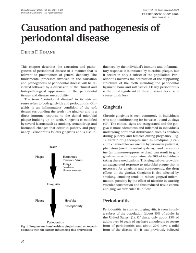
Copyright C Munksgaard 2001 Periodontology 2000, Vol. 25, 2001, 8–20 Printed in Denmark ¡ All rights reserved PERIODONTOLOGY 2000 ISSN 0906-6713 Causation and pathogenesis of periodontal disease D ENIS F. K INANE This chapter describes the causation and pathogenesis of periodontal disease in a manner that is relevant to practitioners of general dentistry. The fundamental processes involved in the causation and pathogenesis of periodontal disease will be reviewed followed by a discussion of the clinical and histopathological appearance of the periodontal tissues and disease susceptibility. The term ‘‘periodontal disease’’ in its strictest sense refers to both gingivitis and periodontitis. Gingivitis is an inflammatory condition of the soft tissues surrounding the teeth (the gingiva) and is a direct immune response to the dental microbial plaque building up on teeth. Gingivitis is modified by several factors such as smoking, certain drugs and hormonal changes that occur in puberty and pregnancy. Periodontitis follows gingivitis and is also in- fluenced by the individual’s immune and inflammatory response. It is initiated by microbial plaque, but it occurs in only a subset of the population. Periodontitis involves the destruction of the supporting structures of the teeth including the periodontal ligament, bone and soft tissues. Clearly, periodontitis is the most significant of these diseases because it causes tooth loss. Gingivitis Chronic gingivitis is seen commonly in individuals who stop toothbrushing for between 10 and 20 days (49). The clinical signs are exaggerated and the gingiva is more edematous and inflamed in individuals undergoing hormonal disturbance, such as children during puberty and females during pregnancy (Fig. 1). Certain drug therapies such as nifedipine (a calcium channel blocker used in hypertensive patients), phenytoin (used to control epilepsy), and cyclosporine (an immunosuppressive drug) can result in gingival overgrowth in approximately 30% of individuals taking these medications. This gingival overgrowth is an exaggerated response to microbial plaque that is necessary for gingivitis and consequently, the drug effects on the gingiva. Gingivitis is also affected by smoking. Smoking tends to reduce gingival inflammation, possibly by the effect of nicotine in causing vascular constriction and thus reduced tissue edema and gingival crevicular fluid flow. Periodontitis Fig. 1. Progression from health to gingivitis and on to periodontitis with the factors influencing this progression 8 Periodontitis, in contrast to gingivitis, is seen in only a subset of the population (about 35% of adults in the United States) (1). Of these, only about 13% of adults over 30 years of age have a moderate or severe form of periodontitis and about 22% have a mild form of the disease (1). It was previously believed Causation and pathogenesis of periodontal disease that all gingivitis progresses to periodontitis. However, the prevailing view is that while gingivitis must precede periodontitis, not all gingivitis progresses to periodontitis (9). Furthermore periodontitis is quite variable in that it does not affect all teeth evenly but has both a subject and site predilection. Since it is currently not feasible to diagnose the specific sites with gingivitis that will progress to periodontitis, a conservative approach is to treat all gingivitis and the corresponding primary preventive approach is to prevent the occurrence of gingivitis. Current concepts regarding the epidemiology of periodontal disease are influenced greatly by studies that suggest that relatively few subjects in each age group suffer from advanced periodontal destruction and that only specific sites in these individuals are affected (35, 63) (Fig. 1). Given the change in attachment level over time, it is also peculiar that only relatively few sites actually undergo extensive periodontal destruction within any given observation period (35, 46). Lindhe et al. (47) reported on a population of 265 subjects monitored over a twoyear period and found that 70% of the sites that deteriorated by 3 mm or more occurred in only 12% of the population. Socransky et al. (76) suggested that periodontitis progresses in episodes of exacerbation and remission and termed this the ‘‘burst hypothesis’’. More recent studies have suggested that progression may actually have a more continuous rather than episodic pattern. Periodontal disease progression can be considered a continuous process that undergoes periods of exacerbation. Therefore, both the continuous and episodic hypotheses of disease progression have merit. In summary, periodontal disease is related to the subject, with only a few individuals experiencing advanced tissue destruction around several teeth. The progression of this disease is probably continuous with brief episodes of localized exacerbation and occasional remission. Role of plaque Experimental short-term clinical studies have shown that microorganisms quickly colonize clean tooth surfaces after ceasing all oral hygiene procedures. Within a few days, microscopic and clinical signs of gingivitis become apparent. These inflammatory changes can be reversed when adequate tooth cleaning methods are resumed (Fig. 2) (49, 84). Microorganisms that form dental plaque and cause gingivitis probably do so by releasing bacterial prod- Fig. 2. Gingivitis (top) before and health (bottom) after the resolution of gingivitis following hygienic phase therapy ucts that induce tissue inflammation. Clinical trials have documented the importance of supragingival and subgingival microbial plaque in the treatment of gingivitis and periodontitis. In a classical long-term study on over 300 subjects, Axelsson et al. (2) demonstrated that preventing plaque accumulation by a variety of professional and home-based techniques was extremely effective in preventing attachment loss over a period of 15 years. Clearly, gingivitis precedes periodontitis and this implies that prevention of gingivitis is also a primary preventive measure for periodontitis (Fig. 1). As stated earlier, not all patients develop periodontitis, and for those who do, it is due to their unique host response to the microbial plaque. This therefore, provides a research challenge for those interested in the pathogenesis of this multifactorial disease. The site specificity and predilection in periodontitis and gingivitis probably relates to the retention of plaque in specific areas, such as in local areas where oral hygiene is impaired or difficult, in areas of calculus accumulation, and in areas of restoration overhangs, poor crown margins. 9 Kinane Fig. 3. Clinically healthy gingiva Overview of the pathogenesis of periodontal disease The normal healthy gingiva is characterized by its pink color and its firm consistency (Fig. 3). Interdentally, the healthy gingival tissues are firm, do not bleed on gentle probing and fill the space below the contact areas between the teeth. Healthy gingiva often exhibits a stippled appearance, and there is a knife-edge margin between the soft tissue and the tooth. In theory, the normal gingiva is free from histological evidence of inflammation, but this ideal condition is very rarely seen in microscopic tissue sections. This is because most human gingival tissues are, no matter how clinically healthy in appearance, slightly inflamed due to the constant presence of microbial plaque. Even in the very healthy state, the gingiva has a leukocyte infiltrate that is predominantly comprised of neutrophils or polymorphonuclear leukocytes. These leukocytes are phagocytes whose primary purpose is to kill bacteria after migrating through and out of the tissues into the gingival crevicular area or the gingival pocket. A subgingival plaque sample, when viewed under the microscope, will invariably show plaque microorganisms and neutrophils (Fig. 4). Neutrophils are recruited to the periodontal pocket or the gingival crevice because of attracting molecules, released by the bacteria, called chemotactic peptides. Furthermore, as bacteria damage the epithelial cells, they cause epithelial cells to release molecules termed cytokines that further attract leukocytes (predominantly neutrophils) to the crevice. The neutrophils within the crevice can phagocytose and digest bacteria and therefore, remove these bacteria from the pocket. If the neutrophil becomes overloaded with 10 bacteria, it degranulates or ‘‘explodes’’. This causes tissue damage from toxic enzymes that are released from the neutrophils. Therefore, neutrophils can be viewed as being both helpful and potentially harmful. The neutrophil defense may in some instances operate well and reduce the bacterial load and can be considered important in preventing the gingivitis lesion from becoming established. If, however, there is an overload of microbial plaque, then the neutrophils and the barrier of epithelial cells will not be sufficient to control the infection. In such instances, the gingival tissue will become very inflamed and this is clinically seen as gingivitis. Most individuals develop clinical signs of gingivitis after 10–20 days of plaque accumulation (84). Gingivitis appears as redness, swelling and an increased tendency of the gingiva to bleed on gentle probing. At this stage, gingival inflammation is reversible if the plaque is removed by effective plaque control measures (49). In the early stages of gingivitis, the clinical changes are quite subtle. However, microscopic examination of the tissues reveals marked histopathological changes. These changes include alterations in the blood vessel network and many capillary beds are open that would otherwise be closed. Serum transudate and proteins from the blood cause swelling and there is an influx of inflammatory cells or leukocytes into the tissue. The inflammatory cells include lymphocytes, macrophages and neutrophils. Macrophages and neutrophils are phagocytic cells that engulf and digest bacteria, whereas lymphocytes are Fig. 4. A gingival crevicular plaque sample viewed under the microscope showing plaque microorganisms and neutrophils Causation and pathogenesis of periodontal disease more involved in mounting an immune response against the microbes. Healthy gingiva that is completely free of histological signs of inflammation is the absolute gold standard of health. However, this standard is clinically very difficult to achieve. Some plaque accumulation occurs in even the most clean and healthy dentitions of people with clinically healthy gingiva. The early gingivitis lesion appears histopathologically to have a heavy inflammatory cell infiltration, but also has plasma cells present in low numbers. The clinically established gingivitis lesion has no bone loss or apical migration of epithelium along the root. In histopathological sections plasma cell density, that is, the density of cells that produce antibodies, is only 10–30% of the total leukocyte infiltration. Periodontitis is characterized clinically by apical migration of epithelium along the root surface or clinical attachment loss, deepened pockets, and crestal bone loss. Histopathologically, it is similar to chronic gingivitis and there is a greater that 50% density of plasma cells. Depending on the individual, progression from gingivitis to periodontitis requires varying amounts of time. Brecx et al. (8) suggests that, in the normal situation, more than 6 months may be needed for the lesion of gingivitis to change to periodontitis. Furthermore, this move from gingivitis to periodontitis only occurs in a subset of the population (1). The reason why some patients develop periodontitis more readily than others is highly elusive and thought to be multifactorial. It should be borne in mind that the changes during gingivitis, even in the prolonged established gingivitis lesion, are to a large extent reversible (Fig. 2), whereas the changes during periodontitis, the bone loss and apical migration of the epithelial attachment, are irreversible. Periodontitis is a cumulative condition, and once bone is lost, it is almost impossible to regain it, and most patients lose tooth-supporting bone incrementally over a period of years. The worst effects of periodontitis are, therefore, commonly seen in older rather than young patients who have yet to develop deep pockets, extensive bone loss, or the associated gingival recession that occurs in periodontitis. The pathogenic processes of the periodontal diseases are largely the result of the host response to microbially induced tissue destruction. These destructive processes are initiated by bacteria but are propagated by host cells. Thus, it is the host response that results in tissue destruction. The host produces enzymes that break down tissue. This is a necessary process that is initiated and controlled by the host in order to allow the tissues to retreat from the destructive lesions initiated by bacteria. The pathogenic processes in periodontal disease can be likened to a situation where the host strategy is to eventually expel the tooth so that the inflammation will be stopped. This is the ultimate tissue retreat from the microbial plaque, and once the tooth has been expelled, the lesion is finally defused. This is because there is no further tooth site for the plaque to build up on and simplistically, there is no periodontium left to be infected. Thus, the exfoliation of teeth during periodontal disease might be considered a host preventive strategy to protect against deeper infection such as osteomyelitis. Microbiology of periodontal disease About 300 or 400 bacterial species are found in the human subgingival plaque samples. Of that number, possibly 10–20 species may play a role in the pathogenesis of destructive periodontal disease (78). These bacterial species must be able to colonize in order to survive and damage the periodontal tissues. To colonize subgingival sites, microbes must be able to 1) attach to periodontal tissues, 2) multiply, 3) compete with other microbes in their habitat and 4) defend themselves from host defense mechanisms. The gingival sulcus area is a lush place for microbial growth, but in order to colonize a subgingival site, a bacterial species must overcome a number of host-derived obstacles. These include nonspecific innate defense mechanisms such as mechanical displacement and the flow of saliva and gingival crevicular fluid. Substances in the saliva and gingival crevicular fluid help to prevent colonization and can block the binding of bacterial cells to tooth and tissue surfaces. Some constituents of saliva and gingival crevicular fluid can even precipitate bacteria and directly kill them. If a bacterium successfully eludes the inhibitory factors in saliva and attaches to a surface in the subgingival area, there are other host mechanisms to overcome. These include such things as sloughing of epithelial cells if bacteria have attached to an epithelial surface, antibodies that prevent bacterial attachment, and phagocytic killing of bacteria by neutrophils. If a species successfully enters the underlying connective tissue, it faces a multitude of host immune cells including B and T lymphocytes, neutrophils, macrophages, and lymphocytes. To be successful, bacteria must have the ability to evade all these host response mechanisms. The microbes involved in periodontal disease are 11 Kinane largely gram negative anaerobic bacilli with some anaerobic cocci and a large quantity of anaerobic spirochetes. The main organisms linked with deep destructive periodontal lesions are Porphyromonas gingivalis, Prevotella intermedia, Bacteroides forsythus, Actinobacillus actinomycetemcomitans and Treponema denticola (88). P. gingivalis is more frequently detected in severe adult periodontitis, in destructive forms of disease and in active lesions than in health or gingivitis or edentulous subjects. They are reduced in numbers in successfully treated sites but are seen in sites with recurrence of disease after therapy (6, 28, 85). It has been shown that P. gingivalis induces elevated systemic and local antibody responses in subjects with various forms of periodontitis (52). P. intermedia levels have been shown to be elevated in certain forms of periodontitis (12, 81). Elevated serum antibodies to this species have been observed in some but not all subjects with refractory periodontitis (29). B. forsythus has been found in higher numbers in sites exhibiting destructive periodontal disease than in gingivitis or healthy sites (41). B. forsythus has also been detected more frequently in active periodontal lesions (13). In localized juvenile periodontitis, A. actinomycetemcomitans appears to be the predominant periodontal pathogen (89). Genco et al. (20) indicated that organisms of the normal flora play a key role in gingivitis while exogenous organisms or more unusual anaerobic organisms seem to be implicated in periodontitis. Although spirochetes such as T. denticola are notoriously difficult to culture in the laboratory, these strict anaerobes may comprise more than 30% of the periodontal subgingival microflora. The above mentioned putative pathogens are always part of a large and varied microflora found in the subgingival plaque. Some are not detected in certain sites with periodontitis and can even be completely absent in cultures from multiple sites in an untreated periodontitis patient. Tissue destruction Periodontal pathogens and other anaerobes produce a variety of enzymes and toxins that can damage the tissues and initiate inflammation (11). They also produce noxious waste products that further irritate the tissue. Their enzymes break down extracellular substances such as collagen and even host cell membranes in order to produce nutrients for their growth. Many of the microbial surface protein molecules are actually capable of inciting an immune response in the host and are also capable of creating local tissue 12 inflammation (11). Therefore, the microbes can damage the host tissues and instigate inflammatory and immune responses, but their main objective is to multiply, grow and survive within the periodontal pocket. Once the immune and inflammatory processes are initiated, various inflammatory molecules such as proteases, cytokines, prostaglandins and host enzymes are released from leukocytes and fibroblasts or structural cells of the tissues (17, 40). Proteases tend to break up the collagen structure of the tissues and thus, create spaces for further leukocyte infiltration to occur (67). The breakdown of tissue proceeds largely under control of the host (71). The periodontal tissues become loosely adapted to the tooth and the tissues become swollen and inflamed. In periodontitis, as the connective tissue attachment to the tooth is destroyed, the epithelial cells proliferate apically along the root surface and the pocket becomes deeper. As the periodontal pocket deepens, so too does the extent of the tissue inflammatory infiltrate. Moreover, osteocytes begin the destruction of bone (71). The accumulation of subgingival plaque increases and, therefore, there is a greater microbial density to further propagate this destructive periodontal lesion. As the pocket deepens, the flora becomes more anaerobic and the host response becomes more destructive and chronic. Eventually, the periodontitis lesion progresses to such an extent that the tooth is lost. Histopathological changes The major pathological features of periodontitis are the accumulation of an inflammatory infiltrate in the tissues adjacent to the periodontal pocket, breakdown of connective tissue fibers anchoring the root to gingival connective tissue and alveolar bone, apical migration of the epithelial attachment or junctional epithelium, and resorption of the marginal portion of the alveolar bone resulting in eventual tooth loss. Page & Schroeder (62) developed a system to categorize the clinical and histopathological stages of periodontal disease and defined four histopathological stages of periodontal inflammatory changes: the initial, early and established gingival lesions and an advanced periodontal lesion. It is important to note that the evidence available at that time largely consisted of animal and human adolescent biopsies. Their description, based on this material, is now considered to not be fully applicable to the normal adult situation (37). Causation and pathogenesis of periodontal disease Fig. 5. The progression from health to periodontitis – clinically and histopathologically Fig. 6. Healthy gingiva. Features of the healthy gingiva. 1) exudation of fluid into tissues and the gingival sulcus. 2) migration of leukocytes into the junctional epithelium and gingival sulcus. Kinane & Lindhe recently classified healthy gingiva into two types (37): O super healthy or ‘‘pristine’’ state, which histologically has little or no inflammatory infiltrate (Fig. 5); and O ‘‘clinically healthy’’ gingiva, which looks similar clinically, but histologically, has features of an inflammatory infiltrate (Fig. 6). In everyday clinical situations one would normally see the healthy gingiva type, and only in exceptional circumstances, such as in a clinical trial with supervised daily cleaning and professional assistance, would one see pristine gingiva. Descriptions of the initial and early lesion of gingivitis reflect the histopathological changes of the early stages of gingivitis, while the established lesion reflects the histopathology of chronic gingivitis. The advanced lesion describes the histopathological features of periodontitis and the progression of gingivitis to periodontitis. The following histopathological description by Kinane & Lindhe (37) is based on the model proposed by Page & Schroeder (62) and is summarized in Table 1. The initial lesion appears within 4 days of plaque accumulation (Fig. 7). It is not clinically visible (49) and is characterized by an acute inflammatory response to plaque accumulation (64, 87). The initial lesion is localized to the region of the gingival sulcus, and the tissues affected include a portion of the junctional epithelium and the most coronal part of the connective tissue. Dilation of the arterioles, capillaries and venules of the dento-gingival plexus is evident histopathologically. An increase in permeability of the microvascular bed also occurs. The major features consist of an increased flow or crevicular fluid, and the migration of neutrophils from the vascular plexus below the junctional and sulcular epithelium into the junctional epithelium and gingival sulcus. The inflammatory infiltrate occupies 5% to 10% of the gingival connective tissue below the epithelium; loss of collagen is localized to the inflammatory infiltrate area. This infiltrated space becomes occupied by fluid, serum proteins and inflammatory cells (64, 70). Table 1. The classification of Kinane & Lindhe (37) Clinical condition Histopathological condition Pristine gingiva Histological perfection (Fig. 6) Normal healthy gingiva Initial lesion of Page & Schroeder (Fig. 7) Early gingivitis Early lesion of Page & Schroeder (Fig. 8) Established gingivitis Established lesion with no bone loss nor apical epithelial migration (plasma cell density between 10% and 30% of leukocyte infiltrate) (Fig. 9) Periodontitis Established lesion with bone loss and apical epithelial migration from the cementoenamel junction (plasma cell density ⬎50%) (Fig. 10) 13 Kinane Fig. 7. The initial lesion. Features of the initial lesion: 1. Vasculitis of vessels below the junctional epithelium. 2. Exudation of fluid into tissues and the gingival sulcus. 3. Increased migration of leukocytes into the junctional epithelium and gingival sulcus. 4. Presence of serum proteins, especially fibrin, extravascularly. 5. Alteration of the most coronal portion of the junctional epithelium. 6. Loss of perivascular collagen. After approximately 7 days of plaque accumulation, an inflammatory infiltrate of mononuclear leukocytes develops at the site of the initial lesion as it progresses to the early lesion (Fig. 8) (7, 64, 69, 70, 74). The vessels below the junctional epithelium remain dilated, but the number increases due to the opening up of previously inactive capillary beds. Lymphocytes and macrophages predominate at the periphery of the lesion with the presence of only a sparse number of plasma cells. At this stage, the infiltrate occupies about 15% of the gingival connective tissue, with collagen destruction in the infiltrated area reaching 60–70%. The infiltrating cells take up the space created by the collagen destruction. Clinically the inflammatory changes are visible and present as edema and erythema (49). After 2 to 3 weeks of plaque accumulation, the early lesion evolves into the established lesion (Fig. 9). Clinically this lesion will exhibit more edematous swelling and could be regarded as ‘‘established’’ gingivitis. This is characterized by a further increase in the size of the affected area and a predominance of plasma cells and lymphocytes at the periphery of the 14 lesion; macrophages and lymphocytes are detectable in the lamina propria of the gingival pocket. Prominent neutrophil infiltration of the junctional and sulcular epithelial is present (8, 45, 48, 59, 74, 75). The junctional and sulcular epithelium may proliferate and migrate deeper into the connective tissue. The gingival sulcus deepens and the coronal portion of the junctional epithelium is converted into pocket epithelium. The pocket epithelium is not attached to the tooth surface and has a heavy leukocyte infiltrate, predominantly of neutrophils that eventually migrate across the epithelium into the gingival crevice or pocket. In human studies of long-term gingivitis that have lasted up to 6 months (8), a predominance of plasma cells is not commonly found. This suggests that this feature of established lesion does not occur in humans as suggested by Page & Schroeder (62), although it may occur in animals. Features of the advanced lesion include periodontal pocket formation, surface ulceration and suppuration, destruction of the alveolar bone and periodontal ligament, tooth mobility and drifting, and eventually tooth loss (Fig. 10). The advanced Fig. 8. The early lesion. Features of the early lesion: 1. Accentuation of the features described for the initial lesion. 2. Accumulation of lymphoid cells immediately below the junctional epithelium. 3. Cytopathic alterations in resident fibroblasts. 4. Further loss of the collagen fiber network of the marginal gingiva. 5. Early proliferation of the basal cells of the junctional epithelium. Causation and pathogenesis of periodontal disease titative differences that may be involved in determining the progression of gingivitis to destructive periodontitis. Seymour et al. (73) hypothesized that a change from T-cell to B-cell dominance causes periodontitis. However, Page (61) has disagreed with this view, as has Gillett et al. (21) who showed that B-cell infiltration was mainly associated with stable, non-progressive lesions in childhood gingivitis. Liljenberg et al. (44) compared plasma cell densities in sites with active progressive periodontitis and in sites with deep pockets and gingivitis but no significant attachment loss over a 2-year period. The density of plasma cells (51.3%) was very much increased in active sites as compared to inactive sites (31.0%). It is now generally accepted that plasma cells are the dominant cell type in the advanced lesion (16). Susceptibility to periodontitis Fig. 9. The established chronic gingivitis lesion. Features of the established lesion: 1. Persistence of inflammatory features. 2. Increased proportion of plasma cells but without bone loss. 3. Presence of immunoglobulins in the connective tissues, junctional epithelium and gingival crevice. 4. Continuing loss of collagen fibers and fibrous connective tissue matrix. 5. Proliferation, apical migration, and lateral extension of the junctional epithelium. 6. Early pocket formation may appear due to gingival swelling (false pocket). Periodontal disease is considered to have multiple risk factors. The term ‘‘risk factor’’ refers to ‘‘an as- lesion is characterized by the same features present in the established gingival lesion, but is accompanied by destruction of the connective tissue attachment to the root surface and the apical migration of the epithelial attachment (45, 48, 72, 73). The progression of gingivitis to periodontitis is marked by the change in T-cell to B-cell predominance. Bone destruction begins along the crest of the interdental septum around the communicating blood vessels. The epithelium proliferates apically along the root surfaces. This leads to extension of finger-like projections of pocket epithelium into the deep connective tissues. The projections are as irregular ridges with a discontinuous basal layer and are not attached to the tooth (57). Differences between chronic gingivitis and periodontitis The similarities in the inflammatory infiltrate between the stable established chronic gingivitis lesion and advanced periodontitis lesions have encouraged many investigators to look for qualitative and quan- Fig. 10. The advanced lesion – periodontitis lesion. Features of the advanced lesion: 1. Persistence of the established lesion features. 2. Extension of the lesion into alveolar bone and periodontal ligament with significant bone loss. 3. Continued loss of collagen fibers and matrix below the pocket epithelium and in the periodontal ligament space. 4. Formation of periodontal pocketing and apical migration of junctional epithelium. 15 Kinane pect of personal behavior or lifestyle, an environmental exposure, or an inborn or inherited characteristics, which on the basis of epidemiological evidence is known to be associated with a health related condition’’ (43). Risk factors are part of the causal chain for a particular disease or can lead to the exposure of the host to a disease (3). The presence of a risk factor implies a direct increase in the probability of a disease occurring. Although specific microorganisms have been considered as potential periodontal pathogens, it has become apparent that pathogens are necessary but not sufficient for disease activity to occur (77). Destructive periodontal disease is a consequence of the interaction of genetic, environmental, host and microbial factors (86). The presence of microorganisms is a crucial factor in inflammatory periodontal disease, but the progression of the disease is related to host based risk factors. Other risk factors include genetics, age, sex, smoking, socioeconomic factors and certain systemic diseases. adulthood. It thus appears that age, per se, is not a risk factor, at least for those under the age of 70 years (20). Socioeconomic status and race Older data from the U.S. Public Health Service (82, 83) indicated that periodontal disease was more severe in individuals of lower socioeconomic status. In a more recent study, when periodontal status was adjusted for oral hygiene and smoking, the association between lower socioeconomic status and more severe periodontal disease was not seen (18, 22, 23). Data from the third National Health and Nutrition Examination Survey (1988–1994) in the United States (NHANES III) indicated that destructive periodontitis was consistently more prevalent in males than females and more prevalent in blacks and Mexican Americans than in whites (1). Indeed, these investigators recommended that primary and secondary preventive measures should be specifically targeted towards these groups (1). Bacterial risk factors Examples of microbes implicated as risk factors in periodontitis are numerous. Carlos et al. (10) found that the presence of P. intermedia, along with gingival bleeding and calculus, was correlated with attachment loss in a group of Navajo adolescents aged 14 to 19 years. Grossi et al. (22) found that P. gingivalis and B. forsythus were associated with increased risk for attachment loss as a measure of periodontal disease after adjustment for age, plaque, smoking, and diabetes. Numerous additional examples exist such that the cumulative risk for a given microflora can be estimated. Age Periodontal disease prevalence is seen to increase with age (1). However, it is not clear if becoming older is related to an increased susceptibility to periodontal disease or if the cumulative effects of disease over a lifetime explain the increased prevalence of disease in older people. Horning et al. (34) stated that age is a risk factor for periodontitis, although loss of attachment and alveolar bone with age is dependent upon the presence of plaque and calculus. Holm-Pederson et al. (33) and Machtei et al. (50) suggest that up until the age of 70 years the rate of periodontal destruction is the same throughout 16 Smoking A consistent, positive association between smoking and loss of periodontal attachment has been reported and confirmed in both cross-sectional and longitudinal studies (5, 25–27, 34, 51, 65, 79). Risk for periodontitis is considerable if an individual uses tobacco products, with estimated ratios being of the order of 2.5 to 7.0 or even greater for smokers as compared with nonsmokers (68). Even when the level of plaque accumulations and gingival inflammation were not significantly different between smokers and nonsmokers, the smokers exhibited an increase prevalence as well as severity of destructive disease (5, 27, 79). Smoking impairs the outcome of both surgical (65) and nonsurgical periodontal therapy (24), and it has been suggested that since the healing and microbial response to treatment is comparable between former smokers and nonsmokers, that smoking cessation may restore normal periodontal healing. The mechanisms by which smoking leads to loss of attachment are not well understood (25). It has been noted that the occurrence, relative frequency, or combinations of microorganisms associated with periodontal destruction did not differ between groups of smokers and nonsmokers (66, 79). However, others have reported that smokers harbored significantly higher levels and were at sig- Causation and pathogenesis of periodontal disease nificantly greater risk for infection with B. forsythus than nonsmokers (90). Smokers with periodontal disease have less clinical inflammation (14) and gingival bleeding (4) compared with nonsmokers. This may be explained by the fact that one of the numerous tobacco smoke by-products, nicotine, exerts local vasoconstriction reducing blood flow, edema and clinical signs of inflammation. Systemic disease Systemic diseases can adversely effect host defense systems and therefore act as risk factors for both gingivitis and periodontitis. Depressed neutrophil number and function (as in neutropenia and ChédiakHigashi syndrome, Down’s syndrome and PapillonLefèvre syndrome) have been reported in association with severe periodontitis (19). There is positive evidence linking diabetes mellitus to increased risk for the inflammatory periodontal diseases (36, 60). All diabetic patients may be at a higher risk for periodontal disease than the population in general. However, certain subgroups including those with those with poor oral hygiene and/or poor diabetic control and diabetic complications appear to be at particularly high risk (33). In early reports, acquired immunodeficiency syndrome (AIDS) appeared to be associated with severe forms of gingivitis and periodontitis, which is often manifested as necrotizing ulcerative periodontitis. More recent cross-sectional studies however have failed to detect any differences in prevalence and severity of periodontal disease in people with AIDS compared to healthy controls (38, 42, 53, 80). Other possible risk factors for periodontal diseases Preliminary studies suggest that stress, distress, and coping behavior (56) are associated with increased severity of destructive periodontal disease. The first links made between stress and periodontal conditions were concerned with acute necrotizing ulcerative gingivitis, also called ‘‘trench mouth’’ after its diagnosis among soldiers on the front lines during the First World War (58). Clearly, acute necrotizing ulcerative gingivitis may have a stress-related causation, but there is insufficient data to substantiate the hypothesis that psychosocial factors are of causative importance in the more common inflammatory periodontal diseases. Genetics There is a growing body of evidence indicating that genetic influences have a prominent role in the periodontal diseases (30, 32, 54). Convincing evidence from twin studies has shown that genetic factors predispose people to periodontal disease (54, 55) and in the rarer and more severe types of periodontitis affecting younger age groups (early-onset periodontitis), family studies provide good evidence for a prominent genetic role (30). Researchers are currently trying to identify the genes that may be involved in the various types of periodontitis. Indeed, a gene mutation for prepubertal periodontitis was recently identified that overlaps the region of chromosome 11q14 that contains the cathepsin C gene responsible for Papillon-Lefèvre and HaimMonk syndromes (31). However, it is likely that the search for genes that are responsible for other forms of periodontitis may be hampered by the lack of efficient methods to accurately diagnose and classify the periodontal diseases and by the strong probability that multiple genes are involved. Recently much attention has been focused on genetic polymorphisms associated with the genes involved in cytokine production that have been linked to an increased risk of adult peridontitis (39). Local risk factors in periodontal disease Although general risk factors are currently gaining much attention it is important to consider that any plaque retentive factor such as restoration overhangs or deficiencies may contribute to the local risk of periodontal disease. Conclusion In adult humans a histopathologically established lesion with plasma cell predominance is likely to undergo bone loss. Susceptibility to periodontitis may be related to whether plasma cells predominate in the tissues of an individual or a site following the microbial challenge from plaque. A tendency for an individual or site to form an extensive plasma cell infiltrate may indicate an inability to defend against periodontopathogenic bacteria and, therefore, a predisposition to periodontitis. Various susceptibility factors may all interact to influence the host response generally and the immune response specifically. 17 Kinane References 1. Albandar JM, Brunelle JA, Kingman A. Destructive periodontal disease in adults 30 years of age and older in the United States, 1988–1994. J Periodontol 1999: 70: 13–29. 2. Axelsson P, Lindhe J, Nyström B. On the prevention of caries and periodontal disease. Results of a 15-year longitudinal study in adults. J Clin Periodontol 1991: 18: 182– 189. 3. Beck J. Methods of assessing risk for periodontitis and developing multifactorial models. J Periodontol 1994: 65: 468– 478. 4. Bergström J, Floderus-Myehed B. Co-twin control study of the relationship between smoking and some periodontal disease factors. Community Dent Oral Epidemiol 1983: 11: 113–116. 5. Bergström J. Cigarette smoking as risk indicator in chronic periodontal disease. Community Dent Oral Epidemiol 1989: 17: 245–247. 6. Bragd L, Dahlén G, Wikström M, Slots J. The capability of Actinobacillus actinomycetemcomitans, Bacteroides gingivalis, Bacteroides intermedius to indicate progressive periodontitis; a retrospective study. J Clin Periodontol 1987: 14: 95–99. 7. Brecx M, Gautchi M, Gehr P, Lang N. Variability of histologic criteria in clinically healthy human gingiva. J Periodontal Res 1987: 22: 468–472. 8. Brecx M, Frölicher I, Gehr P, Lang NP. Stereological observations on long term experimental gingivitis in man. J Clin Periodontol 1988: 15: 621–627. 9. Brown LJ, Löe H. Prevalence, extent, severity and progression of periodontal disease. Periodontol 2000 1993: 2: 57–71. 10. Carlos J, Wolfe M, Zambon J, Kingman A. Periodontal disease in adolescents: some clinical and microbiological correlates of attachment loss. J Dent Res 1988: 66: 1510–1514. 11. Darveau RP, Tanner A, Page RC. The microbial challenge in periodontitis. Periodontol 2000 1997: 14: 12–32. 12. Dzink J, Socransky S, Ebersole J, Frey D. ELISA and conventional techniques for identification of black-pigmented Bacteroides isolated from periodontal pockets. J Periodontal Res 1983: 18: 369–374. 13. Dzink J, Socransky S, Haffajee A. The predominant cultivable microbiota of active and inactive lesions of destructive periodontal diseases. J Clin Periodontol 1988: 15: 316– 323. 14. Feldman R, Bravacos J, Rose C. Association between smoking different tobacco products and periodontal indexes. J Periodontol 1983: 54: 481–487. 15. Friedman R, Gunsolley J, Gentry A, Dinius A, Kaplowitz L, Settle J. Periodontal status of HIV-seropositive and AIDS patients. J Periodontol 1991: 62: 623–627. 16. Garant PR, Mulvihill JE. The fine structure of gingivitis in the beagle. III. Plasma cell infiltration of the subepithelial connective tissue. J Periodontal Res 1972: 7: 161–172. 17. Gemmell E, Marshall RI, Seymour GJ. Cytokines and prostaglandins in immune homeostasis and tissue destruction in periodontal disease. Periodontol 2000 1997: 14: 112–143. 18. Genco R. Current view of risk factors for periodontal disease. J Periodontol 1996: 67: 1041–1049. 19. Genco RJ, Löe H. The role of systemic conditions and disorders in periodontal disease. Periodontol 2000 1993: 2: 98– 116. 18 20. Genco R, Zambon J, Christersson L. The origin of periodontal infections. Adv Dent Res 1988: 2: 245–259. 21. Gillett R, Cruchley A, Johnson NW. The nature of the inflammatory infiltrates in childhood gingivitis, juvenile periodontitis and adult periodontitis: immunocytochemical studies using a monoclonal antibody to HLA Dr. J Clin Periodontol 1986: 13: 281–288. 22. Grossi S, Genco R, Machtei E, Ho A, Koch G, Dunford R, Zambon J, Hausmann E. Assessment of risk for periodontal disease. I. Risk indicators for alveolar bone loss. J Periodontol 1995: 66: 23–29. 23. Grossi S, Zambon J, Ho A, Koch G, Dunford R, Machtei E, Norderyd O, Genco R. Assessment of risk for periodontal disease. I. Risk indicators for attachment loss. J Periodontol 1994: 65: 260–267. 24. Grossi SG, Zambon J, Machtei EE, Schifferle R, Andreana S, Genco RJ, Cummins D, Harrap G. Effects of smoking and smoking cessation on healing after mechanical periodontal therapy. J Am Dent Assoc 1997: 128: 99–607. 25. Haber J. Smoking is a major risk factor for periodontitis. Curr Opin Periodontol 1994: 12–18. 26. Haber J, Kent RL. Cigarette smoking in a periodontal practice. J Periodontol 1992: 63: 100–106. 27. Haber J, Wattles J, Crowley M, Mandell R, Joshipura K, Kent RL. Evidence for cigarette smoking as a major risk factor for periodontitis. J Periodontol 1993: 64: 16–23. 28. Haffajee A, Dzink J, Socransky S. Effect of modified Widman flap surgery and systemic tetracycline on the subgingival microbiota of periodontal lesions. J Clin Periodontol 1988: 15: 255–262. 29. Haffajee A, Socransky S, Dzink J, Taubman M, Ebersole J. Clinical, microbiological and immunological features of subjects with refractory periodontal diseases. J Clin Periodontol 1988: 15: 390–398. 30. Hart TC. Genetic risk factors for early-onset periodontitis. J Periodontol 1996: 67: 355–366. 31. Hart TC, Hart PS, Michalec MD, Zhang Y, Marazita ML, Cooper M, Yassin OM, Nusier M, Walker S. Localisation of a gene for prepubertal periodontitis to chromosome 11q14 and identification of a cathepsin C gene mutation. J Med Genet 2000: 37: 95–101. 32. Hassell TM, Harris EL. Genetic influences in caries and periodontal diseases. Crit Rev Oral Biol Med 1995: 6: 319– 342. 33. Holm-Pederson P, Agerbaek N, Theilade E. Experimental gingivitis in young and elderly individuals. J Clin Periodontol 1975: 2: 14–24. 34. Horning GM, Hatch CL, Cohen ME. Risk indicators for periodontitis in a military treatment population. J Periodontol 1992: 63: 297–302. 35. Jenkins WMM, Kinane DF. The ‘‘high risk’’ group in periodontitis. Br Dent J 1989: 167: 168–171. 36. Katz PP, Wirthlin MR Jr, Szupunar SM, Selby JV, Sepe SJ, Showstack JA. Epidemiology and prevention of periodontal disease in individuals with diabetes. Diabetes Care 1991: 14: 375–385. 37. Kinane DF, Lindhe J. Pathogenesis of periodontal disease. In: Lindhe J, ed. Textbook of periodontology. Copenhagen: Munksgaard, 1997. 38. Klein R, Quart A, Small C. Periodontal disease in heterosexuals with acquired immune deficiency syndrome. J Periodontol 1991: 62: 535–540. 39. Kornman KS, Crane A, Wang HY, di Giovine FS, Newman Causation and pathogenesis of periodontal disease 40. 41. 42. 43. 44. 45. 46. 47. 48. 49. 50. 51. 52. 53. 54. 55. 56. 57. MG, Pirk FW. The interleukin-1 genotype as a severity factor in adult periodontal disease. J Clin Periodontol 1997: 24: 72–77. Kornman KS, Page RC, Tonetti MS. The host response to the microbial challenge in periodontitis: assembling the players. Periodontol 2000 1997: 14: 33–53. Lai C-H, Listgarten M, Shirakawa M, Slots J. Bacteroides forsythus in adult gingivitis and periodontitis. Oral Microbiol Immunol 1987: 2: 152–157. Lamster IB, Begg MD, Michell-Lewis D, Fine JB, Grbic JT, Todak GG, el-Sadr W, Gorman JM, Zambon JJ, Phelan JA. Oral manifestations of HIV infection in homosexual men in intravenous drug users. Study design and relationship of epidemiology, clinical and immunologic parameters to oral lesions. Oral Surg Oral Med Oral Pathol Oral Radiol Endod 1994: 78: 163–174. Last J. A dictionary of epidemiology. 2nd edn. New York: Oxford University Press, 1988: 115–116. Liljenberg B, Lindhe J, Berglundh T, Dahlén G. Some microbiological, histopathological and immunohistochemical characteristics of progressive periodontal disease. J Clin Periodontol 1994: 21: 720–727. Lindhe J, Liljenberg B, Listgarten M. Some microbiological features of periodontal disease in man. J Periodontol 1980: 51: 264–269. Lindhe J, Haffajee AD, Socransky SS. Progression of periodontal disease in adult subjects in the absence of periodontal therapy. J Clin Periodontol 1983: 10: 433–442. Lindhe J, Okamoto H, Yoneyama T, Haffajee A, Socransky SS. Periodontal loser sites in untreated adult subjects. J Clin Periodontol 1989: 16: 671–678. Listgarten M, Helldén L. Relative distribution of bacteria at clinically healthy and periodontally diseased sites in humans. J Clin Periodontol 1978: 5: 115–132. Löe H, Theilade E, Jensen S. Experimental gingivitis in man. J Periodontol 1965: 36: 177–187. Machtei E, Dunford R, Grossi S, Genco R. Cumulative nature of periodontal attachment loss. J Periodontal Res 1994: 29: 361–364. Machtei E, Dunford R, Hausmann E, Grossi SG, Powell J, Cummins D, Zambon JJ, Genco RJ. Longitudinal study of prognostic factors in established periodontitis patients. J Clin Peridontol 1997: 24: 102–109. Mahanonda R, Seymour G, Powell L, Good M, Halliday J. Effect of initial treatment of chronic inflammatory periodontal disease on the frequency of peripheral blood Tlymphocytes specific to periodontopathic bacteria. Oral Microbiol Immunol 1991: 6: 221–227. Masouredis C, Katz M, Greenspan D, Herrera C, Hollander H, Greenspan J, Winkler J. Prevalence of HIV-associated periodontitis and gingivitis in HIV-infected patients attending an AIDS clinic. J Acquir Immune Defic Syndr Hum Retroviral 1992: 5: 479–483. Michalowicz BS. Genetic and heritable risk factors in periodontal disease. J Periodontol 1994: 65: 479–488. Michalowicz BS, Aeppli D, Virag JG, Klump DG, Hinrichs JE, Segal NL, Bouchard TJ Jr, Pihlstrom BL. Periodontal findings in adult twins. J Periodontol 1991: 62: 293–299. Moss M, Beck J, Kaplan B. Exploratory case-control analysis of psychosocial factors and adult periodontitis. J Periodontol 1996: 67(suppl): 1060–1069. Muller-Glauser W, Schroeder H. The pocket epithelium: a light and electron microscopic study. J Periodontol 1982: 53: 133–144. 58. Murayama Y, Kurihara H, Nagai A, Dompkowski D, Van Dyke T.. Acute necrotizing ulcerative gingivitis risk factors involving host defense mechanisms. Periodontol 2000 1994: 6: 116–124. 59. Okada H, Kid T, Yamagami H. Identification and distribution of immunocompetent cells in inflamed gingiva of human chronic periodontitis. Infect Immun 1983: 41: 365. 60. Oliver RC, Tervonen T. Periodontitis and tooth loss: comparing diabetics with the general population. J Am Dent Assoc 1993: 124: 71–76. 61. Page RC. Gingivitis. J Clin Periodontol 1986: 13: 345–355. 62. Page R, Schroeder H. Pathogenesis of inflammatory periodontal disease. A summary of current work. Lab Invest 1976: 33: 235–249. 63. Papapanou PN, Wennström JL, Grondhal K. Periodontal status in relation to age and tooth type. A cross-sectional radiographic study. J Clin Periodontol 1988: 15: 469–478. 64. Payne W, Pafe R, Ogilvie A, Hall W. Histopathologic features of the initial and early stages of experimental gingivitis in man. J Periodontal Res 1975: 10: 51. 65. Preber H, Bergström J. Effect of cigarette smoking on periodontal healing following surgical therapy. J Clin Periodontol 1990: 17: 324–328. 66. Preber H, Bergström J, Linder LE. Occurrence of periopathogens in smoker and non-smoker patients. J Clin Periodontol 1992: 19: 667–671. 67. Reynolds JJ, Meikle MC. Mechanisms of connective tissue matrix destruction in periodontitis. Periodontol 2000 1997: 14: 144–157. 68. Salvi G, Lawrence L, Offenbacher S. Influence of risk factors on the pathogenesis of periodontitis. Periodontol 2000 1997: 14: 173–201. 69. Schroeder H, Munzel-Pedrazzoli S, Page R. Correlated morphometric and biochemical analysis of gingival tissues in early chronic gingivitis in man. Arch Oral Biol 1973: 18: 899. 70. Schroeder H, Graf-de-Beer M, Attström R. Initial gingivitis in dogs. J Periodontal Res 1975: 110: 128. 71. Schwartz Z, Goultschin J, Dean DD, Boyan BD. Mechanisms of alveolar bone destruction in periodontitis. Periodontol 2000 1997: 14: 158–172. 72. Seymour G, Greenspan J. The phenotypic characterization of lymphocyte subpopulations in established human periodontal disease. J Periodontal Res 1979: 14: 39–44. 73. Seymour G, Powell R, Davies W. Conversion of a stable Tcell lesion to a progressive B cell lesion in the pathogenesis of chronic inflammatory periodontal disease: an hypothesis. J Clin Periodontol 1979: 6: 267–275. 74. Seymour G, Powell R, Aitken KJ. Experimental gingivitis in humans. A clinical and histologic investigation. J Periodontol 1983: 54: 522–531. 75. Seymour G, Powell R, Cole K, Aitken J, Brooks D, Beckham I, Zola H, Bradley J, Burns G. Experimental gingivitis in humans. A histochemical and immunological characterization of the lymphoid cell subpopulations. J Periodontal Res 1983: 18: 375–385. 76. Socransky SS, Haffajee AD, Goodson JM, Lindhe J. New concepts of destructive periodontal disease. J Clin Periodontol 1984: 11: 21–32. 77. Socransky S, Haffajee A. The bacterial aetiology of destructive periodontal disease: current concepts. J Periodontol 1992: 63: 332–331. 78. Socransky S, Haffajee A. Evidence of bacterial aetiology: a historical perspective. Periodontol 2000 1994: 5: 7–25. 19 Kinane 79. Stoltenberg Jl, Osborn JB, Pihlstrom BL, Herzberg MC, Aeppli DM, Wolff LF, Fischer GE. Association between cigarette smoking, bacterial pathogens, and periodontal status. J Periodontol 1993: 64: 1225–1230. 80. Swango P, Kleinman D, Konzelman J. HIV and periodontal health. A study of military personnel with HIV. J Am Dent Assoc 1991: 122: 49–54. 81. Tanner ACR, Haffer C, Bratthall GT, Visconti RA, Socransky SS. A study of the bacteria associated with advancing periodontitis in man. J Clin Periodontol 1979: 6: 278–307. 82. U.S. Public Health Service. National Center for Health Statistics. Periodontal Disease in Adults, United States 1960– 1962 PHS Publication no. 1000, Series 11, no. 12. Washington, DC: Government Printing Office, 1965. 83. U.S. Public Health Service. National Center for Health Statistics. Basic data on dental examination findings of persons 1–75 years; United States, 1971–1974. DHEW Publication No. (PHS) 79–1662, Series 11, no. 214. Washington, DC: Government Printing Office, 1979. 84. Van der Weijden GA, Timmerman MF, Danser MM, Nijboer A, Saxton CA, Van der Welden U. Effect of pre-experimental 20 85. 86. 87. 88. 89. 90. maintenance care duration on the development of gingivitis in a partial mouth experimental gingivitis model. J Periodontal Res 1994: 29: 168–173. van Winkelhoff AJ, van der Velden U, de Graaff J. Microbial succession in recolonizing deep periodontal pockets after single course of supra- and subgingival debridement. J Clin Periodontol 1988: 15: 116–122. Wolff L, Dahlen G, Aeppli D. Bacteria as risk markers for periodontitis. J Periodontol 1994: 65: 498–510. Zachrisson B. A histological study of experimental gingivitis in man. J Periodontal Res 1968: 3: 293–298. Zambon JJ. Periodontal diseases: microbial factors. Ann Periodontol 1996: 1: 879–925. Zambon JJ, Christersson LA, Slots J. Actinobacillus actinomycetemcomitans in human periodontal disease. Prevalence in patient groups and distribution of biotypes and serotypes within families. J Periodontol 1983: 54: 707–711. Zambon JJ, Grossi SG, Machtei EE, Ho AW, Dunford R, Genco RJ. Cigarette smoking increases the risk for subgingival infection with periodontal pathogens. J Periodontol 1996: 67(suppl): 1050–1054.
