Cardiogenic unilateral pulmonary edema: an unreported complication of a digestive endoscopic procedure
Anuncio
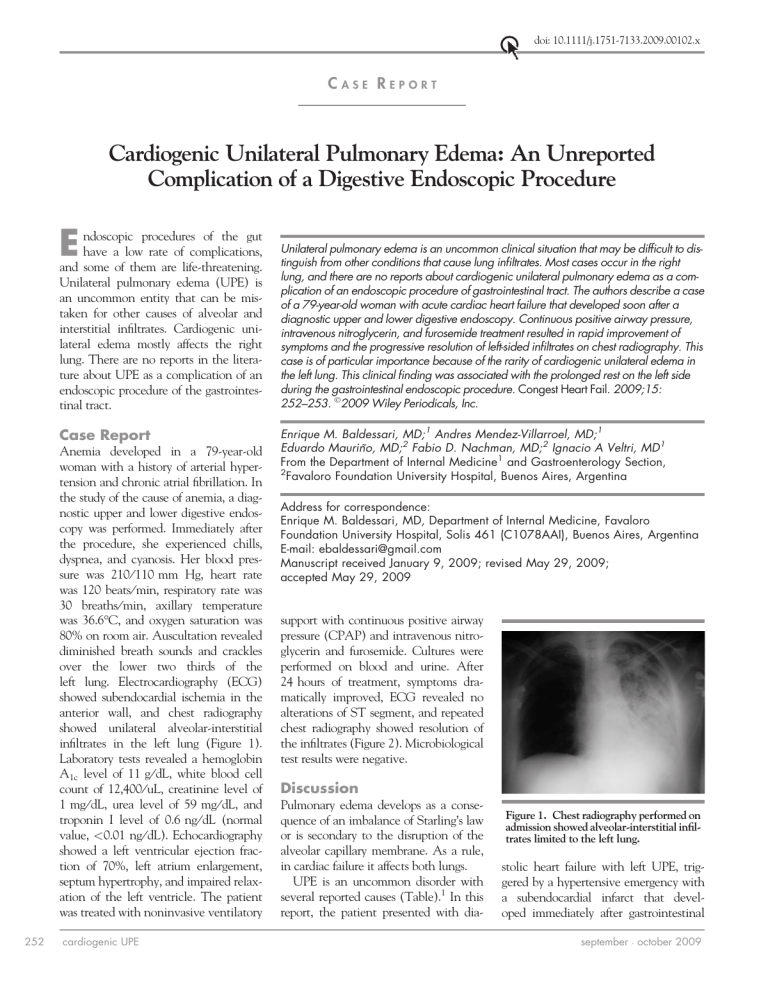
doi: 10.1111/j.1751-7133.2009.00102.x CASE REPORT Cardiogenic Unilateral Pulmonary Edema: An Unreported Complication of a Digestive Endoscopic Procedure E ndoscopic procedures of the gut have a low rate of complications, and some of them are life-threatening. Unilateral pulmonary edema (UPE) is an uncommon entity that can be mistaken for other causes of alveolar and interstitial infiltrates. Cardiogenic unilateral edema mostly affects the right lung. There are no reports in the literature about UPE as a complication of an endoscopic procedure of the gastrointestinal tract. Case Report Anemia developed in a 79-year-old woman with a history of arterial hypertension and chronic atrial fibrillation. In the study of the cause of anemia, a diagnostic upper and lower digestive endoscopy was performed. Immediately after the procedure, she experienced chills, dyspnea, and cyanosis. Her blood pressure was 210 ⁄ 110 mm Hg, heart rate was 120 beats ⁄ min, respiratory rate was 30 breaths ⁄ min, axillary temperature was 36.6C, and oxygen saturation was 80% on room air. Auscultation revealed diminished breath sounds and crackles over the lower two thirds of the left lung. Electrocardiography (ECG) showed subendocardial ischemia in the anterior wall, and chest radiography showed unilateral alveolar-interstitial infiltrates in the left lung (Figure 1). Laboratory tests revealed a hemoglobin A1c level of 11 g ⁄ dL, white blood cell count of 12,400 ⁄ uL, creatinine level of 1 mg ⁄ dL, urea level of 59 mg ⁄ dL, and troponin I level of 0.6 ng ⁄ dL (normal value, <0.01 ng ⁄ dL). Echocardiography showed a left ventricular ejection fraction of 70%, left atrium enlargement, septum hypertrophy, and impaired relaxation of the left ventricle. The patient was treated with noninvasive ventilatory 252 cardiogenic UPE Unilateral pulmonary edema is an uncommon clinical situation that may be difficult to distinguish from other conditions that cause lung infiltrates. Most cases occur in the right lung, and there are no reports about cardiogenic unilateral pulmonary edema as a complication of an endoscopic procedure of gastrointestinal tract. The authors describe a case of a 79-year-old woman with acute cardiac heart failure that developed soon after a diagnostic upper and lower digestive endoscopy. Continuous positive airway pressure, intravenous nitroglycerin, and furosemide treatment resulted in rapid improvement of symptoms and the progressive resolution of left-sided infiltrates on chest radiography. This case is of particular importance because of the rarity of cardiogenic unilateral edema in the left lung. This clinical finding was associated with the prolonged rest on the left side during the gastrointestinal endoscopic procedure. Congest Heart Fail. 2009;15: 252–253. 2009 Wiley Periodicals, Inc. Enrique M. Baldessari, MD;1 Andres Mendez-Villarroel, MD;1 Eduardo Mauriño, MD;2 Fabio D. Nachman, MD;2 Ignacio A Veltri, MD1 From the Department of Internal Medicine1 and Gastroenterology Section, 2 Favaloro Foundation University Hospital, Buenos Aires, Argentina Address for correspondence: Enrique M. Baldessari, MD, Department of Internal Medicine, Favaloro Foundation University Hospital, Solis 461 (C1078AAI), Buenos Aires, Argentina E-mail: [email protected] Manuscript received January 9, 2009; revised May 29, 2009; accepted May 29, 2009 support with continuous positive airway pressure (CPAP) and intravenous nitroglycerin and furosemide. Cultures were performed on blood and urine. After 24 hours of treatment, symptoms dramatically improved, ECG revealed no alterations of ST segment, and repeated chest radiography showed resolution of the infiltrates (Figure 2). Microbiological test results were negative. Discussion Pulmonary edema develops as a consequence of an imbalance of Starling’s law or is secondary to the disruption of the alveolar capillary membrane. As a rule, in cardiac failure it affects both lungs. UPE is an uncommon disorder with several reported causes (Table).1 In this report, the patient presented with dia- Figure 1. Chest radiography performed on admission showed alveolar-interstitial infiltrates limited to the left lung. stolic heart failure with left UPE, triggered by a hypertensive emergency with a subendocardial infarct that developed immediately after gastrointestinal september october 2009 • Figure 2. Chest radiography performed after 24 hours (A) and after 48 hours (B) of treatment showed a progressive clearing of the opacities on the left side. endoscopy. That the left lung infiltrates were cardiogenic is supported by their rapid clearing after diuretic therapy. Most cases of UPE associated with left heart failure affect the right lung.2 A possible explanation is the inferior lymphatic drainage of the right lung by the small-caliber right bronchomediastinal trunk in comparison to the large-caliber thoracic duct in the left lung.3 In patients with mitral regurgitation, the mechanism of right UPE is related to the retrograde flow in the right pulmonary veins during ventricular systole.4–6 The left cardiogenic UPE in this case would be in relation to the left lateral decubitus position adopted by the patient while the procedure was performed in the context of cardiac decompensation. Gravity raises the hydrostatic pressure in the dependent lung, impairing circulation and affecting the produc- tion of surfactant. The lateral decubitus position could compromise lung mechanisms. There is relative hyperperfusion and hypoventilation of the dependent lobes, and when volume overload or heart failure exists, it could result in pulmonary edema of the dependent lung.7 This situation usually develops in patients placed in the lateral decubitus position for a prolonged period. Case reports of left UPE were described in mechanical ventilation patients with left decubitus to promote bronchial drainage, in immobilized individuals because of neurologic deficits, and during transesophageal echocardiography.8 Conclusions Table. Reported Causes of Unilateral Pulmonary Edema Congestive heart failure Acute mitral valve regurgitation Reexpansion after pleurocentesis Prolonged rest on one side (plus cardiac failure or large aumonts of fluids) Chest trauma After talc pleurodesis Epilepsy Upper-airway obstruction Unilateral main stem intubation Neurogenic pulmonary edema Nitrogen mustard use Amiodarone-related and heroin-related pulmonary edema Pregnancy Pulmonary venous obstruction (mediastinal fibrosis or postlobectomy) Pulmonary artery compression from aortic dissection Unilateral pulmonary agenesis UPE due to left heart failure is a distinctly uncommon condition. This case is of particular importance because of the rarity of cardiogenic unilateral edema in the left lung. These clinical findings were associated with prolonged rest on the left side during the gastrointestinal endoscopic procedure. 4 7 REFERENCES 1 2 3 Agarwal R, Aggarwal A, Gupta D. Other causes of unilateral pulmonary edema Agarwal R. et al. Am J Emerg Med. 2007; 25(1):129–131. Brander L, Kloeter U, Henzen C, et al. Rightsided pulmonary oedema. Lancet. 1999; 354:1440. Nitzan O, Saliba WR, Goldstein LH, et al. Unilateral pulmonary edema: a rare presentation of congestive heart failure. Am J Med Sci. 2004;327(6):362– 364. cardiogenic UPE 5 6 Lesieur O, Lorillard R, Thi HH, et al. Unilateral pulmonary oedema complicating mitral regurgitation: diagnosis and demonstration by transoesophageal echocardiography. Intensive Care Med. 2000;26:466–470. Tomcsányi J, Arabadzisz H, Bózsik B. Left sided unilateral pulmonary oedema. Heart. 2005;91:1157. Hassan W, ElShaer F, Eid FawzyM, et al. Cardiac unilateral pulmonary edema: is it really a rare presentation? Congest Heart Fail. 2005;11(4):220–223. 8 Leeming BWA. Gravitational edema of the lungs observed during assisted respiration. Chest. 1973;64:719–722. Stienlauf S, Witzling M, Herling M, et al. Unilateral pulmonary edema during transesophageal echocardiography. J Am Soc Echocardiogr. 1998;11(5):491–493. september october 2009 • 253
