Distal Clavicle Fracture Treatment: K-Wires & Tension Bands
Anuncio
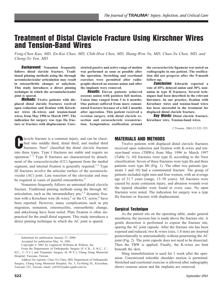
The Journal of TRAUMA威 Injury, Infection, and Critical Care Treatment of Distal Clavicle Fracture Using Kirschner Wires and Tension-Band Wires Feng-Chen Kao, MD, En-Kai Chao, MD, Chih-Hwa Chen, MD, Shang-Won Yu, MD, Chao-Yu Chen, MD, and Cheng-Yo Yen, MD Background: Nonunion frequently follows distal clavicle fracture. Traditional pinning methods using the through acromioclavicular articulation may result in osteoarthritic changes or ankylosis. This study introduces a direct pinning technique in which the acromioclavicular joint is spared. Methods: Twelve patients with displaced distal clavicle fractures received open reduction and fixation with Kirschner wires (K-wires) and tension-band wires, from May 1996 to March 1997. The indication for surgery was type IIa fracture or fracture with displacement. Unre- stricted passive and active range of motion was performed as soon as possible after the operation. Stretching and exertional exercises were permitted after radiographs showed an osseous union and after the implants were removed. Results: Eleven patients achieved osseous union with painless full motion. Union time ranged from 3 to 6 months. One patient suffered from more comminuted fracture because of a fall 2 months after operation. This patient received a revision surgery with distal clavicle resection and coracoclavicle reconstruction. Symptomless ossification around the coracoclavicle ligament was noted on radiographs in one patient. The ossification did not progress after the 9-month follow-up. Conclusion: Edwards reported a rate of 45% delayed union and 30% nonunion in type II fractures. Several techniques had been described in the relevant literature. In our practice, fixation with Kirschner wires and tension-band wires has been successful in the treatment for displaced distal clavicle fracture. Key Words: Distal clavicle fracture, Kirschner wire, Tension-band wires. J Trauma. 2001;51:522–525. C lavicle fracture is a common injury, and can be classified into middle third, distal third, and medial third fractures. Neer1 classified the distal clavicle fracture into three types. Type I fractures are stable and require no operation.1–3 Type II fractures are characterized by detachment of the coracoclavicular (CC) ligament from the medial segment, and internal fixation is generally required.1,3 Type III fractures involve the articular surface of the acromioclavicular (AC) joint. Late resection of the clavicular end may be required in cases of painful osteoarthritis.4 Nonunion frequently follows an untreated distal clavicle fracture. Traditional pinning methods using the through AC articulation, such as the intramedullary pin,1,3 dynamic fixation with a Kirschner wire (K-wire),5 or the CC screw,6 have been reported. However, many complications such as pin migration, nonunion, osteomyelitis, osteoarthritic change, and ankylosing have been noted. Plate fixation is often impractical for the small distal segment. This study introduces a direct pinning technique in which the AC joint is spared. Submitted for publication January 27, 2000. Accepted for publication May 14, 2001. Copyright © 2001 by Lippincott Williams & Wilkins, Inc. From the Departments of Orthopaedic Surgery (F.-C.K., E.-K.C., C.H.C., C.-Y.C.) and Traumatic Surgery (S.-W.Y.), Chang Gung Memorial Hospital, Taoyuan, Taiwan. Address for reprints: Chao-Yu Chen, MD, Department of Orthopaedic Surgery, Chang Gung Memorial Hospital, No. 5, Fu-Hsing St., Kweishan, Taoyuan 333, Taiwan; email: [email protected]. 522 MATERIALS AND METHODS Twelve patients with displaced distal clavicle fractures received open reduction and fixation with K-wires and tension-band wires (TBWs) from May 1996 to March 1997 (Table 1). All fractures were type II, according to the Neer classification. Seven of these fractures were type IIa and three patients were type IIb (Fig. 1). The other two patients (patients 3 and 10) had a comminuted fracture. The group of patients included eight men and four women, with an average age of 31.7 years (range, 13–58 years). All fractures were caused by acute contusion injury, and abrasion wounds over the injured shoulder were found in every case. No open fractures were noted. The indication for surgery was a type IIa fracture or fracture with displacement. Surgical Technique As the patient sits on the operating table, under general anesthesia, the incision line is made above the fracture site. A gentle dissection is performed to expose the fracture site, sparing the AC joint capsule. After the fracture site has been exposed and reduced, two K-wires (size, 1.8 mm) are inserted posterolaterally to anteromedially without penetrating the AC joint (Fig. 2). The joint capsule does not need to be dissected. Then the TBW is applied. Finally, the K-wires are bent beneath the skin. Sling immobilization is used for 1 week after the operation. Unrestricted tolerable shoulder motion is permitted. Stretched and exertional exercise is allowed after radiography shows osseous union and the implants are removed. September 2001 Treatment of Distal Clavicle Fracture Table 1 Patient Data Patient Age Type Sex (y) (Neer) Mechanism Bony Union (mo) 1 2 3 4 5 6 45 F 27 F 30 M 58 M 13 M 26 F IIa IIa II IIa IIa IIb MVC MCC MCC MCC Falling down MCC 4 3 3 5 6 3 7 8 9 16 M 46 M 23 F IIb IIa IIb MCC MCC MCC 5 4 4 10 11 32 M 22 F II IIa MVC MVC 6 — 12 34 M IIa Falling down 6 Complication — — — — — Heterotopic ossification — — Persistent AC joint pain — More comminuted fracture — MVC, motor vehicle crash; MCC, motorcycle crash. RESULTS The twelve patients were examined every month with radiography. Eleven of the 12 patients (92%) attained a bony union with a range of painless motion after an average of 4 months (range, 3– 6 months). Patient 11 suffered from comminution of the distal fragment, which was repaired by distal clavicle resection and CC reconstruction. Fig. 2. (A) Patient 9: A 23-year-old woman suffered from a right shoulder injury by falling down. The radiograph shows a right distal clavicle fracture. (B) The postoperative radiograph showing fracture fixed with K-wires and TBWs. (C) The radiograph showing a bony union of the right clavicle 6 months postoperatively. The implant had been removed 2 months previously. Fig. 1. (A) Type IIa fracture. (B) Type IIb fracture. Volume 51 • Number 3 After operation, 11 patients had a full range of motion of the shoulder joint without tenderness. One patient (patient 9) suffered from persistent pain over the AC joint, although radiographs showed bony union. Heterotopic ossification 523 The Journal of TRAUMA威 Injury, Infection, and Critical Care Fig. 4. (A) Patient 10: A 32-year-old man incurred a left shoulder injury in a traffic accident. The radiograph shows a left distal clavicle fracture with three part fragments. (B) Postoperative radiograph showing the fracture fixed with K-wires, TBWs, and an encircling wire. DISCUSSION Fig. 3. (A) Patient 6: A 26-year-old woman suffered from a right shoulder injury because of a motorcycle crash. The radiograph shows a right distal clavicle fracture. (B) The postoperative radiograph showing a fracture fixed with K-wires and TBWs. (C) The implant had been removed 9 months postoperatively. The radiograph shows heterotopic ossification around the CC ligament. around the CC ligament (Fig. 3) was noted in another patient (patient 6). This patient showed no clinical complaint. None suffered from osteoarthritis of the AC joint, wound infection, pin irritation, or pin migration. 524 Fracture of the distal end of the clavicle is not an infrequent shoulder injury,7,8 which may result either from a direct blow to the clavicle, or indirectly from a fall onto the acromion. Our patients’ fractures were all caused by acute injury, mostly from motorcycle crashes. Abrasion wounds were also noted around the fracture site. All fracture lines were oblique from the posterior side of the distal part to the anterior side of the proximal part (Fig. 2). We postulate that this type of injury is caused by a direct force, rather than by an indirect force on the acromion. This study involved eight men and four women with a mean age of 31.7 years (range, 13–58 years). Most patients were young and experienced injury because of a direct blow during a fall. In Taiwan, this type of injury happens frequently in younger motorcycle riders. Distal clavicle fractures are divided into three types according to the Neer classification.1 Many authors (e.g., Neer,2,3 Neviaser,9 Zenni et al.,6 Jager and Breither,10 Eskola September 2001 Treatment of Distal Clavicle Fracture et al.,11 and Post12) have preferred open reduction and internal fixation for type II fractures. An intramedullary pin,1,3 dynamic fixation with a K-wire,5 and the coracoclavicular screw6 have been reported to show good results. However, complications such as nonunion, osteomyelitis of the clavicle, pin migration, osteoarthritic change, and ankylosis have also been noted. Edwards13 reports 45% delayed union and 30% nonunion in type II fractures. Nordqvist14 reports 22% nonunion in type II fractures with conservative treatment. In our series, the indication of surgery is a type IIa fracture or a fracture with displacement. In the Neer series,2 10 nonunions occurred after open reduction with various techniques. Most of those needed extensive soft tissue stripping, procedures that were often complicated by infection. In our series, the use of K-wires and TBWs required only the exposure of the fracture site. The soft tissue around the clavicle incurred little damage, leading to a lower infection rate. No infections occurred in our patients. In addition, the use of K-wires and TBWs can provide a more rigid fixation than K-wires only. Rigid fixation with little complication contributes to good results. Eleven of the 12 patients (92%) in our series attained a bony union after an average of 4 months (range, 3–5 months). Only one patient suffered from loss of reduction (patient 11) because of an accident. Distal clavicle resection and CC ligament reconstruction were performed on this patient. Furthermore, our method requires no dissection of the AC joint during operation, and the K-wires are inserted without penetrating the AC joint. Therefore, postoperative osteoarthritis did not occur. During follow-up, only one patient suffered from persistent AC joint pain (patient 9). The range of motion of her shoulder joint was unlimited, no radiographic change was noted, and her daily activity was not affected. Although K-wire migration has been reported,15,16 our series showed no migration. This benefit may be because of bending of the K-wires at the distal end17 and TBWs providing better stability to prevent the K-wires from migrating. The convex appearance of the displaced fracture can be reduced and stabilized by applying TBWs on the tension side, resulting in a cosmetic appearance better than that obtained with conservative treatment alone. Volume 51 • Number 3 Traditional types of IIa and IIb fractures can be treated by K-wires and TBWs. The comminuted fracture reported by Parkes and Deland4 in 1983 requires the use of additional wire (Fig. 4). We think that this latter type of fracture requires a new classification and a different treatment. In conclusion, fixation with K-wires and TBWs is a reliable means of treating displaced distal clavicle fractures. Clinical results are encouraging. REFERENCES 1. 2. 3. 4. 5. 6. 7. 8. 9. 10. 11. 12. 13. 14. 15. 16. 17. Neer CS II. Fractures of the distal third of the clavicle. Clin Orthop. 1968;58:43–50. Neer CS II. Nonunion of the clavicle. JAMA. 1960;172:1006 –1011. Neer CS II. Fracture of the distal clavicle with detachment of the coracoclavicular ligaments in adults. J Trauma. 1963;3:79 –110. Parkes JC, Deland JT. A three-part distal clavicle fracture. J Trauma. 1983;23:437– 438. Katznelson A, Nerubay J, Oliver S. Dynamic fixation of the avulsed clavicle. J Trauma. 1976;16:841– 844. Zenni EJ Jr, Krieg JK, Rosen MJ. Open reduction and internal fixation of clavicular fractures. J Bone Joint Surg Am. 1981;63:147– 151. Bailey RW, Metten CF, O’Connor GA, Titus PD, Baril JD, Moosman DA. A dynamic method of repair for acute and chronic acromioclavicular disruption. Am J Sports Med. 1976;4:58 –71. Katznelson A, Nerubay J, Oliver S, et al. Dynamic repair of acromioclavicular dislocation. Acta Orthop Scand. 1975;46:199 –204. Neviaser JS. The treatment of fractures of the clavicle. Surg Clin North Am. 1963;43:1555–1563. Jager M, Breitner S. Therapy related classification of lateral clavicular fracture [in German]. Unfallheilkunde. 1984;87:467– 473. Eskola A, Vainionpaa S, Patiala H, Rokkanen P. Outcome of operative treatment in fresh lateral clavicular fracture. Ann Chir Gynaecol. 1987;76:167–169. Post M. Current concepts in the treatment of fractures of the clavicle. Clin Orthop. 1989;245:89 –101. Edwards DJ. Fractures of the distal clavicle: a case for fixation. Injury. 1992;23:44 – 46. Nordqvist A. The natural course of lateral clavicle fracture: 15 (11– 21) year follow-up of 110 cases. Acta Orthop Scand. 1993;64:87–91. Lyons FA, Rockwood CA. Migration of pins used in operations on the shoulder. J Bone Joint Surg Am. 1990;72:1262–1267. Nordqvist A. Shortening of clavicle after fracture: incidence and clinical significance—a 5-year follow-up of 85 patients. Acta Orthop Scand. 1997;68:349 –351. Lyons FA, Rockwood CA Jr. Current concepts review migration of pins used in operations on the shoulder. J Bone Joint Surg Am. 1990;72:1262–1267. 525
