Nature2019 Elemental signatures of Australophitecus africanus theeth reveal seasonal dietary stress
Anuncio
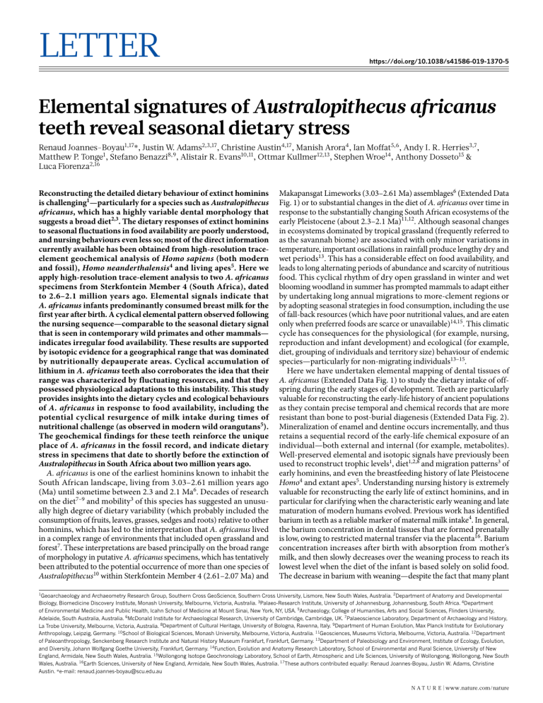
Letter https://doi.org/10.1038/s41586-019-1370-5 Elemental signatures of Australopithecus africanus teeth reveal seasonal dietary stress Renaud Joannes-Boyau1,17*, Justin W. Adams2,3,17, Christine Austin4,17, Manish Arora4, Ian Moffat5,6, Andy I. R. Herries3,7, Matthew P. Tonge1, Stefano Benazzi8,9, Alistair R. Evans10,11, Ottmar Kullmer12,13, Stephen Wroe14, Anthony Dosseto15 & Luca Fiorenza2,16 Reconstructing the detailed dietary behaviour of extinct hominins is challenging1—particularly for a species such as Australopithecus africanus, which has a highly variable dental morphology that suggests a broad diet2,3. The dietary responses of extinct hominins to seasonal fluctuations in food availability are poorly understood, and nursing behaviours even less so; most of the direct information currently available has been obtained from high-resolution traceelement geochemical analysis of Homo sapiens (both modern and fossil), Homo neanderthalensis4 and living apes5. Here we apply high-resolution trace-element analysis to two A. africanus specimens from Sterkfontein Member 4 (South Africa), dated to 2.6–2.1 million years ago. Elemental signals indicate that A. africanus infants predominantly consumed breast milk for the first year after birth. A cyclical elemental pattern observed following the nursing sequence—comparable to the seasonal dietary signal that is seen in contemporary wild primates and other mammals— indicates irregular food availability. These results are supported by isotopic evidence for a geographical range that was dominated by nutritionally depauperate areas. Cyclical accumulation of lithium in A. africanus teeth also corroborates the idea that their range was characterized by fluctuating resources, and that they possessed physiological adaptations to this instability. This study provides insights into the dietary cycles and ecological behaviours of A. africanus in response to food availability, including the potential cyclical resurgence of milk intake during times of nutritional challenge (as observed in modern wild orangutans5). The geochemical findings for these teeth reinforce the unique place of A. africanus in the fossil record, and indicate dietary stress in specimens that date to shortly before the extinction of Australopithecus in South Africa about two million years ago. A. africanus is one of the earliest hominins known to inhabit the South African landscape, living from 3.03–2.61 million years ago (Ma) until sometime between 2.3 and 2.1 Ma6. Decades of research on the diet7–9 and mobility3 of this species has suggested an unusually high degree of dietary variability (which probably included the consumption of fruits, leaves, grasses, sedges and roots) relative to other hominins, which has led to the interpretation that A. africanus lived in a complex range of environments that included open grassland and forest7. These interpretations are based principally on the broad range of morphology in putative A. africanus specimens, which has tentatively been attributed to the potential occurrence of more than one species of Australopithecus10 within Sterkfontein Member 4 (2.61–2.07 Ma) and Makapansgat Limeworks (3.03–2.61 Ma) assemblages6 (Extended Data Fig. 1) or to substantial changes in the diet of A. africanus over time in response to the substantially changing South African ecosystems of the early Pleistocene (about 2.3–2.1 Ma)11,12. Although seasonal changes in ecosystems dominated by tropical grassland (frequently referred to as the savannah biome) are associated with only minor variations in temperature, important oscillations in rainfall produce lengthy dry and wet periods13. This has a considerable effect on food availability, and leads to long alternating periods of abundance and scarcity of nutritious food. This cyclical rhythm of dry open grassland in winter and wet blooming woodland in summer has prompted mammals to adapt either by undertaking long annual migrations to more-clement regions or by adopting seasonal strategies in food consumption, including the use of fall-back resources (which have poor nutritional values, and are eaten only when preferred foods are scarce or unavailable)14,15. This climatic cycle has consequences for the physiological (for example, nursing, reproduction and infant development) and ecological (for example, diet, grouping of individuals and territory size) behaviour of endemic species—particularly for non-migrating individuals13–15. Here we have undertaken elemental mapping of dental tissues of A. africanus (Extended Data Fig. 1) to study the dietary intake of offspring during the early stages of development. Teeth are particularly valuable for reconstructing the early-life history of ancient populations as they contain precise temporal and chemical records that are more resistant than bone to post-burial diagenesis (Extended Data Fig. 2). Mineralization of enamel and dentine occurs incrementally, and thus retains a sequential record of the early-life chemical exposure of an individual—both external and internal (for example, metabolites). Well-preserved elemental and isotopic signals have previously been used to reconstruct trophic levels1, diet1,2,8 and migration patterns3 of early hominins, and even the breastfeeding history of late Pleistocene Homo4 and extant apes5. Understanding nursing history is extremely valuable for reconstructing the early life of extinct hominins, and in particular for clarifying when the characteristic early weaning and late maturation of modern humans evolved. Previous work has identified barium in teeth as a reliable marker of maternal milk intake4. In general, the barium concentration in dental tissues that are formed prenatally is low, owing to restricted maternal transfer via the placenta16. Barium concentration increases after birth with absorption from mother’s milk, and then slowly decreases over the weaning process to reach its lowest level when the diet of the infant is based solely on solid food. The decrease in barium with weaning—despite the fact that many plant 1 Geoarchaeology and Archaeometry Research Group, Southern Cross GeoScience, Southern Cross University, Lismore, New South Wales, Australia. 2Department of Anatomy and Developmental Biology, Biomedicine Discovery Institute, Monash University, Melbourne, Victoria, Australia. 3Palaeo-Research Institute, University of Johannesburg, Johannesburg, South Africa. 4Department of Environmental Medicine and Public Health, Icahn School of Medicine at Mount Sinai, New York, NY, USA. 5Archaeology, College of Humanities, Arts and Social Sciences, Flinders University, Adelaide, South Australia, Australia. 6McDonald Institute for Archaeological Research, University of Cambridge, Cambridge, UK. 7Palaeoscience Laboratory, Department of Archaeology and History, La Trobe University, Melbourne, Victoria, Australia. 8Department of Cultural Heritage, University of Bologna, Ravenna, Italy. 9Department of Human Evolution, Max Planck Institute for Evolutionary Anthropology, Leipzig, Germany. 10School of Biological Sciences, Monash University, Melbourne, Victoria, Australia. 11Geosciences, Museums Victoria, Melbourne, Victoria, Australia. 12Department of Paleoanthropology, Senckenberg Research Institute and Natural History Museum Frankfurt, Frankfurt, Germany. 13Department of Paleobiology and Environment, Institute of Ecology, Evolution, and Diversity, Johann Wolfgang Goethe University, Frankfurt, Germany. 14Function, Evolution and Anatomy Research Laboratory, School of Environmental and Rural Science, University of New England, Armidale, New South Wales, Australia. 15Wollongong Isotope Geochronology Laboratory, School of Earth, Atmospheric and Life Sciences, University of Wollongong, Wollongong, New South Wales, Australia. 16Earth Sciences, University of New England, Armidale, New South Wales, Australia. 17These authors contributed equally: Renaud Joannes-Boyau, Justin W. Adams, Christine Austin. *e-mail: [email protected] N A t U r e | www.nature.com/nature Ab ou Ab t 6–9 ou mo t1 3 m nths on t hs gp ea k e 1 mm din red om c 7Li/43Ca stf ee ilk p b a 0 Br ea fm do En Gr ow th d ire cti on ina nc e a Ab ou t Ab 12 ou mo t 9 nt m hs on th s RESEARCH Letter d 0 0.14 f as of m B re End b 138Ba/43Ca 0.005 ed ilk tfe pe min ak do pre ing e anc Low Ena Lower canine StS 51 Barium High 1 mm Upper first molar StS 28 Enamel mel 1 mm 7Li/43Ca 1 mm 0 0.004 138Ba/43Ca 0 0.12 Fig. 1 | Breastfeeding period of A. africanus. a, Section of enamel of the lower canine of StS 51, and associated 138Ba/43Ca elemental mapping. b, Section of the enamel of the upper first molar of StS 28, and associated 138 Ba/43Ca elemental mapping. The dotted lines indicate the beginning of enamel calcification, the time at which the breastfeeding peaked and the date at which the infant A. africanus breast milk intake decreased with respect to solid food intake. A period of approximately 12 and 13 months for StS 51 and StS 28, respectively, for predominant breastfeeding was estimated using the distance between the identified lines and the average rate of calcification of the species (5.5 μm d−1)16–18. Fig. 2 | Elemental mapping of A. africanus StS 28 and StS 51 fossil teeth. a, b, Micrograph (10× magnification) of StS 28C permanent first molar (a) and StS 51A permanent first premolar (P3) (b). c–f, Associated elemental mapping of 7Li/43Ca StS 28 (c) and StS 51 (d) and 138Ba/43Ca StS 28 (e) and StS 51 (f). Assuming similar timings for tooth development in A. africanus and Homo, the crown formation of the permanent first molar would correspond to an early-life record from birth to about 7 years of age, and the crown formation of the permanent first premolar would correspond to an early-life record from 1.5 years to about 5 years of age. foods having higher barium concentrations than are present in maternal milk—has previously been attributed to differences in the bioavailability of barium in milk compared with non-milk foods4. Biochemical processes that increase the bioavailability of calcium in milk probably also affect barium (because of the chemical similarity between the two elements17), which would lead to greater absorption of barium from milk compared to non-milk foods. We estimate that the mineralization of the upper and lower A. africanus first molars (M1 and M1, respectively) from specimen StS 28 and a lower canine from specimen StS 51 started soon after and about three months after birth, respectively18. To preserve these samples, thin sections of the teeth were not created and developmental timing of the tooth samples were estimated using known values18,19. Both teeth show an increasing Ba/Ca ratio from the start of mineralization, which peaks at six and nine months for StS 28 M1 molar and StS 51 lower canine, respectively, on the basis of crown formation times18,19. This indicates a diet predominated by breast milk (Fig. 1) for a minimum of 6–9 months, followed by increased supplementation with non-milk foods (which peak at around 12 months). After the initial deposition at a high Ba/Ca ratio, the elemental ratio increases and decreases in a cyclical pattern that has a period of about 4–6 months for StS 51 (Figs. 1–3, Extended Data Fig. 4) and 6–9 months for StS 28 (Figs. 1, 2, Extended Data Fig. 3). The cyclical signal is clearly visible in all of the A. africanus teeth that we analysed, with multiple occurrences from cusp to root that follow a pattern that is associated with tooth growth rather than diagenesis, and which are consistent across teeth from the same individual. Uranium (a marker of diagenesis) shows a diffuse pattern that does not follow tooth growth patterns, which is markedly different from the pattern of the Ba/Ca ratio. Although the presence of uranium indicates some post-burial alteration, the presence of barium, strontium and lithium banding that follows the developmental architecture of dentinogenesis and amelogenesis, and the repetition of the pattern across teeth from the same individual, confirms that the observed cyclical patterns are biogenic (Extended Data Figs. 3, 4, Supplementary Discussion). The highly cyclical Ba/Ca pattern observed in permanent teeth from StS 28 and StS 51 indicates a repeated behaviour over time until at least about 4–5 years of age (Figs. 1–3, Extended Data Figs. 3, 4). This pattern is reinforced by Sr/Ca and Li/Ca signals, which also occur cyclically along the growth axis of the tooth. A similar recurring pattern in Li/Ca, Ba/Ca and Sr/Ca ratios has previously been observed in modern wild orangutans (both Pongo abelii and Pongo pygmaeus)5 up to nine years of age. This pattern has been interpreted as seasonal dietary adaptation, in which Ba/Ca ratios in teeth increased when infants relied more heavily on their mother’s milk during periods of low food availability. To investigate further, we analysed teeth from several modern mammals from a similar ecological landscape (Extended Data Fig. 5) to that of A. africanus. All of these teeth also showed cyclical Ba/Ca signals. Although no reliable crown formation times could be sourced for these mammals (Supplementary Table 1), they are reported to wean at a young age (between 2 and 9 months, or up to 12 months for baboons)15. This suggests that the cyclical pattern shown in the teeth of mammals living in grassland-dominated ecosystems reflects seasonal dietary adaptations; later periods of higher Ba/Ca ratios may indicate increased consumption of another, non-milk source of highly bioavailable barium. The recurring pattern was least noticeable in carnivores, which suggests that these animals show less variation in their diet. A cycle of about eight months was estimated in the baboon, which is notably similar to that observed in A. africanus (Extended Data Fig. 6a). Additionally, the Ba/Ca value measured in the modern baboon first molar is higher than for the third molar, which may reflect a mixture of the seasonal dietary oscillation and milk intake signals in the first molar compared to only the seasonal dietary oscillation signal in the third molar (Extended Data Fig. 6b). However, the possibility that differences in mineralization and the physiological cycle during amelogenesis had a role in generating the difference in values cannot be excluded. Although the Ba/Ca banding in A. africanus appears to have greater regularity than that in the modern mammals (in particular for the StS 28 M1 molar), the number of samples and lack of accurate tooth-age estimates limits further analysis of any variation in seasonal regularity between modern environments and those of the early Pleistocene epoch. In general, Sr/Ca banding was clearer across the teeth in A. africanus and showed additional narrow lines that were not observable in the Ba/ Ca images (Extended Data Figs. 3, 4). However, the Sr/Ca and Ba/Ca patterns were largely synchronous, which indicates a common source of exposure (Extended Data Figs. 5, 6). This is in contrast to modern human samples, which often show asynchronous Ba/Ca and Sr/Ca patterns (Extended Data Fig. 7). Li/Ca banding in teeth from A. africanus N A t U r e | www.nature.com/nature Letter RESEARCH a b c 1 Transect 2 3 4 7Li/43Ca 1 mm 0 1 2 3 0.1 4 7Li/43Ca 138Ba/43Ca d 138Ba/43Ca 0 0.0035 Distance transect (mm) Fig. 3 | Elemental mapping of StS 51 A. africanus canine. a, Micrograph (10× magnification) of StS 51B permanent lower canine. b, c, Associated elemental mapping of 7Li/43Ca (b) and 138Ba/43Ca (c). Permanent canine teeth develop only a few months after birth, with completion of the crown between three and four years of age18. d, Comparison of the temporal periodicity of Ba/Ca (black line and diamonds) and Li/Ca (red line and crosses) accentuated lines (labelled 1, 2, 3 and 4), along the transect illustrated in a. Li/Ca recurrence shows a slight temporal offset compared to Ba/Ca periodicity. was less frequent than the other elemental patterns that we observed, but occurred predominantly just before the highest-amplitude Ba/Ca intake episodes (Fig. 2, Extended Data Fig. 8). Furthermore, this relationship between the two cycles was not observed in the non-primate mammals that we analysed (Extended Data Fig. 5), but was detected in both modern baboons and modern orangutans5. The presence of highly cyclical Ba/Ca and Li/Ca banding in A. africanus probably reflects seasonal dietary shifts, and perhaps also physiological responses similar to those of modern wild orangutans5. It appears that A. africanus underwent seasonal food stress, and had to adapt to changing resources and food access20. The analysis of the strontium isotope ratio (87Sr/86Sr) of A. africanus from Sterkfontein shows that some individuals lived principally on the dolomite karst, which was dominated by more-open bushland and grassland (instead of the closed woodland characteristic of surrounding lithologies)3,10,11. Our strontium isotope data (Extended Data Fig. 9) are consistent with the scenario in which both of the individuals that we analysed spent the majority of their time in the Malmani Dolomite Subgroup during amelogenesis. This specific geological setting is severely depleted in many nutrients, which limits plant growth8. Some primate species opportunistically adapted their physiology to cope with seasonal food availability due to specific environmental pressures21. Immature baboons in high-altitude environments—a challenging setting for offspring—have been reported to extend their nursing cycles, decrease their foraging time, wean later and engage in lower-energy activity than lowland baboons21. Varying milk intake can compensate for periods of extreme, and unpredictable, oscillations in food availability. This adaptation enables the survival of immature individuals, who are particularly vulnerable to fluctuations in food accessibility because of their lower fat reserves and weaker muscle tissues. During periods of abundance the infant can rely more heavily on solid food, which allows the mother to replenish her energetic and calcium reserves to support an increase in lactation during periods of food scarcity. Previous reports have suggested that wild orangutan (P. abelii and P. pygmaeus) females have adapted to seasonal variation by increasing the weaning period until an offspring age of 8–9 years old5. This ecological exigency has forced orangutan females to lower their metabolic requirements20, increase their ability to rapidly build energetic reserves22 and to catabolize fat reserves and muscle tissue faster during periods of nutrient insufficiency23. Elemental mapping of a tooth from a captive orangutan that received a constant supply of food showed a pattern that lack imprints of cyclical dietary intake, unlike the patterns of teeth from wild orangutans (Extended Data Fig. 10). Narrow bands of increased barium and strontium were observed in this captive orangutan; we attribute them to acute stress events24. Intense narrow barium and strontium bands were also observed in teeth from A. africanus, baboons and other savannah mammals; however, these bands overlap with broader dietary bands, which indicates a co-occurrence of stress and seasonal dietary changes. The overlap of possible cyclical nursing and a stressful environment due to changing food availability make it difficult to accurately interpret the underlying cause of the banding pattern (Extended Data Fig. 6a). Similarly, the Li/Ca banding pattern—which is also found in modern orangutans and (to a lesser extent) baboons, but is rarely observed in modern Homo samples (Extended Data Fig. 7) or in the non-primate mammals that we analysed—suggests complex physiological adaptations to cyclical periods of abundance and starvation. Although lithium is not directly incorporated within the fatty tissues, this element has been shown to vary in concentration with body mass25. However, the exact relationship between weight gain and lithium storage remains unclear; in modern humans, there is a possible link between lithium storage, weight gain and psychological factors (Supplementary Discussion). Another explanation could be the role of lithium in preventing protein deficiency during periods of low caloric intake26. Primates are known to switch to fall-back foods with a higher protein content during seasonal resource scarcity, to maintain their strength20,22. Moreover, lithium concentration can vary greatly between individual plants, or between parts of the same plant, and has been reported to transfer to breast milk (Supplementary Discussion). Although evidence for an adaptive shift to fall-back resources in A. africanus has been questioned8,9,27, the periodic lithium signal (which is compatible with seasonal changes28) might suggest the existence of such an adaptive trait. During periods of severe food shortage, immature australopithecines might have developed physiological adaptations to compensate for low caloric intake from fall-back resources, which perhaps included a long weaning sequence. Undoubtedly, the high Ba/Ca and Li/Ca ratio bands in A. africanus dental tissues attest to a strong seasonal oscillation in food access, which would have had a substantial effect on australopithecine development. This interpretation is reinforced by the high frequency of developmental defects in the enamel of this species, which was the result of nutritional deficiencies29. Our elemental analysis suggests A. africanus had a short period (not exceeding a year) during which breastfeeding was predominant, which is a very different sequence to that of extant great apes and instead represents a timing that is comparable with modern Homo species4,30. Because nursing and seasonal dietary banding cannot be precisely disentangled, it remains possible that the species retained a weaning sequence well into advanced offspring age to overcome seasonal food shortages, similar to modern great apes5,30. Our results have identified important dietary cycles and physiological adaptations in response to food access, which would have had considerable repercussions for the social structures and ecological behaviours adopted by groups of A. africanus. These adaptations in response to seasonal variability and resource scarcity would have extracted a toll on the resilience to other environmental pressures, and thus possibly had a role in the disappearance of the genus from the fossil record at about 2 Ma6. Online content Any methods, additional references, Nature Research reporting summaries, source data, extended data, supplementary information, acknowledgements, peer review information; details of author contributions and competing interests; and statements of data and code availability are available at https://doi.org/10.1038/s41586019-1370-5. Received: 3 October 2018; Accepted: 7 June 2019; Published online xx xx xxxx. 1. Balter, V., Braga, J., Télouk, P. & Thackeray, J. F. Evidence for dietary change but not landscape use in South African early hominins. Nature 489, 558–560 (2012). N A t U r e | www.nature.com/nature RESEARCH Letter 2. 3. 4. 5. 6. 7. 8. 9. 10. 11. 12. 13. 14. 15. 16. 17. Ungar, P. S. & Sponheimer, M. The diets of early hominins. Science 334, 190–193 (2011). Copeland, S. R. et al. Strontium isotope evidence for landscape use by early hominins. Nature 474, 76–78 (2011). Austin, C. et al. Barium distributions in teeth reveal early-life dietary transitions in primates. Nature 498, 216–219 (2013). Smith, T. M., Austin, C., Hinde, K., Vogel, E. R. & Arora, M. Cyclical nursing patterns in wild orangutans. Sci. Adv. 3, e1601517 (2017). Herries, A. I. R. et al. in The Paleobiology of Australopithecus (Vertebrate Paleobiology and Paleoanthropology Series) (eds Reed, K. E. et al.) 21–40 (Springer, Dordrecht, 2013). Peterson, A., Abella, E. F., Grine, F. E., Teaford, M. F. & Ungar, P. S. Microwear textures of Australopithecus africanus and Paranthropus robustus molars in relation to paleoenvironment and diet. J. Hum. Evol. 119, 42–63 (2018). Sponheimer, M. & Lee-Thorp, J. A. Isotopic evidence for the diet of an early hominid, Australopithecus africanus. Science 283, 368–370 (1999). Strait, D. S. et al. The feeding biomechanics and dietary ecology of Australopithecus africanus. Proc. Natl Acad. Sci. USA 106, 2124–2129 (2009). Dupont, L. M., Donner, B., Vidal, L., Pérez, E. M. & Wefer, G. Linking desert evolution and coastal upwelling: Pliocene climate change in Namibia. Geology 33, 461–464 (2005). Pickering, R. & Herries, A. I. R. in Hominin Postcranial Remains from Sterkfontein, South Africa (eds Ward, C. V. & Zipfel, B.) (Oxford Univ. Press, Oxford, in the press). Clarke, R. in The Paleobiology of Australopithecus (Vertebrate Paleobiology and Paleoanthropology Series) (eds Reed, K. et al.) 105–123 (Springer, Dordrecht, 2013). Ecker, M., Brink, J., Kolska-Horwitz, L., Scott, L. & Lee-Thorp, J. A. A. 12,000 years record of changes in herbivore niche separation and palaeoclimate (Wonderwerk Cave, South Africa). Quat. Sci. Rev. 180, 132–144 (2018). Caley, T. et al. A two-million-year-long hydroclimatic context for hominin evolution in southeastern Africa. Nature 560, 76–79 (2018). Shorrocks, B. The Biology of African Savannas (Oxford Univ. Press, New York, 2007). Krachler, M., Rossipal, E. & Micetic-Turk, D. Concentrations of trace elements in sera of newborns, young infants, and adults. Biol. Trace Elem. Res. 68, 121–135 (1999). Bouhallab, S. & Bouglé, D. Biopeptides of milk: caseinophosphopeptides and mineral bioavailability. Reprod. Nutr. Dev. 44, 493–498 (2004). N A t U r e | www.nature.com/nature 18. Smith, T. M. et al. Dental ontogeny in Pliocene and Early Pleistocene hominins. PLoS ONE 10, e0118118 (2015). 19. Lacruz, R. S., Dean, M. C., Ramirez-Rozzi, F. & Bromage, T. G. Megadontia, striae periodicity and patterns of enamel secretion in Plio-Pleistocene fossil hominins. J. Anat. 213, 148–158 (2008). 20. Pontzer, H., Raichlen, D. A., Shumaker, R. W., Ocobock, C. & Wich, S. A. Metabolic adaptation for low energy throughput in orangutans. Proc. Natl Acad. Sci. USA 107, 14048–14052 (2010). 21. van Noordwijk, M. A., Willems, E. P., Utami Atmoko, S. S., Kuzawa, C. W. & van Schaik, C. P. Multi-year lactation and its consequences in Bornean orangutans (Pongo pygmaeus). Behav. Ecol. Sociobiol. 67, 805–814 (2013). 22. Vogel, E. R. et al. Bornean orangutans on the brink of protein bankruptcy. Biol. Lett. 8, 333–336 (2012). 23. Harrison, M. E., Morrogh-Bernard, H. C. & Chivers, D. J. Orangutan energetics and the influence of fruit availability in the nonmasting peat-swamp forest of Sabangau, Indonesian Borneo. Int. J. Primatol. 31, 585–607 (2010). 24. Austin, C. et al. Uncovering system-specific stress signatures in primate teeth with multimodal imaging. Sci. Rep. 6, 18802 (2016). 25. Gitlin, M. Lithium side effects and toxicity: prevalence and management strategies. Int. J. Bipolar Disord. 4, 27 (2016). 26. Tandon, A., Bhalla, P., Nagpaul, J. P. & Dhawan, D. K. Effect of lithium on rat cerebrum under different dietary protein regimens. Drug Chem. Toxicol. 29, 333–344 (2006). 27. Wood, B. & Schroer, K. Reconstructing the diet of an extinct hominin taxon: the role of extant primate models. Int. J. Primatol. 33, 716–742 (2012). 28. Potts, R. Paleoenvironmental basis of cognitive evolution in great apes. Am. J. Primatol. 62, 209–228 (2004). 29. Guatelli-Steinberg, D. Macroscopic and microscopic analyses of linear enamel hypoplasia in Plio-Pleistocene South African hominins with respect to aspects of enamel development and morphology. Am. J. Phys. Anthropol. 120, 309–322 (2003). 30. Kennedy, G. E. From the ape’s dilemma to the weanling’s dilemma: early weaning and its evolutionary context. J. Hum. Evol. 48, 123–145 (2005). Publisher’s note: Springer Nature remains neutral with regard to jurisdictional claims in published maps and institutional affiliations. © The Author(s), under exclusive licence to Springer Nature Limited 2019 Letter RESEARCH Methods Geochemical mapping. The fossil teeth StS 28 (n = 3) and StS 51 (n = 2) (and all other specimens presented in this paper) were sectioned with a high-precision diamond saw at the apex of the cusp, then surface-polished to 10-μm smoothness (Extended Data Figs. 2, 3). Each tooth was then introduced into the laser-ablation inductively coupled-plasma mass spectrometry (LA-ICP-MS) chamber for analysis (Extended Data Fig. 4). LA-ICP-MS was used for trace-element mapping analyses of the sample, according to a published protocol previously used for Neanderthal breastfeeding analyses4. SOLARIS at Southern Cross University (ESI NWR213 coupled to an Agilent 7700 ICP-MS), and a separate system at Icahn School of Medicine at Mount Sinai in New York (ESI NWR193 ArF excimer laser coupled to an Agilent 8800 ICP-MS), were used to map the samples, with the laser beam rastered along the sample surface in a straight line. The laser spot size was 40 μm or 35 μm, the laser scan speed was 80 μm s−1 or 70 μm s−1, and the laser intensity was 80% or 60%, respectively, and an ICP-MS total integration time of 0.50 s produced data points that corresponded to a pixel size of approximately 40 × 40 μm or 35 × 35 μm, respectively. NIST610, NIST612 and NIST614 (certified standard reference materials) were used to assess signal drift. Isotopic profiling. 87Sr/86Sr was measured by a laser-ablation multi-collector inductively coupled plasma mass spectrometer (LA-MC-ICP-MS) at the University of Wollongong (WIGL laboratory) (Thermo Neptune Plus connected to an ESI NWR193 ArF excimer laser ablation system). Both enamel and dentine of StS 28 and StS 51 were analysed with nine and six 150-μm ablation spots (respectively), a dwell time of 50 s, 20-Hz frequency and 40% laser intensity. Sample aerosol was carried to the ICP-MS using a mixture of He and N2. The following isotopes were collected in static mode: 82Kr, 83Kr, 84Sr, 85Rb, 86Sr, 172Yb2+, 87Sr, 88Sr and 177Hf2+. Each isotope was collected using cycles of 1 s each. Data reduction (including a background subtraction, mass bias, Rb/Sr and Ca argide/dimer corrections) were performed using Iolite31 with the Ca–Ar interference correction Sr isotope scheme32. A seal tooth and a clam shell with modern seawater 87Sr/86Sr value (0.7092)33 were analysed four times at the start and end of each session and used as a matrix-matched primary standard to further correct the 87Sr/86Sr ratios. Image processing. For elemental maps obtained by LA-ICP-MS, ablation lines were extracted in the form of .csv files generated by the MassHunter Workstation software (Agilent). Each file was then imported into the interactive R Shiny application ‘shinyImaging’ (the application can be accessed by contacting the corresponding author or at http://labs.icahn.mssm.edu/lautenberglab/), which transforms each isotope into a separate file that contains the counts per second values of one element and is organised as a matrix (number of ablation lines multiplied by the number of ablation spots per ablation line). For each element, gas blank counts per second (median value in pixels per ablation spot) collected during the first 10 s of each analysis (gas blank) were used as background and subtracted from the rasterstack. Background signal around the teeth (arising from the encasing resin) was converted to white coloration (no intensity) to increase clarity of the figures, by isolating the dental tissue from its surroundings. Colour scales were applied using the linear blue–red Lookup Table. For Fig. 3c, the enamel and dentine of the StS 51 canine were set at a slightly different scale to enable clearer identification of the different bands, and the sliding coloured scale was adjusted accordingly. Reporting summary. Further information on research design is available in the Nature Research Reporting Summary linked to this paper. Data availability The datasets generated during and/or analysed during the current study are available from the corresponding author on reasonable request. 31. Paton, C., Hellstrom, J., Paul, B., Woodhead, J. & Hergt, J. Iolite: Freeware for the visualisation and processing of mass spectrometric data. J. Anal. Atom. Spectrom. 26, 2508 (2011). 32. Woodhead, J. D., Swearer, S., Hergt, J. & Maas, R. In situ Sr-isotope analysis of carbonates by LA-MC-ICP-MS: interference corrections, high spatial resolution and an example from otolith studies. J. Anal. Atom. Spectrom. 20, 22 (2005). 33. Veizer, J. Strontium isotopes in seawater through time. Annu. Rev. Earth Planet. Sci. 17, 141–167 (1989). 34. Kuman, K. & Clark, R. J. Stratigraphy, artefact industries and hominid associations for Sterkfontein, Member 5. J. Hum. Evol. 38, 827–847 (2000). Acknowledgements Part of this study was funded by Monash University seed grant to L.F., J.W.A., A.R.E., A.I.R.H., S.W., S.B., O.K. and R.J.-B. A.I.R.H., J.W.A. and R.J.-B. received funding from the Australian Research Council Discovery Grant DP170100056. C.A. is supported by NICHD award R00HD087523. I.M. is supported by an Australian Research Council DECRA Fellowship (DE160100703), a Commonwealth Rutherford Fellowship from the Commonwealth Scholarship Commission and a Research Associate position from Homerton College. M.A. is supported by the US National Institutes of Environmental Health Sciences Grants U2CES026561 and DP2ES025453. We thank the South African Heritage Resources Agency (SAHRA), B. Zipfel from the Evolutionary Studies Institute of the University of Witwatersrand and M. Tawane from the Ditsong National Museum of Natural History for granting the export permit for the samples for analyses; C. Lawrence and K. Simon-Menasse from Perth Zoo, who provided access to modern orangutan dental material; and K. Schultz for comments on this manuscript. Author contributions R.J.-B., J.W.A., C.A., I.M., M.A., A.I.R.H., L.F., S.B., A.R.E., O.K. and S.W. selected the samples, designed the project and/or received funding for the research. R.J.-B., C.A. and I.M. conceived and designed the experiments. R.J.-B., C.A., I.M., M.P.T. and A.D. conducted the analysis and interpreted the data. R.J.-B. prepared the figures and wrote the manuscript, with extensive contribution from C.A., M.A., I.M., L.F., A.I.R.H., J.W.A., M.P.T., S.B., A.R.E., O.K., A.D. and S.W. Competing interests The authors declare no competing interests. Additional information Supplementary information is available for this paper at https://doi.org/ 10.1038/s41586-019-1370-5. Correspondence and requests for materials should be addressed to R.J.-B. Reprints and permissions information is available at http://www.nature.com/ reprints.
