Effect of exhaustive exercise on the immune system, measured
Anuncio
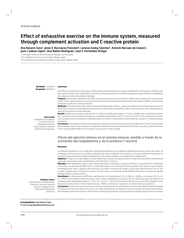
Artículo original Effect of exhaustive exercise on the immune system, measured through complement activation and C-reactive protein Ana Navarro Sanz1, Jesús E. Barruecos Francioni2, Lorenzo Godoy Sánchez3, Antonio Narvaez de Linares2, Juan L Galeas-López2, Ana Robles Rodríguez2, Juan F. Fernández Ortega2 1 Center Sport Medicine. Área de Deporte. Málaga City Hall. Spain. ICU. Hospital Universitario Carlos Haya. Málaga. Spain. 3 Clinical Laboratory. Hospital Universitario Carlos Haya. Málaga. Spain. 2 Recibido: 12.06.2013 Aceptado: 30.08.2013 Key words: Complement activation. C-reactive protein. Exhaustive exercise. Lactic acidosis. Systemic inflammatory response. Summary The influence of exercise in the systemic inflammatory response has been a subject of debate for over ten years. However very few original studies have analyzed their activation and are practically nonexistent translational studies that have extrapolated the analysis results to the reality of athletes. Purpose: We aimed to determine the effect of exhaustive exercise on two known inflammatory markers, the complement system and C-reactive protein, as a result of exhaustive exercise consisting of three intermittent bouts of 800 m at max speed interspersed with 30-s recovery intervals. Methods: Ten healthy volunteers were recruited. Anthropometric and V02max data were obtained in the laboratory and venous parameters of basal and immediate post-exercise CK, pH, lactate, complement and C-reactive protein were obtained on an open-air running track three days later. Results: Significant increases in plasma CK (114.7±80.3, p=0.005) and lactate (17.27±5.5, p=0.005) were noted. Both parameters, increased CK and lactate post-exercise, correlated significantly (r=0.751, r2=0.574, p=0.012). The complement system was activated, with the increase in CK predicting the increase in C3 (p=0.003). No post-exercise increase in C-reactive protein was found. Conclusion: The systemic inflammatory response measured in terms of increases of the complement C3 and 4 was activated by exhaustive exercise. New studies are required to determine the physiological benefit of this activation, and discriminate it from the prejudicial effects this activation could cause in other settings. Efecto del ejercicio intenso en el sistema inmune, medido a través de la activación del complemento y de la proteína C-reactiva Resumen Palabras clave: Activación del Complemento. Proteína C-reactiva. Ejercicio Intenso. Acidosis láctica. Respuesta inflamatoria sistémica. La influencia del ejercicio en la respuesta inflamatoria sistémica ha sido objeto de debate desde hace más de diez años, sin embargo son muy escasos los estudios originales que hayan analizado su activación y son practicamente inexistentes los estudios translacionales que hayan extrapolado los resultados analíticos a la realidad de los deportistas. Objetivo: El objetivo de este trabajo ha sido determinar el efecto del ejercicio intenso sobre dos marcadores inflamatorios conocidos: la activación del Complemento y de la Proteína C-reactiva. Métodos: Diez deportistas sanos y bien entrenados fueron sometidos, después de firmar el consentimiento informado, a tres series consecutivas de 800 metros a velocidad máxima con intervalos de 60 segundos entre series en pista abierta. Inmediatamente antes y después del ejercicio se tomaron muestras de sangre venosa para determinación de CK, pH, Lactato, Complemento y Proteína C-reactiva. Tres días antes, en el Laboratorio de Medicina Deportiva se realizó un estudio Antropométrico y cálculo de V02max. Resultados: Se obtuvieron incrementos significativos en CK plasmática (114,7 ± 80,3, p = 0,005) y en lactato (17,27 ± 5,5, p = 0.005) tras la realización de la prueba en pista. Ambos parámetros, el aumento de CK y lactato después del ejercicio, se correlacionaron significativamente (r = 0,751, r2 = 0,574, p = 0,012). El incremento de CK actuó como factor predictor de la elevación de la fracción C3 del Complemento (p = 0,003). La Proteína C-reactiva no se incrementó tras el ejercicio. Conclusión: El ejercicio intenso funcionó como factor de activación inmediato de la respuesta inflamatoria sistémica medida en términos de elevación de las fracciones C3 y 4 del complemento. Se requieren nuevos estudios para determinar el beneficio fisiológico de esta activación y discriminar los efectos perjudiciales que esta activación podría causar en otros entornos. Correspondencia: Ana Navarro-Sanz E-mail: [email protected] 348 Arch Med Deporte 2013;30(6):348-353 Effect of exhaustive exercise on the immune system, measured through complement activation and C-reactive protein Introduction Procedures The immune system is a complex network distributed throughout the body and composed of several cell lines and over a hundred watersoluble molecules, all intrinsically linked1, whose initial function is the restoration of homeostatic conditions and the elimination of agents that could alter homeostasis2. Despite this healing role, changes in its own self-control mechanisms have led to situations of aggression to the organism itself, as in certain autoimmune diseases3,4 or in critically ill patients in situations of multiple organ failure5, where an exaggerated response or a response maintained over time may lead to metabolic autophagy disorders or indiscriminate harm to the body's own tissues. Currently various associations are known between exercise and the immune response6. Strenuous exercise could cause activation of the immune response by cellular destruction of ischemic or traumatic origin, the purpose of which is to restore homeostasis through the recovery of injured cells and cell removal from irreversibly injured tissues7. This mechanism may activate the immune system, via the classic complement (C) pathway and other acute phase proteins involved in the immune response, such as C-reactive protein (CRP). As there is involvement of heterogeneous cell lines, with diverse molecules with a different halflife and an irregular distribution, various different measurements have been used to measure the inflammatory response to exercise. However, the various studies involve different exercise patterns and very diverse and heterogeneous populations, all of which hinder comparison of the analytical results obtained in the various clinical trials of immune activation secondary to strenuous exercise8-10. We report a study involving a group of elite, middle distance runners who ran three consecutive 800-meter series on an outdoor track at full speed, after which we analyzed muscle damage inflicted by Creatine kinase (CK) elevation and the immune response against this damage by determining the activation of the complement system and the CRP activity. Material and methods Subjects Ten healthy sports volunteers were selected for the study (5 men, 5 women). All were elite middle distance runners. Their mean age was 28.7 (4.7) years, the body mass index (BMI) was 20.87 (2.27) kg/m2, the muscle percentage was estimated at 44.76 (3.39), and the fat percentage at 13.85 (3.67). The muscle and fat percentages were estimated using the Statement of the Spanish Group of Kinanthropometry of the Spanish Federation of Sports Medicine11 and ISAK (International Society for the Advancement of Kinanthropometry)12. All the subjects had been involved in aerobic activities for at least three years, six times per week, 120-150 minutes per day. At the time of the study the subjects were at the end of the training season. All the participants were informed of the potential risks and discomfort associated with the exercise testing protocols and blood drawing, and subsequently provided written informed consent. The study comprised two phases. The first phase took place in the laboratory and the second phase took place 72 hours later on an openair running track. All the subjects already had previous experience with treadmill exercises. At the laboratory visit, an anthropometric study was made at rest and measurements were taken of blood pressure, heart rate and maximal cardiopulmonary exercise during a treadmill test. This test was performed on a motorized treadmill (PowerJog Version 5, Serie J Mod J200. Birminghan, UK). During the test a complete 12-lead ECG monitoring was done (Norav Medical Ltd 1200s v 5.0.1 Wiesbaden, Germany). The ambient temperature during the study ranged from 19 to 20ºC and the relative humidity from 55-60%. For the 24 h prior to the study the participant was required to abstain from physical exercise and the consumption of alcohol, caffeine and soft drinks. The maximum heart rate (HRmax) and maximum oxygen consumption (V02max) were determined using a ramp protocol. The treadmill inclination was set at 3% throughout the test. A three-minute warm-up period was performed at 4 km h-1 and the initial test workload was 6 km h-1. The speed was increased 2 km h-1 every two minutes until exhaustion. The tests were considered as maximal if the subjects satisfied at least one of the following two criteria: (a) maximum voluntary exhaustion defined by attaining a 10 on the Borg CR-10 scale; (b) 90% of the predicted HRmax or presence of a heart rate plateau between two consecutive work rates. The absolute V02max values were obtained by applying the ACSM equation13: 3.5 (ml kg-1 min-1) + 0.2 (speed m min-1) + 0.9 (speed m min-1) x 0.03 At the open-air running track 72 hours after the laboratory study the participants ran three bouts of 800 meters at maximum velocity, interspersed with 30-s recovery intervals. Previously they had performed a 20-minute warming period. Twenty ml of venous blood were drawn from an antecubital vein at baseline and immediately after finishing the last bouts. Whole venous blood was collected in serum-gel vacutainer tubes and allowed to clot for ~30 min. After centrifugation at 4000 rpm, one sample of serum was aliquoted, frozen and stored at −80°C for later analysis, and another sample was immediately analyzed for creatin kinase (CK), C-reactive protein (CRP), and C3 and C4. Two final samples, taken at baseline and after the test, were used for blood gases and lactate measurement in venous blood with 80 IU electrolyte-balanced heparin The CK concentration was measured by an enzymatic reaction. C3, C4 and CRP were measured by immunonephelometry (Dimension Vista, Siemens Healthcare, Diagnostic Products GmbH Marburg, Germany). The pH and pCO2 were measured by potentiometry with selective electrodes and the pO2 and lactate by amperometry with selective electrodes (both techniques from ABL Flex Radiometer, Copenhagen, Denmark). The results are expressed as –log [H+] (normal range 7.36-7.46), lactate as mM/L (normal range <2), CK as U/L (normal range <300), C3/C4 as mg/dL (normal range 60-140 and 10-40, respectively) and PCR as mg/L (normal range <2.9). Data analysis The numerical values were analyzed calculating the mean ± standard deviation (SD). Comparison of related means (SD) was done with Arch Med Deporte 2013;30(6):348-353 349 Ana Navarro Sanz, et al. the Wilcoxon non-parametric test. The correlation between variables was determined using the Pearson linear correlation test, obtaining the correlation coefficient or r regression and the r2 coefficient of determination. The Pearson and Wilcoxon tests were considered significant if the p<0.05. To study the relation of causality a multiple linear regression model was used, including the increase in C3 as the dependent variable and the percentage increase in CK, the reduction in pH and the postexercise lactate value as independent variables. The statisitcal analyses were done with SPSS v 19.0.01. Results Table 1 shows the epidemiologic results concerning age and sex distribution, expressed as individual and mean (SD), as well as the anthropometric results relating to BMI, muscle and fat percentage expressed as individual and mean (SD), as well as V02max values expressed in ml kg-1 min-1 as individual and mean (SD), and finally the individual and mean (SD) increase in heart rate and systolic blood pressure at resting and after the treadmill test. These data were all obtained in the laboratory. Table 2 shows the individual and mean (SD) results of CK and pH before and after the intermittent exercise bouts and lactate after the exercise. CK and pH variations showed significant variations before and after the intermittent exercise bouts (pH = 0.005). Lactate showed an important increase. Table 3 shows the individual and mean (SD) results of C3, C4 and CRP before and after the intermittent exercise bouts. C3 and C4 were significantly elevated after the intermittent exercise bouts (p=0.005 and p=0.014 respectively). CRP showed no increase. There is a correlation between CK percentage increase vs. lactate results after exercise, with a Pearson correlation coefficient (r) of 0.751 and a coefficient of determination (r2) of 0.574 (p=0.012) Figure 1 shows the correlation between CK percentage increase vs lactate results after the exercise, giving a Pearson correlation coefficient (r) of 0.751 and a coefficient of determination (r2) of 0.574 (p=0.012) Figure 2 shows the correlation between CK percentage increase vs C3 increase, giving a Pearson correlation coefficient (r) of 0.847 and coefficient of determination (r2) of 0.717 (p=0.002). Similar results were found after correlating the post-exercise lactate with C4 variation (r=0.689), p=0.028 No significant results were found with C4 variation vs CK percentage increase or post-exercise lactate, though there was a trend towards a positive correlation. Table 1. Epidemiological, anthropometric and clinical findings. Case Sex Age BMI % Fat % Musc V02max HR SBP M M F F M M F F M F 24 24 31 36 32 24 24 34 26 26 28.7 (4.7) 20.31 21.55 21.3 19.28 25.79 19.30 18.22 19.95 23.5 19.87 20.87 (2.2) 10.7 11.1 16.2 16.2 19.48 9.91 12.27 18.66 9.49 14.5 13.8 (3.6) 47.36 46.33 43.62 42.67 41.30 46.44 46.06 39.68 51.31 42.89 44.76 (3.4) 64.8 54.1 62.3 51.8 54 62.7 63.4 63.8 61.4 57.5 59.8 (4.7) 120 111 113 120 116 120 98 103 111 112 112 (4.1) 65 55 85 65 70 50 65 65 70 60 60 (5,5) 1 2 3 4 5 6 7 8 9 10 Mean (SD) BMI: Body Mass Index; %: muscle percentage; V02max: maximum oxygen consumption;HR: increased heart rate; SBP: increased systolic blood pressure. Table 2. Absolute values and variations in CK, pH and lactate levels before and after the three exercise bouts, expressed as mean (SD). Case 1 2 3 4 5 6 7 8 9 10 Mean (SD) CK-pre (*) CK-post (*) 281 380 124 106 105 376 92 308 676 97 254.5 (189.8) 460 588 205 167 140 577 126 410 883 116 369.2 (260.9) CK 179 208 81 61 35 201 34 102 207 19 114.7 (80.3) pH-pre (*) 7.40 7.39 7.39 7.42 7.34 7.36 7.34 7.38 7.34 7.33 7.37 (0.02) *statistically significant 350 Arch Med Deporte 2013;30(6):348-353 pH-post (*) 7.00 7.06 7.10 7.16 7.27 7.08 7.15 7.21 7.04 7.10 7.10 (0.03) pH Lact-post 0.40 0.33 0.29 0.26 0.07 0.28 0.19 0.17 0.30 0.23 0.27 (0.09) 22 22 17 14.4 12.2 21 12.3 10.3 18 13.5 17.27 (5.5) Effect of exhaustive exercise on the immune system, measured through complement activation and C-reactive protein Table 3. C3, C4 and CRP before and after the test, plus the increase from the first to the second measurement, expressed as mean ± standard deviation. Case C3-pre(*) 1 2 3 4 5 6 7 8 9 10 Mean (SP) 87 80 88 93 88 103 86 72 84 96 87,70 (±8,52) C3-post(*) C3 156 69 114 34 154 66 161 68 110 22 124 21 101 15 76 4 109 25 110 14 124,5 (±28,16) 36,8 (±26,41) C4-pre(*) C4-post(*) C4 PCR- pre PCR- post 16 21 30 24 20 12 18 17 27 13 19,8 (±5.84) 25 33 46 39 23 13 20 18 34 11 25,44 (±11,9) 9 12 16 15 3 1 2 1 7 -2 5,77 (±6,41) <2,9 <2,9 <2,9 <2,9 <2,9 <2,9 <2,9 <2,9 <2,9 <2,9 <2,9 <2,9 <2,9 <2,9 <2,9 <2,9 <2,9 <2,9 <2,9 <2,9 <2,9 <2,9 C3-pre: C3 before the test; C3-post: C3 after the test; C3: increase in C3; C4-pre: C4 before the test; C4-post: C4 after the test; C4: increase in C4; PCR-pre: PCR before the test; CRP-post: CRP after the test; *significant pre-post difference (p<0.05). Figure 1. Correlation coefficient between the percentage increase in CK and the post-exercise lactate values. There was a significant correlation between the two variables, with a Pearson correlation coefficient = 0.75 and a determination coefficient = 0.574 (p = 0.012). Figure 2. Correlation coefficient between the percentage increase in CK and the variation in C3 after the exercise. There was a significant correlation between the two variables, r = 0.847 and r2 = 0.717, (p = 0.002). The multiple linear regression model showed that the percentage increase in CK predicted the percentage increase in C3 (p = 0.003). No other results are associated. do not necessarily have a clinical impact in healthy athletes with a normal baseline immune system. Traditionally, the difference has been attributed to whether the exercise is performed in exhaustive conditions or aerobic conditions for a non-competitive purpose, which have very different metabolic and hormonal environments. Our study population comprised healthy, young, well-trained runners in an excellent physical condition, as can be seen from the anthropometric and maximal oxygen consumption reported in Table 1. The runners exercised strenuously, evidenced by the remarkable change in blood pH (resulting from the increased lactic acid) and the post-exercise elevation in CK (see Table 2). Other authors have tried to correlate decreases in pH with the ability to recover, using this decrease as an important indicator to avoid Discussion The effect of strenuous exercise on the immune system has been of great interest for over a decade. An activating as well as a depressing effect of the immune response has been attributed to exercise activity, without clarifying which is predominant14. Extrapolating the experimental findings to clinical experience is even more difficult because significant changes in the immune response found in the laboratory Arch Med Deporte 2013;30(6):348-353 351 Ana Navarro Sanz, et al. overtraining or excessive exercise-induced muscle damage, leading to a lower athletic performance in subsequent sessions15. However, as pH measurement is an invasive procedure requiring drawing of blood immediately after the exhaustive exercise, noninvasive determinations, such as near-infrared reflectance spectroscopy, have been sought, with some authors finding good correlations16. Nevertheless, we believe that these new tests require further validation since their methodology is complex, making them difficult to perform on an outdoor track and in the laboratory. Some authors have studied the behavior of the pH within the exercise-induced damaged muscle, in which even more severe decreases in pH have been observed than those found by us in venous blood17. However, comparisons cannot be made between the two measurments (intracellular and extracellular) as it has long been known that intracellular pH levels are lower than extracellular pH levels, even at rest18. Other authors have observed changes in the plasma concentration of CK after strenuous exercise19-21, results that are consistent with ours. In our study, the increase in CK would have been more marked if serial measurements had been made during the first 24 hours, given the kinetics of CK in the presence of muscular damage of any origin22. In some extreme cases, the CK increase has resulted in rhabdomyolysis complicated by acute renal failure23. Anyway, the increase in CK in our participants was sufficient to show muscle damage secondary to exercise. The correlation found between the percent increase in CK and degree of metabolic acidosis, measured in terms of pH and lactic acid, suggests that the muscular damage may result from the hypoxia and anaerobic metabolism to which the muscle is exposed, and not just from the local traumatic damage resulting from stroke. However, we have found no publications examining the relations between CK and pH comparing types of exercises with (running) and without (cycling) direct muscle trauma. Our results showed a significant correlation between the percent increase in CK and post-exercise lactate, though it is not possible to determine whether the CK increase was secondary to lactic acidosis or to the exercise-induced trauma. The inflammatory response is made up of a dense network of substances, mostly hepatic synthetic polypeptides1, with multiple modulating and overlapping functions. It is to notable that such different processes as are strenuous exercise or a serious systemic infection both activate the same mediators, with sometimes paradoxical results. Thus, a prolonged inflammatory response over time can cause tissue damage that magnifies and perpetuates the inflammatory response and may finally lead to the systemic inflammatory response syndrome (SIRS), multiple organ failure (MOF) and even death of the individual24 whereas the body's inflammatory response should restore normal conditions. Understanding the intensity of the response and its evolution over time could be helpful in directing the body's inflammatory response to various insults25, and such knowledge could lead to lines of treatment of certain diseases caused by external aggression, as well as to better comprehension of the role of the different components of the inflammatory response26. The CRP levels before and after the test were <2.9 mg/L, compatible with the normal reference values in our laboratory. CRP was first isolated in 1930, in response to pneumococcal polysaccharide C27, and it has 352 been implicated in many proinflammatory functions, especially at the beginning of the chain of recognition of aggressions28 and activation of the complement system and other proinflammatory cytokines29. It is striking that no increase was detected in our population, suggesting that our model of aggression does not activate CRP production or that its elevation takes place later. Other authors have measured its concentration several hours after the conclusion of vigorous exercise and found it significanty elevated30 or slightly elevated31. In our series the rise in the complement system without an increase in CRP suggests independent CRP pathways of complement activation. The C system is a complex network of about 30 substances, most of them soluble and some membrane-bound macrophages32, whose function is the recognition of foreign substances, opsonization of the macrophage-lymphocyte system and the destruction of invading microorganisms or the damaged cells themselves. Three activation pathways are recognized: (a) the classical pathway, activated by immunocomplexes, (b) the alternative pathway, which does not require the formation of immunecomplexes and can be activated by viruses, cell fractions, or other acute phase proteins, and (c) the pathway initiated by the mannose-binding lectin33. The C3 and C4 fractions of the complement system act as opsonizing and activating agents of the cellular and molecular pathway of inflammation. We chose to study the C3 and C4 fractions because they are readily measurable in the clinical laboratory, they are part of the immune response activated by attacks of various kinds, not just microbial, and because they are the acute-phase proteins that have been best studied. All our patients had normal baseline C3 and C4 values. An increase >25%, which is the minimum increase to be considered as an acutephase protein34, was observed after the three intermittent bouts. C activation after exercise has been considered an inductor of the chronic fatigue syndrome35, though it has been found to be increased in healthy youngs males after prolonged exercise36. Our results suggest the C system is one of the activation pathways of immunity, aimed at restoring homeostasis after strenuous exercise. However, further studies are needed to understand the chronological evolution of C activation and its definitive role in repairing damaged tissues. A correlation was observed between the elevations of CK and C3, suggesting that the intensity of the C system activation is correlated with the intensity of muscle damage, as measured in terms of CK elevation. Furthermore, the multiple regression model showed a significant correlation, indicating that the increase in CK is causing the activation of C3. The relation between CK and C4, however, was not significant, though the trend was similar. We believe that inclusion of more cases would have resulted in a significant association. A limitation of our study is that the measurements were not repeated at different times. This would have helped to know the chronological intensity of the activation, but this would require serial extraction of blood. It should be noted, though, that the participants undertook strenuous exercise every day, so the baseline values reported were also the values found within 24 hours of completing the exercise of the day before, by which time they had returned to normal, suggesting the response is transient. What we do have, however, is a reflection of the activated immune system immediataly after exhaustive exercise. Arch Med Deporte 2013;30(6):348-353 Effect of exhaustive exercise on the immune system, measured through complement activation and C-reactive protein Conclusions 14. Shephard RJ. Development of the discipline of exercise immunology. Exerc Immunol Rev. 2010;16:194-222 Exhaustive exercise triggers the immune response, expressed as C system activation. A correlation exists between this activation and the degree of intensity of effort, measured by increases in lactate and CK. Further studies are necessary to determine the chronological course of this activation, and translational studies are required to study the role of C activation and other inflammatory markers in homeostasis recovery, or whether in fact they increase the muscle damage and are detrimental for exercise performance. Determining the inflammatory activation in response to a serious injury in our group of sportspersons could in the future help to discriminate between the positive and the negative effects of complement activation in other setting of patients. Conflict of interest 15. Del Coso J, Hamouti N, Aguado-Jimenez R, Mora-Rodriguez R. Restoration of blood pH between repeated bouts of high-intensity exercise: effects of various active-recovery protocols. Eur J Appl Physiol. 2010;108:523-32 16. Yang Y, Soyemi OO, Landry MR, Soller BR. Noninvasive in vivo measurement of venous blood pH during exercise using near-infrared reflectance spectroscopy. Applied Spectroscopy. 2007;61:223-29. 17. Davies RC, Eston RG, Fulford J, Rowlands AV, Jones AM. Muscle damage alters the metabolic response to dynamic exercise in humans: P-MRI study. J of Applied Physiology 2011;111:782-90. 18. Adler S. The simultaneous determination of muscle cell pH using a weak acid and weak base. J Clin Invest. 1972;51:256-65. 19. Bruunsgaard H, Galbo H, Halkjaer-Kristensen J, Johansen TL, McLean DA, Pedersen BK. Exercise induced increase in serum interleukine-6 in humans is related to muscle damage. J of Physiology.1997;499:833-41. 20. Pournot H, Bieuzen F, Louis J, Fillard JR, Barbiche E, Hausswirth C. Time-Course of Changes in Inflammatory Response after Whole-Body Cryotherapy Multi Exposures following Severe Exercise. PLoS ONE 2011; http://www.plosone.org/article/ info:doi/10.1371/journal.pone.0022748 21. Schiff HB, MacSearraigh ET, Kallmeyer JC. Myoglobinuria, rhabdomiolysis and marathon running. Q J Med. 1978;47:463-72. The authors have no conflicts of interest that are directly or indirectly relevant to the content of this paper. 22. Byrne Ch, Eston R. The effects of exercise-induced muscle damage on isometric and dynamic knee extensor strength and vertical jump performance. J of Sports Sciences. 2002;20:417-25. 23. Moghtader J, Brady WJ, Bradio WW. Exertional rhabdomyolysis in an adolescent athlete. Ped Emerg Care. 1997;13:382-5. References 1. Gabay C, Kushner I. Acute-Phase Proteins and Other Systemic Responses to Inflammation. The New Eng J of Med. 1999;340:448-54. 24. Sakr Y, Reinhart K, Bloos F, Marx G, Russwurm S, Bauer M, et al. Time course and relationship between plasma selenium concentrations, systemic inflammatory response, sepsis, and multiorgan failure. Br J Anaesth. 2007;78:775-84. 2. Abbas AK, Lichtman AH, Pillai S. Cellular and Molecular Immunology. 6th ed. St Louis: Saunders; 2007. 25. Kushner I. Semantics, inflammation, cytokines and common sense. Cytokine Growth Factor Rev. 1998;9:191-6. 3. Mondgil KD, Choubey D. Cytokines in Autoimmunity: Role in Induction, Regulation and Treatment. J of Interferon and Cytokine Research. 2011;31:695-703. 26. Neubauer O, Reichhold S, Nersesyan A, Konig D, Wagner KH. Exercise-induced DNA damage: Is there a relationship with inflammatory responses?. Exerc Immunol Rev. 2008;14:51-72. 4. Kollias G, Doumi E, Kassiotis G, Kontoyannis D. On the role of Tumor Necrosis Factor and receptors in models of Multiorgan failure, Rheumatoid arthritis, multiple sclerosis and inflammatory bowell disease. Immunol Rev. 1999;169:175-94. 27. Tillett WS, Francis T Jr. Serological reactions in pneumonia with nonprotein somatic fraction of pneumoccoccus. J Exp Med. 1930;52:561-71. 5. Bone RC. Towards a theory regarding the pathogenesis of the systemic inflammatory response syndrome: what we do and don`t know about cytokine regulation. Crit Care Med. 1996;24:163-7. 28. Volanakis JE. Acute phase proteins in rheumatic disease. In: Koopman WJ, ed. Arthritis and allied conditions: a textbook of rheumatology, 13th ed. Baltimore, Williams & Wilkins; 1997: 505-14. 6. Walsh NP, Gleeson M, Shephard R, Woods JA, Bishop NC, Fleshner M, et al. Position Statement Part One: Immune function and exercise. Exerc Immunol Rev. 2011;17:6-63. 29. Ballou SP, Lozanski G. Induction of inflammatory cytokine release from cultured human monocytes by C-reactive protein. Cytokine. 1992;4:361-8. 7. Peake J, Nosaka K, Suzuki K. Characterization of inflammatory responses to eccentric exercise in humans. Exercise Immunol Rev. 2005;11:64-85. 30. Castell LM, Poortmans JR, Leclercq R, Brasseur M, Duchateau J, Newsholme EA. Some aspects of the Acute Phase Response after a marathon race, and the effects of glutamine supplementation. Eur J Appl Physiol Occup Physiol. 1997;75:47-53. 8. Bortolon JR, Silva Junior AJ, Murata GM, Newsholme P, Curi R, Pithon-Curi TC, et al. Persistence of inflammatory response to intense exercise in diabetic rats. Exp Diabetes Res. (Epub 2012 Aug 13) DOI 10.1155/2012/213986. 9. Lu J, Zhang HL, Yin ZZ, Tu Y, Li ZG, Zhao BX, et al. Moxibustion attenuates inflammatory response to chronic exhaustive exercise in rats. Int J Sports Med. 2012;33:580-5. 10. Nieman DC, Konrad M, Henson DA, Kennerly K, Shanely RA, Wallner-Liebmann SJ. J Interferon Cytokine Res. 2012;32:12-7. 11. Grupo Español de Cineantropometria (GREC). Body composition assessment in Sports Medicine. Statement of Spanish group of kinanthropometry of Spanish federation of sports medicine. Arch Med Deporte. 2009;131:166-79. 12. Stewart A, Marfell-Jones, M OldsT, De Ridder H. International standards for anthropometric assessment ISAK: Lower Hutt, New Zealand, 2011. 13. ACSM (2009) ACSM´s guidelines for exercise testing and prescription. Baltimore: Lippincott, 2009 31. Skarpanska-Stejnborn A, Pilaczynska-Szczesniak L, Basta P, Deskur-Smielecka E, Slubowska Woitas Z. Effects of oral supplementation with plant extract superoxide dismutase redox on selected parameters and an inflammatory marker in a 2000-mrowing ergometer test. Int J Sport Nutr Exerc Metab. 2011;21:124-34. 32. Frank MM, Fries LF. The role of complement in inflammation and phagocytosis. Immunol Today. 1991;12:322-6. 33. Gasque Ph. Complement: a unique innate immune sensor for danger signals. Molecular Immunology. 2004;41:1089-98. 34. Morley JJ, Kushner I. Serum C-reactive protein levels in disease. Ann NY Acad Sci. 1982;389:406-18. 35. Sorensen B, Jones JF, Vernon SD, Rageevan MS. Transcriptional control of complement activation in an exercise model of chronic fatigue syndrome. Mol Med. 2009;15:34-42. 36. Dufaux B, Order U. Complement activation after prolonged exercise. Clin Chim Acta. 1989;179:45-9. Arch Med Deporte 2013;30(6):348-353 353
