Cuerpo extraño órbito
Anuncio
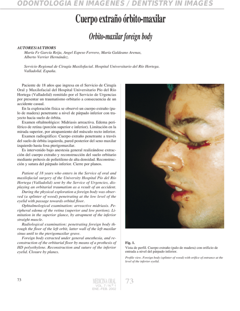
ODONTOLOGÍA EN IMÁGENES / DENTISTRY IN IMAGES Cuerpo extra–o —rbito-maxilar Orbito-maxilar foreign body AUTORES/AUTHORS Mar’a Fe Garc’a Reija, Angel Espeso Ferrero, Mar’a Galdeano Arenas, Alberto Verrier Hern‡ndez. Servicio Regional de Cirug’a Maxilofacial. Hospital Universitario del R’o Hortega. Valladolid. Espa–a.Universidad de Santiago de Compostela. Espa–a. Paciente de 18 a–os que ingresa en el Servicio de Cirug’a Oral y Maxilofacial del Hospital Universitario P’o del R’o Hortega (Valladolid) remitido por el Servicio de Urgencias por presentar un traumatismo orbitario a consecuencia de un accidente casual. En la exploraci—n f’sica se observ— un cuerpo extra–o (palo de madera) penetrante a nivel de p‡rpado inferior con trayecto hacia suelo de —rbita. Examen oftalmol—gico: Midriasis arreactiva. Edema perifŽrico de retina (porci—n superior e inferior). Limitaci—n en la mirada superior, por atrapamiento del mœsculo recto inferior. Examen radiogr‡fico: Cuerpo extra–o penetrante a travŽs del suelo de —rbita izquierda, pared posterior del seno maxilar izquierdo hasta fosa pterigomaxilar. Es intervenido bajo anestesia general realiz‡ndose extracci—n del cuerpo extra–o y reconstrucci—n del suelo orbitario mediante pr—tesis de polietileno de alta densidad. Reconstrucci—n y sutura del p‡rpado inferior. Cierre por planos. Patient of 18 years who enters in the Service of oral and maxilofacial surgery of the University Hospital P’o del R’o Hortega (Valladolid) sent by the Service of Urgencies, displaying an orbitarial traumatism as a result of an accident. During the physical exploration a foreign body was observed (a splinter of wood) penetrating at the low level of the eyelid with passage towards orbital floor. Ophtalmological examination: arreactive midriasis. Peripheral edema of the retina (superior and low portion). Limitation in the superior glance, by atrapment of the inferior straight muscle. Radiological examination: penetrating foreign body through the floor of the left orbit, latter wall of the left maxilar sinus until to the pterigomaxilar grave. Foreign body extracted under general anesthesia, and reconstruction of the orbitarial floor by means of a prothesis of HD polyethylene. Reconstruction and suture of the inferior eyelid. Closure by planes. Fig. 1. Vista de perfil. Cuerpo extra–o (palo de madera) con orificio de entrada a nivel del p‡rpado inferior. Profile view. Foreign body (splinter of wood) with orifice of entrance at the level of the inferior eyelid. 73 MEDICINA ORAL VOL. 7 / N.o 1 ENE.-FEB. 2002 73 GARCêA A, y cols. Fig. 2. Vista de frente. La direcci—n del cuerpo extra–o es hacia el suelo de —rbita. Fig. 3. Exploraci—n oftalmol—gica: midriasis arreactiva y atrapamiento del recto inferior. Front view. Foreign body towards the orbit floor. Ophtalmologic exploration: arreactive midriasis and atrapament of the inferior rectum muscle. Fig. 4. Tomograf’a computerizada: Cuerpo extra–o penetrante a travŽs del suelo de —rbita izquierda, pared posterior del seno maxilar izquierdo hasta fosa pterigomaxilar. Figs. 5 Cuerpo extra–o que mide 20 cm de largo. Foreign body of 20 cm length. Computerised tomography: penetrating foreign body through the floor of the left orbit, later wall of the left maxilar sinus until to the pterigomaxilar grave. 74 MEDICINA ORAL VOL. 7 / N.o 1 ENE.-FEB. 2002 74
