hyperresponsiveness is only resolved after many months. In most
Anuncio
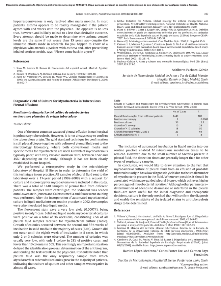
Documento descargado de http://www.archbronconeumol.org el 18/11/2016. Copia para uso personal, se prohíbe la transmisión de este documento por cualquier medio o formato. 307 Letters to the Editor / Arch Bronconeumol. 2009;45(6):306-310 hyperresponsiveness is only resolved after many months. In most patients, asthma appears to be readily manageable if the patient agrees with and works with the physician. The opposite is no less true, however, and is likely to lead to a less than desirable outcome. Every attempt should be made to determine why asthma control levels are the same if not worse than 15 years ago—despite the immense efforts made in research. Do you happen to know of a physician who attends a patient with asthma and, after prescribing inhaled corticosteroids, says, “Please come back in a year?” References 1. Seco M, Andrés O, Ramos G. Diccionario del español actual. Madrid: Aguilar; 1999. 2. Barnes PJ, Woolcock AJ. Difficult asthma. Eur Respir J. 1999;12:1209-18. 3. Rabe KF, Vermeire PA, Soriano JB, Maier WC. Clinical management of asthma in 1999: the Asthma Insights and Reality in Europe (AIRE) study. Eur Respir J. 2000; 16:802-7. Diagnostic Yield of Culture for Mycobacteria in Tuberculous Pleural Effusions Rendimiento diagnóstico del cultivo de micobacterias en derrames pleurales de origen tuberculoso To the Editor: One of the most common causes of pleural effusion in our hospital is pulmonary tuberculosis. However, it is not always easy to confirm the tuberculous origin. The gold standard technique for confirmation is still pleural biopsy together with culture of pleural fluid sent to the microbiology laboratory, where both conventional media and specific media for mycobacteria are used. Growth of mycobacteria is typically poor,1 with very variable isolation rates, between 8.5%2 and 35%,3 depending on the study, although it has not been clearly established in our hospital. We performed a retrospective study in the microbiology laboratory of Hospital El Bierzo in order to determine the yield of this technique in our practice. All samples of pleural fluid sent to the laboratory over a 17-year period (1992-2008) with a request for culture and microscopy for mycobacteria were included in the study. There was a total of 1440 samples of pleural fluid from different patients. The samples were centrifuged; the sediment was seeded onto Lowenstein-Jensen and Coletsos media and fluorescent staining was performed. After the incorporation of automated mycobacterial culture in liquid media into our routine practice in 2002, the samples were also inoculated into liquid media. The fluorescent stain gave a very low yield (0.0007%), being positive in only 1 case. Solid and liquid media mycobacterial cultures were positive on a total of 36 occasions, constituting 2.5% of all pleural fluid samples received for mycobacterial culture (Table). Growth of colonies occurred between the second and fifth weeks of incubation in solid media in the majority of cases (64%). Growth did not occur until the eighth week of incubation in 5 cases, in which only 2 or 3 colonies were observed. The number of colonies was usually very low, with only 1 colony in 28% of positive cases, and fewer than 10 colonies in 50%. This seemingly unimportant situation delayed the identification process, determination of the antibiogram, and sending the tests results to the physician. It should be noted that pleural fluid was the only respiratory sample from which Mycobacterium tuberculosis colonies grew in the majority of patients, indicating that culture of sputum and other samples was negative in almost all cases. 4. Global Initiative for Asthma. Global strategy for asthma management and prevention. NHLBI/WHO workshop report. National Institutes of Health, National Heart, Lung and Blood Institute. January 1995. NIH publication 95-3659. 5. Plaza V, Bolívar I, Giner J, Lauger MA, López-Viña A, Quintano JA, et al. Opinión, conocimientos y grado de seguimiento referidos por los profesionales sanitarios españoles de la Guía Española para el Manejo del Asma (GEMA). Proyecto GEMATEST. Arch Bronconeumol. 2008;44:245-51. 6. Barnes PJ. Achieving asthma control. Curr Med Res Opin. 2005;21 Suppl 4:S5-9. 7. Cazzoletti L, Marcon A, Janson C, Corsico A, Jarvis D, Pin I, et al. Asthma control in Europe: a real-world evaluation based on an international population-based study. J Allergy Clin Immunol. 2007;120:1360-7. 8. Wolfenden L, Diette GB, Krishnan JA, Skinner EA, Steinwachs DM, Wu AW. Lower physician estimate of underlying asthma severity leads to undertreatment. Arch Intern Med. 2003;163:231-6. 9. Pacheco-Galván A. Asma y tabaco, una conexión inmunológica. Med Clin (Barc). 2007;129:213-5. Adalberto Pacheco-Galván Servicio de Neumología, Unidad de Asma y Tos de Difícil Manejo, Hospital Ramón y Cajal, Madrid, Spain E-mail address: [email protected] Table Results of Culture and Microscopy for Mycobacterium tuberculosis in Pleural Fluid Samples Received in Hospital El Bierzo Over a 17-Year Period (1992-2008) Pleural fluid samples from different patients Positive microscopy Positive cultures Growth of 1 colony Growth of <10 colonies Growth between weeks 2 and 5 Growth during week 8 No. of Cases % 1440 1 36 10 18 23 5 100 0.0007 2.5 28 50 64 14 The inclusion of automated incubation in liquid media into our routine practice enabled M tuberculosis incubation times to be reduced. However, due to the small number of bacteria present in pleural fluid, the detection times are generally longer than for other types of respiratory samples. In conclusion, we would like to draw attention to the fact that mycobacterial culture of pleural fluid from an effusion of probable tuberculous origin has a low diagnostic yield due to the small number of mycobacteria present in the fluid. Whenever possible, it should be associated with image-guided pleural biopsy, which achieves higher percentages of mycobacterial isolation.4 Although other parameters— determination of adenosine deaminase or interferon in the pleural fluid—are more useful for the initial diagnostic and therapeutic decisions, culture is the only method that will confirm the diagnosis and enable the sensitivity of the isolated strains to antituberculosis drugs to be determined. References 1. Villena V, Ferrer J, Hernández L, de Pablo A, Pérez E, Rodríguez F, et al. Diagnóstico y tratamiento del derrame pleural. Arch Bronconeumol. 2006;42:349-72. 2. Valdés L, Álvarez D, San José E, Penela P, Valle JM, García-Pazos JM, et al. Tuberculous pleurisy: a study of 254 patients. Arch Intern Med. 1998;158:1967-83. 3. Moreno R. Manejo del derrame pleural tuberculoso. Boletín de la Escuela de Medicina de la Universidad Católica de Chile [revista electrónica]. 1996;26(2) [cited 03/05/2008]. Available from: http://escuela.med.puc.cl/pub/boletin/ PatologiaPleural/ProblemasAtencion.html 4. Caminero JA, Casal M, Ausina V, Pina JM, Sauret J. Diagnóstico de la tuberculosis. Normativas de la Sociedad Española de Patología Respiratoria (SEPAR). [cited 03/05/2008]. Available from: http://www.separ.es/normativas. Ramiro López-Medrano, * Carlos Fuster Foz, and Carmen Raya Fernández Sección de Microbiología, Hospital El Bierzo, Ponferrada, León, Spain * Corresponding author. E-mail address: [email protected] (R. López-Medrano).
