arches transfers the compression forces to the bony supports at
Anuncio
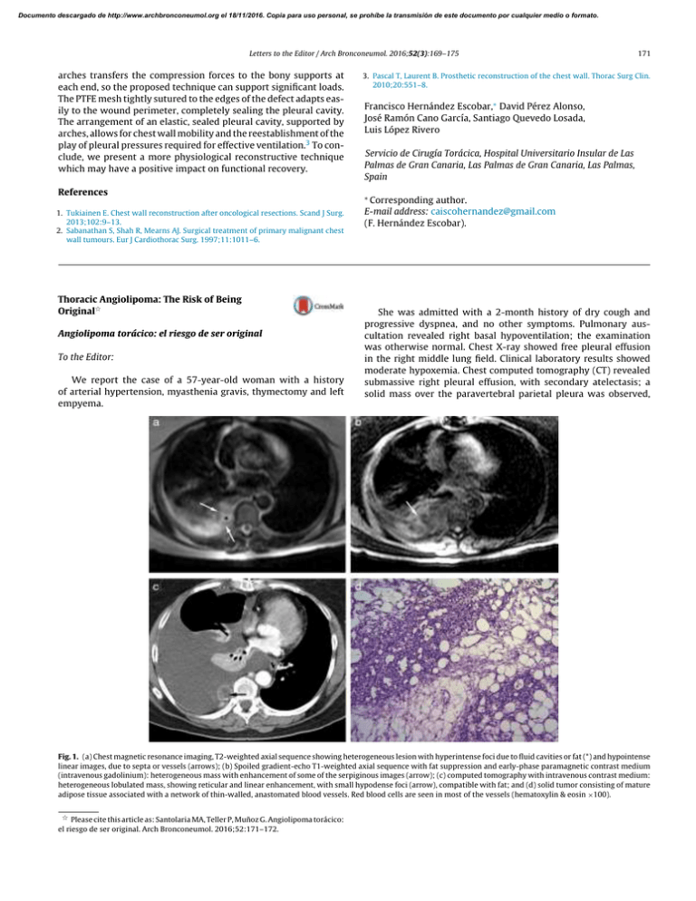
Documento descargado de http://www.archbronconeumol.org el 18/11/2016. Copia para uso personal, se prohíbe la transmisión de este documento por cualquier medio o formato. Letters to the Editor / Arch Bronconeumol. 2016;52(3):169–175 arches transfers the compression forces to the bony supports at each end, so the proposed technique can support significant loads. The PTFE mesh tightly sutured to the edges of the defect adapts easily to the wound perimeter, completely sealing the pleural cavity. The arrangement of an elastic, sealed pleural cavity, supported by arches, allows for chest wall mobility and the reestablishment of the play of pleural pressures required for effective ventilation.3 To conclude, we present a more physiological reconstructive technique which may have a positive impact on functional recovery. References 1. Tukiainen E. Chest wall reconstruction after oncological resections. Scand J Surg. 2013;102:9–13. 2. Sabanathan S, Shah R, Mearns AJ. Surgical treatment of primary malignant chest wall tumours. Eur J Cardiothorac Surg. 1997;11:1011–6. Thoracic Angiolipoma: The Risk of Being Original夽 Angiolipoma torácico: el riesgo de ser original To the Editor: We report the case of a 57-year-old woman with a history of arterial hypertension, myasthenia gravis, thymectomy and left empyema. 171 3. Pascal T, Laurent B. Prosthetic reconstruction of the chest wall. Thorac Surg Clin. 2010;20:551–8. Francisco Hernández Escobar,∗ David Pérez Alonso, José Ramón Cano García, Santiago Quevedo Losada, Luis López Rivero Servicio de Cirugía Torácica, Hospital Universitario Insular de Las Palmas de Gran Canaria, Las Palmas de Gran Canaria, Las Palmas, Spain ∗ Corresponding author. E-mail address: [email protected] (F. Hernández Escobar). She was admitted with a 2-month history of dry cough and progressive dyspnea, and no other symptoms. Pulmonary auscultation revealed right basal hypoventilation; the examination was otherwise normal. Chest X-ray showed free pleural effusion in the right middle lung field. Clinical laboratory results showed moderate hypoxemia. Chest computed tomography (CT) revealed submassive right pleural effusion, with secondary atelectasis; a solid mass over the paravertebral parietal pleura was observed, Fig. 1. (a) Chest magnetic resonance imaging, T2-weighted axial sequence showing heterogeneous lesion with hyperintense foci due to fluid cavities or fat (*) and hypointense linear images, due to septa or vessels (arrows); (b) Spoiled gradient-echo T1-weighted axial sequence with fat suppression and early-phase paramagnetic contrast medium (intravenous gadolinium): heterogeneous mass with enhancement of some of the serpiginous images (arrow); (c) computed tomography with intravenous contrast medium: heterogeneous lobulated mass, showing reticular and linear enhancement, with small hypodense foci (arrow), compatible with fat; and (d) solid tumor consisting of mature adipose tissue associated with a network of thin-walled, anastomated blood vessels. Red blood cells are seen in most of the vessels (hematoxylin & eosin ×100). 夽 Please cite this article as: Santolaria MA, Teller P, Muñoz G. Angiolipoma torácico: el riesgo de ser original. Arch Bronconeumol. 2016;52:171–172. Documento descargado de http://www.archbronconeumol.org el 18/11/2016. Copia para uso personal, se prohíbe la transmisión de este documento por cualquier medio o formato. 172 Letters to the Editor / Arch Bronconeumol. 2016;52(3):169–175 suggestive of tumor disease (Fig. 1). Thoracentesis was performed and a serofibrinous fluid was drained. This was considered exudate in view of the protein ratio of 0.8, although pH and glucose were normal and the leukocyte count was very low (140 mm3 ). Adenosine deaminase levels were normal. Sputum smears and cultures were negative. Cytology revealed inflammation and reactive mesothelial hyperplasia. Fiberoptic bronchoscopy was normal. Positron emission tomography (PET) was negative for malignancy. Magnetic resonance imaging (MRI) identified a poorly delimited, heterogeneous paraspinal lesion, with no vertebral involvement. Possible pleural infiltration with a vascular component and pleural effusion was observed, the origin of which was located in the paraspinal white tissue or parietal pleura (Fig. 1). Rapid pleural filling required frequent drainage by thoracentesis. Finally, videoassisted thoracoscopy was performed and a solid paravertebral and intravertebral tumor was resected. Gross examination revealed an uneven, purplish 4 cm × 2 cm × 2 cm tumor with hemorrhagic areas, partially enveloped in pleura. It was identified microscopically as a fragment of parietal pleura, with transmural thickening as a result of a poorly delimited, unencapsulated solid tumor, consisting of mature adipocytes intermixed with abundant vessels of varying sizes, and no endothelial cell atypia. The mesothelial sheath showed mild hyperplastic reactive changes (Fig. 1d). Immunohistochemistry showed: CD-34, CD-31 and factor VIII: intense, diffuse positivity in the vascular areas; calretinin, pankeratin, and Ck 5/6: positivity in the mesothelial sheath; Ki57: low proliferative index (<3%). The definitive diagnosis was mesenchymal tumor, consistent with paravertebral chest wall angiolipoma. One year after resection and talc pleurodesis, the patient remains without relapse. Angiolipomas are benign tumors, generally located under the skin, most often in the trunk or the limbs, although they have very occasionally been described in the thoracic cavity.1 This is the first report of an intrapleural location with associated effusion. In anatomical pathology terms, these tumors are formed of mature adipocytes and numerous vessels in varying proportions.2 They Antineutrophil Cytoplasmic Antibodies (ANCA)-Negative Vasculitis in a Patient With Alpha-1-Antitrypsin Deficiency夽 Vasculitis con Anticuerpos anticitoplasma de neutrófilos (ANCA) negativos en paciente con déficit de alfa-1 antitripsina To the Editor We report the case of a 62-year-old man, former smoker of 20 pack-years, with a history of arterial hypertension, diabetes mellitus and previous ictus with no neurological sequelae. He was referred from the respiratory medicine clinic with dyspnea on moderate effort (mMRC 2). Lung function tests showed FEV1 /FVC 47%; FEV1 1.4 l (44%); FVC 3.3 l (82%); VR 4.4 l (182%); TLC 8.0 l (119%); DLCO 49% and KCO 59%. Chest computed tomography showed severe panacinar emphysema, primarily in the lower lobes. Severe alpha-1-antitrypsin (AAT) deficiency (28.4 mg/dl) associated with ZZ phenotype was observed. A diagnosis of COPD with severe airflow obstruction and type ZZ alpha-1-antitrypsin deficiency (AATD) was made, and after abdominal ultrasound confirmed chronic liver disease, the decision was made to administer 夽 Please cite this article as: Gonçalves JMF, D’amato R. Vasculitis con Anticuerpos anticitoplasma de neutrófilos (ANCA) negativos en paciente con déficit de alfa-1 antitripsina. Arch Bronconeumol. 2016;52:172–173. tend to appear benign on PET imaging.3 CT may reveal heterogeneity with areas of fat attenuation and enhancement in vascular areas, but differences in the ratios of each type of tissue make it difficult to make an accurate diagnosis. Differential diagnoses to consider include infiltrating hemangioma, neuroendocrine tumors, or other mesenchymal tumors. In view of the difficulty of achieving diagnosis before surgery, MRI may be the gold standard imaging test, as it reveals isointense images in T1 (lipomatous component) and hyperintense images in T2 (vascular component).4 Acknowledgments We thank Dr José Antonio Fernández Gómez for his collaboration. References 1. Hamano A, Suzuki K, Saito T, Kuwatsuru R, Oh S, Suzuki K. Infiltrating angiolipoma of the thoracic wall: a case report. Open J Clin Diag. 2013;3:19–22. 2. Choi JY, Goo JM, Chung MJ, Kim HC, Im JG. Angiolipoma of the posterior mediastinum with extension into the spinal canal: a case report. Korean J Radiol. 2000;1:212–4. 3. Jiang L, Wang YL, Zhou YM, Xie BX, Wang L, Ding JA, et al. Bronchial angiolipoma. Ann Thorac Surg. 2009;88:300–2. 4. Fujiwara H, Kaito T, Takenaka S, Makino T, Yonenobu K. Thoracic spinal epidural angiolipoma: report of two cases and review of the literature. Turk Neurosurg. 2013;23:271–7. Miguel Angel Santolaria,a,∗ Pablo Teller,a Guillermo Muñozb a Servicio de Neumología, Hospital Clínico Universitario Lozano Blesa, Zaragoza, Spain b Servicio de Anatomía Patológica, Hospital Clínico Universitario Lozano Blesa, Zaragoza, Spain ∗ Corresponding author. E-mail address: [email protected] (M.A. Santolaria). replacement AAT. Before initiation of treatment, the patient had an episode of epistaxis associated with purplish lesions in the lower limbs, tending to converge, with no blanchability on diascopy. Pathology study results showed leukocytoclastic vasculitis in small and medium caliber vessels, associated with elevated IgA (754 mg/dl), microhematuria and proteinuria suggestive of nephritis. Together, these signs were consistent with a diagnosis of adult Henoch–Schönlein purpura (HSP). ANCA antibodies (MPO<0.8 IU/ml and anti-PR3<0.4 IU/ml) and ENA were negative; ANA were positive with a titer of 1/160 in a fine speckled pattern. On the basis of this diagnosis, treatment began with oral corticosteroids (0.5 mg/kg/day) in a tapering regimen. One month after beginning this treatment, the patient suffered a fall at home and injured his left arm, with subsequent development of diffuse arthralgia and asthenia. Magnetic resonance imaging revealed cellulitis, arthritis and synovitis in the distal radioulnar and carpometacarpal joints of the upper left limb. Synovial biopsy and fluid culture were performed, confirming synovitis with positive multi-resistant Pseudomonas aeruginosa culture. Despite the administration of wide-spectrum antibiotics and systemic corticosteroids, the patient developed multiorgan failure and died in the intensive care unit. A review of AATD and concomitant necrotizing vasculitis shows that microscopic polyangiitis and Wegener’s granulomatosis are the most common forms, while HSP is an unusual manifestation. The mean age for presentation is generally around 48 years, and
