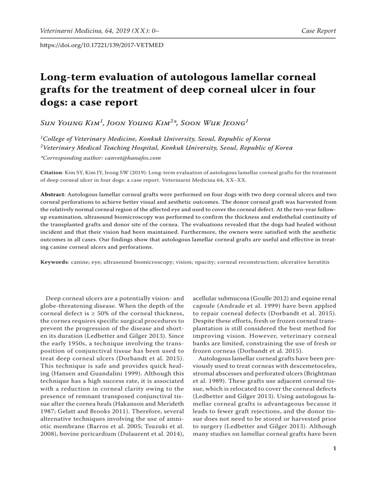
Veterinarni Medicina, 64, 2019 (X X ): 0– Case Report https://doi.org/10.17221/139/2017-VETMED Long-term evaluation of autologous lamellar corneal grafts for the treatment of deep corneal ulcer in four dogs: a case report Sun Young Kim1, Joon Young Kim2*, Soon Wuk Jeong1 1 2 College of Veterinary Medicine, Konkuk University, Seoul, Republic of Korea Veterinary Medical Teaching Hospital, Konkuk University, Seoul, Republic of Korea *Corresponding author: [email protected] Citation: Kim SY, Kim JY, Jeong SW (2019): Long-term evaluation of autologous lamellar corneal grafts for the treatment of deep corneal ulcer in four dogs: a case report. Veterinarni Medicina 64, XX–XX. Abstract: Autologous lamellar corneal grafts were performed on four dogs with two deep corneal ulcers and two corneal perforations to achieve better visual and aesthetic outcomes. The donor corneal graft was harvested from the relatively normal corneal region of the affected eye and used to cover the corneal defect. At the two-year followup examination, ultrasound biomicroscopy was performed to confirm the thickness and endothelial continuity of the transplanted grafts and donor site of the cornea. The evaluations revealed that the dogs had healed without incident and that their vision had been maintained. Furthermore, the owners were satisfied with the aesthetic outcomes in all cases. Our findings show that autologous lamellar corneal grafts are useful and effective in treating canine corneal ulcers and perforations. Keywords: canine; eye; ultrasound biomicroscopy; vision; opacity; corneal reconstruction; ulcerative keratitis Deep corneal ulcers are a potentially vision- and globe-threatening disease. When the depth of the corneal defect is ≥ 50% of the corneal thickness, the cornea requires specific surgical procedures to prevent the progression of the disease and shorten its duration (Ledbetter and Gilger 2013). Since the early 1950s, a technique involving the transposition of conjunctival tissue has been used to treat deep corneal ulcers (Dorbandt et al. 2015). This technique is safe and provides quick healing (Hansen and Guandalini 1999). Although this technique has a high success rate, it is associated with a reduction in corneal clarity owing to the presence of remnant transposed conjunctival tissue after the cornea heals (Hakanson and Merideth 1987; Gelatt and Brooks 2011). Therefore, several alternative techniques involving the use of amniotic membrane (Barros et al. 2005; Tsuzuki et al. 2008), bovine pericardium (Dulaurent et al. 2014), acellular submucosa (Goulle 2012) and equine renal capsule (Andrade et al. 1999) have been applied to repair corneal defects (Dorbandt et al. 2015). Despite these efforts, fresh or frozen corneal transplantation is still considered the best method for improving vision. However, veterinary corneal banks are limited, constraining the use of fresh or frozen corneas (Dorbandt et al. 2015). Autologous lamellar corneal grafts have been previously used to treat corneas with descemetoceles, stromal abscesses and perforated ulcers (Brightman et al. 1989). These grafts use adjacent corneal tissue, which is relocated to cover the corneal defects (Ledbetter and Gilger 2013). Using autologous lamellar corneal grafts is advantageous because it leads to fewer graft rejections, and the donor tissue does not need to be stored or harvested prior to surgery (Ledbetter and Gilger 2013). Although many studies on lamellar corneal grafts have been 1 Case Report Veterinarni Medicina, 64, 2019 (X X ): 0– https://doi.org/10.17221/139/2017-VETMED Figure 1. The initial ophthalmic examination (A) and the surgical results one year postoperatively (B) in Case 1. (A) A corneal perforation in the right eye. (B) The opacity of the corneal graft was almost completely clear in the corneas (A) published (Jensen 1963; Kim et al. 2016), to the best of our knowledge, only a few studies have employed autologous lamellar corneal grafts in dogs (Brightman et al. 1989). Hence, the purpose of this case report is to describe the clinical presentations, ultrasound biomicroscopy (UBM) findings and surgical management strategies in four dogs with severe corneal defects that were treated with autologous lamellar corneal grafts. Case description Case 1. A nine-year-old castrated male Yorkshire Terrier was referred to the Veterinary Medical Teaching Hospital of Konkuk University with a corneal defect in the right eye. One week prior to referral, the dog received medical treatment and a third eyelid flap to treat a corneal ulcer, but no improvement in the clinical signs or in the defect itself was observed. The menace response, dazzle reflex and direct and indirect pupillary light reflex (PLR) were all present. Schirmer tear test (STT) values Figure 2. The initial ophthalmic examination (A) and the surgical results one year postoperatively (B) in Case 2. (A) Case 2 had a corneal perforation in the right eye and the defect was sealed with a prominent fibrin and blood plug. (B) Cornea showed severe fibrosis of the graft cornea owing to the development of severe keratitis and uveitis in the eye at the initial examination 2 (B) were normal (18 mm/min) at measured time points. Intraocular pressure (IOP) could not be measured due to perforation of the cornea. Ophthalmic examinations with slit-lamp biomicroscopy (Hawk eye®, Dioptrix, France) revealed corneal perforation with a 2-mm-diameter defect (Figure 1A). Corneal oedema and neovascularisation were also observed around the defect (Figure 1A). Autologous lamellar corneal grafting was performed to reconstruct the corneal perforation. Case 2. An 11-year-old intact female Shih Tzu presented to our hospital with a corneal perforation in the right eye. The corneal defect was identified two days before admission. During the ophthalmic examination, a 3-mm-diameter fibrin clot was observed (Figure 2A). Although the defect was sealed with a prominent fibrin and blood plug, a small amount of aqueous leakage was detected. Moderate conjunctival hyperaemia and diffuse corneal oedema were observed in the affected eye. The PLR could not be assessed because of corneal opacity. The menace response and dazzle reflex were negative. We were unable to measure the STT and IOP owing to the corneal (A) (B) Veterinarni Medicina, 64, 2019 (X X ): 0– Case Report https://doi.org/10.17221/139/2017-VETMED Figure 3. The initial ophthalmic examination (A) and the surgical results one year postoperatively (B) in Case 3. (A) Deep corneal ulcer in the left eye. (B) Corneal ulcer was healed but showed pigmented keratitis because of keratoconjunctivitis sicca (KCS) (A) perforation. Although the recommended course of action was to perform a conjunctival graft to treat the perforation, at the insistence of the dog owner an autologous lamellar corneal grafting was performed instead. Case 3. A 15-year-old castrated male Shih Tzu had been treated for a corneal ulcer in the left eye using topical and systemic drugs for two weeks at a local animal hospital. Ophthalmic examinations revealed the presence of a deep corneal ulcer that was 3 mm in diameter (Figure 3A). The menace response and dazzle reflex were not assessed. We were also unable to assess the direct and indirect PLRs due to corneal oedema and severe aqueous flare. The STT was low (9 mm/min), and the IOP was slightly low (11 mm Hg), as measured by tonometry (Tono-Pen, Reichert, USA). Autologous lamellar corneal grafting was performed to prevent progression of the corneal ulceration. Case 4. A two-year-old male Maltese dog with cataracts presented to our hospital with a corneal ulcer in the right eye. Four weeks previously, the dog had been scratched in the right eye by a cat, resulting in a corneal laceration. Despite undergo- Figure 4. The initial ophthalmic examination (A) and the surgical results one year postoperatively (B) in Case 4. Cornea had a > 2/3 deep corneal ulcer in the right eye. (B) The opacity of the corneal graft was almost completely clear in the corneas (B) ing four weeks of medical treatment, the corneal defect deteriorated in the local hospital. Upon ophthalmic examination, a 3-mm oval-shaped corneal defect was found (Figure 4A). Although the menace response and dazzle reflex were positive, the direct and indirect PLRs were negative. The STT value was normal at 17 mm/min and the IOP was slightly low in the right eye at 11 mm Hg. We believe this eye showed anterior uveitis secondary to corneal ulceration. Surgical procedures. Topical medications, including ofloxacin (Tarivid ®, Santen Pharm., Ltd., Republic of Korea) and atropine sulphate (Isopto Atropine® 1% Eye Drop, Alcon®, USA), were initiated preoperatively. Cefazolin (30 mg/kg intravenously; Chongkundang Pharm. Co., Ltd., Republic of Korea) was administered as a prophylactic antibiotic prior to the induction of anaesthesia with propofol (6 mg/kg intravenously; Provive 1% ® , Myungmoon Pharm. Co., Ltd., Republic of Korea). Maintenance of anaesthesia was achieved using 1.5–2% isoflurane (Forane®, Choongwae, Republic of Korea) and oxygen. Intraoperative fluid therapy was performed using 0.9% normal saline. The dog (A) (B) 3 Case Report Veterinarni Medicina, 64, 2019 (X X ): 0– https://doi.org/10.17221/139/2017-VETMED was placed in the dorsal recumbent position, and the eye with the corneal defect was clipped and prepared for surgery. After cleaning the periocular skin with a 1 : 10 dilution of povidone-iodine solution, the conjunctival sac was cleaned with a sterile cotton tip that was soaked in a 1 : 50 dilution of povidone-iodine solution. Following this, the cornea and conjunctiva were carefully flushed using Hartmann’s solution (Daihan Hartmann’s Sol., Dai Han Pharm. Co., Ltd., Republic of Korea) to remove any mucus, debris or antiseptics. The devitalised tissue or fibrin plug around the corneal defect was debrided to prepare the recipient graft site. If the eye was perforated, then the anterior chamber was filled with sodium hyaluronate (Healon®, AMO Uppsala AB, Sweden). The donor corneal graft was harvested from the normal corneal region of the affected eye using a skin biopsy punch that was 1 mm larger than the diameter of the corneal defect (Figure 5A). A one-half thickness keratectomy was performed with a crescent blade knife (Ophthalmic crescent knife®, Alcon®) and corneal section scissors (Figure 5B). The lamellar corneal graft covered the recipient site. It was secured at the 12, 6, 3 and 9 o’clock positions with simple interrupted sutures (Figure 5C) and then (A) (D) 4 with a simple continuous pattern over the top of the interrupted sutures using a 10-0 polyamide monofilament (Dafilon ®, B. Braun, Spain) (Figure 5D). A partial temporary tarsorrhaphy was performed with a 4-0 polyamide monofilament (Dafilon®, B. Braun, Spain) to protect the cornea for two weeks postoperatively. Postoperatively, ofloxacin (eye drops, four times daily for two weeks, and then three times daily for two weeks) and cefazolin (systemic medication for two weeks) were administered. In addition, 0.05% cyclosporine (Cyporin N ®, Taejoon Pharm. Co., Ltd., Republic of Korea; three times daily beginning ten days postoperatively and then gradually reduced to once daily for two months) and 1% prednisolone (Pred Forte®, Allergan, Ireland; three times daily administered ten days postoperatively and then gradually reduced to every other day for three months) eye drops were administered to reduce neovascularisation and fibrosis at the recipient site. Surgical outcomes and follow-up. No evidence of aqueous humour leakage was observed on the second postoperative day. The menace response and dazzle reflex were present, with no blepharospasm, within five days following surgery in all four cases. Seven days after surgery, all graft sites were (B) (C) Figure 5. Surgical procedures for placing the autologous lamellar corneal graft in the dog. (A) The donor corneal graft was harvested from the normal corneal region of the affected eye using a skin biopsy punch 1 mm greater than the diameter of the corneal defect. (B) A one-half thickness keratectomy was performed with a crescent blade knife and corneal section scissor. (C) The lamellar corneal graft covered the recipient site and was secured at 12, 6, 3 and 9 o’clock by simple interrupted suture patterns. (D) The corneal graft was sewn over with a simple continuous suture Veterinarni Medicina, 64, 2019 (X X ): 0– Case Report https://doi.org/10.17221/139/2017-VETMED (B) (A) Figure 6. Postoperative photographs of the dog’s right eye in Case 4. (A) To assess the eye on day 7 postoperatively, the partial tarsorrhaphy was removed. Oedema of the corneal graft was observed, and neovascularisation extended to the graft margin. Subsequently, we re-sutured the eye under local anaesthesia. (B) After removing the partial tarsorrhaphy on day 14 postoperatively, the oedema was reduced, and vascularisation was less prominent fluorescein dye-negative and all sutures remained intact. Because a partial tarsorrhaphy involving 1/3 or 1/4 of the eyelid was performed, the operated area could be observed without removing the sutures. Oedema from the corneal graft and neovascularisation extending to the graft margin were observed in the early postoperative phase (Figure 6A). However, the oedema resolved, and the vascularisation became less prominent (Figure 6B). One month after surgery, the vessels were attenuated, and the grafts became translucent, thereby allowing us to scrutinise the anterior chamber. The suture materials were removed a year later, and the cyclosporine eye drops were continuously used after the suture materials were removed. All transplanted grafts and corneal donor sites were intact, and the owners of all dogs were satisfied with the aesthetics. Vision was satisfactorily maintained in all cases, although the eyesight of Case 2 was suboptimal. The opacity of the corneal graft was almost clear (minimally opaque) in the corneas of Cases 1 and 4 (Figures 1B and 4B, respectively). However, Case 2 showed severe fibrosis of the graft cornea (Figure 2B) owing to the development of severe keratitis and uveitis in the eye at the initial examination. The cornea of Case 3 showed pigmented keratitis because of keratoconjunctivitis sicca (Figure 3B). One month after surgery, the STT value of this eye had decreased, and at three months postoperatively the STT value was almost 0 mm/min. Two years postoperatively, ultrasound biomicroscopy (MD-320W, MEDA Co., Ltd., China) was performed to determine the thickness, echogenicity and endothelial continuity of the transplanted grafts and corneal donor site. The UBM evaluations revealed that the thickness of the transplanted grafts and corneal donor sites were similar to those observed for the normal corneal layer and that the grafts were connected to the corneal endothelium continuously in Cases 1 and 4 (Figure 7). DISCUSSION Several surgical techniques have been developed for repairing, replacing and regenerating corneal defects in veterinary ophthalmology, including tissue adhesives (Refojo et al. 1968), conjunctival pedicle grafts (Hakanson and Merideth 1987), lamellar/penetrating keratoplasty (Hansen and Guandalini 1999) and biomaterial grafts (Goulle 2012; Dulaurent et al. 2014). However, new techniques aimed at maintaining corneal transparency and ensuring rapid and easy recoveries continue to be researched and developed. In human ophthalmology, the corneal graft technique is the preferred treatment for corneal defects, as it provides transparency and tectonic support to the cornea (Moffatt et al. 2005). In veterinary ophthalmology, although conjunctival grafting is a reliable technique that has been used for over 60 years to treat deep or perforating corneal wounds, the use of conjunctival tissue can reduce corneal clarity owing to fibrosis (Dorbandt et al. 2015). Therefore, corneal transplantation, which is effective, stable 5 Case Report Veterinarni Medicina, 64, 2019 (X X ): 0– https://doi.org/10.17221/139/2017-VETMED (A) (B) (C) Figure 7. Case 1 (A), Case 2 (B), Case 4 (C). The epithelial and endothelial continuity of the transplanted graft sites are intact in (A) and (C), but not in (B, Case 2), and the thicknesses of the transplanted grafts were almost the same as in the normal corneal layer and can provide corneal transparency, is preferred over conjunctival grafting (Lacerda et al. 2017). Corneoconjunctival transposition (CCT) is another alternative method (Andrew et al. 2001) that has been used to treat deep corneal ulcerations, descemetoceles and feline corneal sequestra involving the deeper stroma (Gelatt and Brooks 2011). Although CCT is a good treatment option for corneal ulcer, it can create a scar in the peripheral cornea. Corneal graft rejection is one of the main complications in corneal transplantation and the most common cause of graft failure postoperatively. Additionally, graft rejection can result in the dehiscence of sutures and corneal opacity. This reaction occurs when the host’s immune system recognises the foreign donor-graft antigens. A major risk factor for graft rejection is host corneal vascularisation (Laguna et al. 2015), which allows antigens to leave 6 the donor graft via blood vessels, thus permitting host antigen-presenting cells to move to the graft (Brooks et al. 2008). In a recent study by Lacerda et al. (2017), the rejection rates of corneal grafts and lamellar grafts in canines were 56% and 55%, respectively. However, in a recent human study by Barut Selver et al. (2018), the incidence of graft failure and repeat penetrating keratoplasty among 1613 penetrating keratoplasties was 9.23%. In contrast, in another human study by Coster (1997), the success rate of keratoplasties was only 62%. Therefore, a better understanding of the risk factors, causes and complications of graft failures can help increase the success rate of keratoplasties in veterinary ophthalmology. One way to reduce graft rejections is to use autologous lamellar corneal grafts. In the present report, neovascularisation was observed after surgery; however, the formation of new blood vessels was attenuated via the administration of low-dose cyclosporine eye drops. Although the opacity was not perfectly clear, keratitis almost disappeared. However, Case 2 showed severe fibrosis of the corneal graft. This eye had corneal perforation with severe keratitis and diffuse corneal oedema at the initial examination. Although the perforation was well covered with an autologous lamellar corneal graft, fibrosis remained. As the graft was harvested from the affected eye, we believe that it was not sufficiently healthy to maintain the corneal clearance. Case 3 showed pigmented keratitis with keratoconjunctivitis sicca. This made the corneal surface very irregular. The pigmented area of the cornea was slightly thicker than other areas, although the ulcer had healed well. The donor corneal graft was harvested from the normal corneal region of the affected eye using a skin biopsy punch that was 1 mm larger than the diameter of the corneal defect. Autologous lamellar corneal grafting also obviates the need to obtain donor material from another source. This is important because, despite the advancements in corneal grafting surgery and postoperative care in veterinary medicine, corneal bank facilities are not yet available worldwide. Due to the shortage in facilities, some biomaterials that can serve as substitutes for the cornea have been developed in veterinary ophthalmology. However, as autologous lamellar corneal grafts are obtained from the affected animal, no outside donor material is needed. Whilst there are some limitations, Veterinarni Medicina, 64, 2019 (X X ): 0– Case Report https://doi.org/10.17221/139/2017-VETMED in general, we have found it quite easy to obtain a graft from the affected eye. In our experience, ulceration does not occur in the entire cornea except in a melting ulcer; hence, even in severe cases of ulceration, most affected corneas will have a potential graft site. In the present report, even when corneal perforation was present, a graft could be harvested from the affected cornea. In addition, a graft can be obtained from the healthy eye if it cannot be obtained from the affected cornea. Interestingly, a study by Brightman et al. (1989) revealed that healthy endothelial cells adjacent to the graft site become elongated and increase their surface area as they migrate, eventually covering the endothelium-deficient lamellar graft. Such findings support the notion that autologous lamellar corneal grafts can be used even in a perforated cornea. After autologous lamellar corneal grafting, however, the donor site in the cornea is thin, and whether normal healing occurs following harvesting of the donor cornea remained unclear. To determine if the donor site undergoes healing, UBM was utilised. This non-invasive technique employs highfrequency (50 MHz) ultrasound probes and permits the microscopic visualisation of the structures between living tissues in vivo (Bentley et al. 2003). UBM can be used to acquire images of the anterior chamber structures with higher resolution than those obtained with high-resolution ultrasound (frequencies of 20 MHz). The level of tissue penetration and image resolution of UBM is adequate for assessing the anterior segment structures in vivo. Additionally, UBM examinations can be performed without anaesthesia in most cases. UBM may provide additional information regarding the pathogenesis and prognosis and can be performed on an opaque or oedematous cornea (Gibson et al. 1998). In this study, UBM examinations revealed that the thicknesses of the transplanted grafts and the donor sites of the cornea were almost the same when compared to the normal corneal layer and that the endothelium was continuously connected with the normal cornea. Although the opacity of the transplanted graft was not perfectly clear, the cosmetic results are better than those of conjunctival grafts, leading to improved owner satisfaction. In conclusion, autologous lamellar corneal grafting is an effective surgical method for severe corneal defects that require immediate treatment, owing to its feasibility and easy application in practice. The graft provides sufficient ocular tectonic support and provides good optical and cosmetic outcomes. REFERENCES Andrade AL, Laus JL, Figueiredo F, Batista CM (1999): The use of preserved equine renal capsule to repair lamellar corneal lesions in normal dogs. Veterinary Ophthalmology 2, 79–92. Andrew SE, Tou S, Brooks DE (2001): Corneoconjunctival transposition for the treatment of feline corneal sequestra: a retrospective study of 17 cases (1990–1998). Veterinary Ophthalmology 4, 107–111. Barros PS, Safatle AM, Godoy CA, Souza MS, Barros LF, Brooks DE (2005): Amniotic membrane transplantation for the reconstruction of the ocular surface in three cases. Veterinary Ophthalmology 8, 189–192. Barut Selver O, Karaca I, Palamar M, Egrilmez S, Yagci A (2018): Graft failure and repeat penetrating keratoplasty. Experimental and Clinical Transplantation, doi: 10.6002/ ect.2017.0165. Bentley E, Miller PE, Diehl KA (2003): Use of high-resolution ultrasound as a diagnostic tool in veterinary ophthalmology. Journal of the American Veterinary Medical Association 223, 1617–1622. Brightman AH, McLaughlin SA, Brogdon JD (1989): Autogenous lamellar corneal grafting in dogs. Journal of the American Veterinary Medical Association 195, 469–475. Brooks DE, Plummer C, Kallberg M, Barrie K, Ollivier FJ, Hendrix DV, Baker A, Scotty NC, Utter ME, Blackwood SE, Nunnery CM, Ben-Shlomo G, Gelatt KN (2008): Corneal transplantation for inflammatory keratopathies in the horse: visual outcome in 206 cases (1993–2007). Veterinary Ophthalmology 11, 123–133. Coster DJ (1997): Evaluation of corneal transplantation. British Journal of Ophthalmology 81, 618–619. Dorbandt DM, Moore PA, Myrna KE (2015): Outcome of conjunctival flap repair for corneal defects with and without an acellular submucosa implant in 73 canine eyes. Veterinary Ophthalmology 18, 116–122. Dulaurent T, Azoulay T, Goulle F, Dulaurent A, Mentek M, Peiffer RL, Isard PF (2014): Use of bovine pericardium (Tutopatch ®) graft for surgical repair of deep melting corneal ulcers in dogs and corneal sequestra in cats. Veterinary Ophthalmology 17, 91–99. Gelatt KN, Brooks DE (2011): 8. Surgery of the cornea and sclera. In: Gelatt KN, Gelatt JP (eds): Veterinary Ophthalmic Surgery. Saunders. 191–236. Gibson T, Roberts SM, Severin GA, Steyn PH, Wrigley RH (1998): Comparison of gonioscopy and ultrasound biomi- 7 Case Report Veterinarni Medicina, 64, 2019 (X X ): 0– https://doi.org/10.17221/139/2017-VETMED croscopy for evaluating the iridocorneal angle in dogs. Journal of the American Veterinary Medical Association 213, 635–638. Goulle F (2012): Use of porcine small intestinal submucosa for corneal reconstruction in dogs and cats: 106 cases. Journal of Small Animal Practice 53, 34–43. Hakanson NE, Merideth RE (1987): Conjunctival pedicle grafting in the treatment of corneal ulcers in the dog and cat. Journal of the American Animal Hospital Association 23, 641. Hansen PA, Guandalini A (1999): A retrospective study of 30 cases of frozen lamellar corneal graft in dogs and cats. Veterinary Ophthalmology 2, 233–241. Jensen EC (1963): Experimental corneal transplantation in the dog. Journal of the American Veterinary Medical Association 142, 11–22. Kim S, Kwak JY, Jeong M, Seo K (2016): Deep anterior lamellar keratoplasty of dog eyes using the big-bubble technique. Journal of Veterinary Science 17, 347–352. Lacerda RP, Pena Gimenez MT, Laguna F, Costa D, Rios J, Leiva M (2017): Corneal grafting for the treatment of full-thickness corneal defects in dogs: a review of 50 cases. Veterinary Ophthalmology 20, 222–231. Laguna F, Leiva M, Costa D, Lacerda R, Pena Gimenez T (2015): Corneal grafting for the treatment of feline corneal sequestrum: a retrospective study of 18 eyes (13 cats). Veterinary Ophthalmology 18, 291–296. Ledbetter EC, Gilger BC (2013): 18. Diseases and surgery of the canine cornea and sclera. In: Gelatt KN, Gilger BC, Kern TJ (eds): Veterinary Ophthalmology. 5th edn. WileyBlackwell. 976–1049. Moffatt SL, Cartwright VA, Stumpf TH (2005): Centennial review of corneal transplantation. Clinical & Experimental Ophthalmology 33, 642–657. Refojo MF, Dohlman CH, Ahmad B, Carroll JM, Allen JC (1968): Evaluation of adhesives for corneal surgery. Archives of Ophthalmology 80, 645–656. Tsuzuki K, Yamashita K, Izumisawa Y, Kotani T (2008): Microstructure and glycosaminoglycan ratio of canine cornea after reconstructive transplantation with glycerinpreserved porcine amniotic membranes. Veterinary Ophthalmology 11, 222–227. Received: October 25, 2017 Accepted after corrections: December 6, 2018 8
