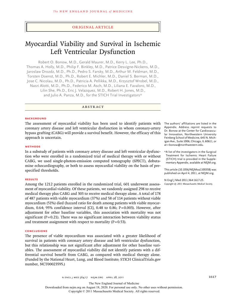
The n e w e ng l a n d j o u r na l of m e dic i n e original article Myocardial Viability and Survival in Ischemic Left Ventricular Dysfunction Robert O. Bonow, M.D., Gerald Maurer, M.D., Kerry L. Lee, Ph.D., Thomas A. Holly, M.D., Philip F. Binkley, M.D., Patrice Desvigne-Nickens, M.D., Jaroslaw Drozdz, M.D., Ph.D., Pedro S. Farsky, M.D., Arthur M. Feldman, M.D., Torsten Doenst, M.D., Ph.D., Robert E. Michler, M.D., Daniel S. Berman, M.D., Jose C. Nicolau, M.D., Ph.D., Patricia A. Pellikka, M.D., Krzysztof Wrobel, M.D., Nasri Alotti, M.D., Ph.D., Federico M. Asch, M.D., Liliana E. Favaloro, M.D., Lilin She, Ph.D., Eric J. Velazquez, M.D., Robert H. Jones, M.D., and Julio A. Panza, M.D., for the STICH Trial Investigators* A bs t r ac t Background The assessment of myocardial viability has been used to identify patients with coronary artery disease and left ventricular dysfunction in whom coronary-artery bypass grafting (CABG) will provide a survival benefit. However, the efficacy of this approach is uncertain. Methods The authors’ affiliations are listed in the Appendix. Address reprint requests to Dr. Bonow at the Center for Cardiovascular Innovation, Northwestern University Feinberg School of Medicine, 645 N. Michigan Ave., Suite 1006, Chicago, IL 60611, or at [email protected]. In a substudy of patients with coronary artery disease and left ventricular dysfunction who were enrolled in a randomized trial of medical therapy with or without CABG, we used single-photon-emission computed tomography (SPECT), dobutamine echocardiography, or both to assess myocardial viability on the basis of prespecified thresholds. * A list of the investigators in the Surgical Treatment for Ischemic Heart Failure (STICH) trial is provided in the Supplementary Appendix, available at NEJM.org. Results N Engl J Med 2011;364:1617-25. Among the 1212 patients enrolled in the randomized trial, 601 underwent assessment of myocardial viability. Of these patients, we randomly assigned 298 to receive medical therapy plus CABG and 303 to receive medical therapy alone. A total of 178 of 487 patients with viable myocardium (37%) and 58 of 114 patients without viable myocardium (51%) died (hazard ratio for death among patients with viable myocardium, 0.64; 95% confidence interval [CI], 0.48 to 0.86; P = 0.003). However, after adjustment for other baseline variables, this association with mortality was not significant (P = 0.21). There was no significant interaction between viability status and treatment assignment with respect to mortality (P = 0.53). This article (10.1056/NEJMoa1100358) was published on April 4, 2011, at NEJM.org. Copyright © 2011 Massachusetts Medical Society. Conclusions The presence of viable myocardium was associated with a greater likelihood of survival in patients with coronary artery disease and left ventricular dysfunction, but this relationship was not significant after adjustment for other baseline variables. The assessment of myocardial viability did not identify patients with a differential survival benefit from CABG, as compared with medical therapy alone. (Funded by the National Heart, Lung, and Blood Institute; STICH ClinicalTrials.gov number, NCT00023595.) n engl j med 364;17 nejm.org april 28, 2011 The New England Journal of Medicine Downloaded from nejm.org on August 18, 2020. For personal use only. No other uses without permission. Copyright © 2011 Massachusetts Medical Society. All rights reserved. 1617 The n e w e ng l a n d j o u r na l C oronary artery disease is an important contributor to the rise in the prevalence of heart failure and in associated mortality and morbidity.1-4 It has not been clearly established whether coronary-artery bypass grafting (CABG) has a role in improving the symptoms and the rate of survival of patients with coronary artery disease and heart failure. We conducted the multicenter Surgical Treatment for Ischemic Heart Failure (STICH) trial5,6 to examine two hypotheses, one of which (hypothesis 1) compared the efficacy of medical therapy alone with that of medical therapy plus CABG in patients with coronary artery disease and left ventricular dysfunction. Left ventricular dysfunction in patients with coronary artery disease is not always an irreversible process related to previous myocardial infarction, since left ventricular function improves substantially in many patients and may even normalize after CABG.7-9 The assessment of myocardial viability with the use of single-photon-emission computed tomography (SPECT) or low-dose dobutamine echocardiography is commonly performed to predict improvement in left ventricular function after CABG, and numerous studies have suggested that the identification of viable myocardium with the use of such methods also predicts improved survival after CABG.8,10-33 However, previous studies that have suggested an association between myocardial viability and outcome have been retrospective in nature, and it is uncertain in most of these studies whether the decision to perform CABG may have been driven by the results of the tests, whether adjustment for key baseline variables was adequate, and whether patients who did not undergo CABG received aggressive medical therapy for heart failure.34 In this substudy, we report the outcome of patients who were randomly assigned to receive medical therapy alone or medical therapy plus CABG in the hypothesis 1 component of STICH study and who also underwent assessment of myocardial viability. Me thods Study Design The rationale and design of the STICH trial have been described previously.5 We conducted a multicenter, nonblinded, randomized trial that was funded by the National Heart, Lung, and Blood 1618 n engl j med 364;17 of m e dic i n e Institute. The hypothesis 1 comparison involved 99 sites in 22 countries. Details of the study design for the hypothesis 1 comparison are reported in the study by Velazquez et al. in this issue of the Journal.6 The trial protocol, which was approved by the ethics committee at each study center, is available with the full text of this article at NEJM.org. The authors of this report assume responsibility for the completeness and accuracy of the data and the analyses and for the fidelity of the study to the trial protocol. Study Patients Patients with angiographic documentation of coronary artery disease amenable to surgical revascularization and with left ventricular systolic dysfunction (ejection fraction, ≤35%) were eligible for enrollment. Exclusion criteria included left main coronary artery stenosis of more than 50%, cardiogenic shock, myocardial infarction within 3 months, and a need for aortic-valve surgery. All patients provided written informed consent. Patients were randomly assigned to receive medical therapy alone or medical therapy plus CABG. A “risk at randomization” (RAR) score was calculated for each patient with the use of an equation derived in an independent data set from multiple variables with a known power to predict the 5-year risk of death without CABG.35 Details of the RAR score are provided in the Supplementary Appendix, available at NEJM.org. Study Procedures In the initial design of the STICH trial, viability testing with SPECT was required for the enrollment of patients.5 However, this requirement proved to be an impediment to enrollment. Therefore, the protocol was subsequently revised to make viability testing optional and to allow the use of either SPECT or dobutamine echocardiography for viability testing. Investigators at all study centers were strongly encouraged to perform viability testing in every patient, but the decision to perform the test was left up to the recruiting investigators. Therefore, only a subgroup of patients in the hypothesis 1 component of the trial underwent viability testing. Details regarding the selection of patients for imaging are described in the Supplementary Appendix. Independent core laboratories that were funded by the National Heart, Lung, and Blood Institute in which investigators were unaware of study- nejm.org april 28, 2011 The New England Journal of Medicine Downloaded from nejm.org on August 18, 2020. For personal use only. No other uses without permission. Copyright © 2011 Massachusetts Medical Society. All rights reserved. Myocardial Viability in Ischemic Ventricular Dysfunction group assignments and the individual characteristics of patients coordinated data collection and analysis for the SPECT and dobutamine echocardiography studies. Details of the imaging protocols for identifying and quantifying the extent of viable myocardium are provided in the Supplementary Appendix. Briefly, thresholds of the extent of viable myocardium were prespecified to classify patients in a binary fashion as either having or not having substantial myocardial viability. For SPECT, patients with viability were defined as those with 11 or more viable segments on the basis of relative tracer activity. For dobutamine echocardiography, patients with viability were defined as those with 5 or more segments with abnormal resting systolic function but manifesting contractile reserve during dobutamine administration. Core laboratory measurements were submitted to the Duke Clinical Research Institute, which performed all statistical analyses. Patient Follow-Up and Outcomes After trial enrollment, patients were followed every 4 months for the first year and every 6 months thereafter. The primary outcome was death from any cause. Secondary end points included death from cardiovascular causes and a composite of death from any cause or hospitalization for cardiovascular causes. Definitions of the trial end points are provided in the report on the main study by Velazquez et al.6 All end points were adjudicated by an independent clinical events committee.6 The comparisons of outcomes that were related to treatment were based on intentionto-treat analyses. Analyses that were based on actual treatment received were also performed to account for crossovers.6 Statistical Analysis We used means and standard deviations to summarize the baseline clinical characteristics of patients unless otherwise specified. We used the Wilcoxon rank-sum test, the Chi-square test, or Fisher’s exact test, as appropriate, to assess baseline differences in individual variables between patients who underwent viability testing and those who did not undergo such testing and between patients who underwent testing who met the prespecified criteria for viability and those who did not meet such criteria. To further describe and characterize these differences, we den engl j med 364;17 veloped multivariable propensity models using logistic regression to identify the key baseline clinical characteristics that distinguished patients who underwent viability testing from those who did not undergo such testing. Among patients who underwent viability testing, we also used these models to distinguish patients with viable myocardium from those without viable myocardium. We used the Cox proportional-hazards model to assess the relationship between the presence of viable myocardium and the end point of death from any cause. We compared the strength of the univariate relationship between viability and the rate of death with the strength of other known prognostic factors, including the left ventricular ejection fraction, the left ventricular end-systolic and end-diastolic volume indexes, and the RAR score. Finally, we used multivariable Cox model analyses to assess the relationship between viability and each end point with adjustment for other known prognostic factors, including age, sex, race or ethnic group, heart failure class at baseline, history of myocardial infarction, previous revascularization, ejection fraction, number of diseased vessels, chronic renal insufficiency, mitral regurgitation, history of stroke, and history of atrial fibrillation. To test whether patients in the CABG group had a better outcome than those in the medicaltherapy group among patients with viable myocardium, as compared with those without viable myocardium, we used Kaplan–Meier curves to examine the outcomes in each study group, according to viability status. We used the Cox regression model to test for an interaction between treatment and viability status. We used three separate prespecified analyses to assess the association between myocardial viability and outcome according to study-group assignment (for details, see the Supplementary Appendix). R e sult s Patients Of 1212 patients enrolled in the hypothesis 1 comparison, 601 who underwent assessment of myocardial viability were included in the analysis reported here. The numbers of patients undergoing each viability test, the timing of the tests, and baseline characteristics of the patients are detailed in the Supplementary Appendix. Among the 601 patients, 487 were found to have viable nejm.org april 28, 2011 The New England Journal of Medicine Downloaded from nejm.org on August 18, 2020. For personal use only. No other uses without permission. Copyright © 2011 Massachusetts Medical Society. All rights reserved. 1619 The n e w e ng l a n d j o u r na l myocardium on the basis of the prespecified criteria, and 114 were found not to have viable myocardium. In the subgroup of 487 patients with myocardial viability, 244 were assigned to receive medical therapy plus CABG, and 243 were assigned to receive medical therapy alone. Likewise, in the subgroup of 114 patients without myocardial viability, 54 were assigned to receive medical therapy plus CABG, and 60 were assigned to receive medical therapy alone. The baseline characteristics of patients who were assigned to undergo CABG or to receive medical therapy were similar in each subgroup (Table 1, and Table S6 in the Supplementary Appendix). of m e dic i n e a continuous model of viability and risk also revealed no significant interactions. This was the case whether the results were examined for all patients who had undergone viability testing, those with SPECT data alone, or those with dobutamine echocardiography data alone (Table S8 in the Supplementary Appendix). Analysis of outcomes on the basis of the treatment received rather than that assigned showed similar trends, again reflecting no interaction between viability status and treatment with respect to death from any cause (P = 0.96), death from cardiovascular causes (P = 0.26), or death or hospitalization for cardiovascular causes (P = 0.98) (Fig. S3 and Table S9 in the Supplementary Appendix). Outcomes During a median of 5.1 years of follow-up of 601 patients, there were 236 deaths (39%). These deaths included 58 of 114 patients without myocardial viability (51%) and 178 of 487 patients with myocardial viability (37%). Patients with viable myocardium had lower overall rates of death than those without viable myocardium (hazard ratio among patients with viable myocardium, 0.64; 95% confidence interval [CI], 0.48 to 0.86; P = 0.003) (Fig. 1). However, after adjustment for other significant baseline prognostic variables in a multivariable model, the prespecified viability status was no longer significantly associated with the rate of death (P = 0.21) (Supplementary Appendix). Patients with myocardial viability also had lower rates of the secondary end points of death from cardiovascular causes (hazard ratio, 0.61; 95% CI, 0.44 to 0.84; P = 0.003) and a composite of death or hospitalization for cardiovascular causes (hazard ratio, 0.59; 95% CI, 0.47 to 0.74; P<0.001) (Fig. S1 in the Supplementary Appendix). The relationship between myocardial viability and death from cardiovascular causes was not significant on multivariable analysis (P = 0.34), but the relationship with the composite of death or hospitalization for cardiovascular causes remained significant (P = 0.003). There was no significant interaction between myocardial viability and study-group assignment with respect to death (P = 0.53) (Fig. 2), death from cardiovascular causes (P = 0.70), or the composite of death or hospitalization for cardiovascular causes (P = 0.39) on the basis of Kaplan–Meier and Cox model analyses. Additional prespecified analyses that were based on median viability scores or on 1620 n engl j med 364;17 Discussion In this substudy of the STICH trial, in which all patients were eligible for CABG as well as optimal medical therapy, we analyzed patients for whom data were available with respect to myocardial viability in order to determine whether the presence of viable myocardium had an influence on the outcome. On univariate analysis, there was a significant association between myocardial viability and outcome. However, this association was not significant on multivariable analysis that included other prognostic variables. The findings of this multivariable analysis do not necessarily indicate that myocardial viability does not have pathophysiological importance in patients with coronary artery disease and left ventricular dysfunction. Instead, it is likely that some of the other variables in the analysis (e.g., left ventricular volumes and ejection fraction) are causally determined by the extent of viable myocardium. A second, and more important, objective of this substudy was to determine whether the presence of substantial myocardial viability influenced the likelihood of benefit from medical therapy plus CABG, as compared with medical therapy alone. We did not find a significant interaction between myocardial viability and medical versus surgical treatment with respect to the rates of death from any cause or from cardiovascular causes or the rate of death or hospitalization for cardiovascular causes. This was true whether patients were grouped according to the assigned treatment (i.e., intention-to-treat analysis) or to the treatment actually received. nejm.org april 28, 2011 The New England Journal of Medicine Downloaded from nejm.org on August 18, 2020. For personal use only. No other uses without permission. Copyright © 2011 Massachusetts Medical Society. All rights reserved. Myocardial Viability in Ischemic Ventricular Dysfunction Table 1. Baseline Characteristics of Patients Who Underwent Assessment of Myocardial Viability.* All Patients (N = 601) Characteristic Patients with Myocardial Viability (N = 487) Medical Therapy (N = 243) CABG (N = 244) Patients without Myocardial Viability (N = 114) P Value Medical Therapy (N = 60) CABG (N = 54) P Value Age — yr 60.7±9.4 60.0±9.7 61.5±9.2 0.05 61.6±8.5 60.0±9.2 0.34 Male sex — no. (%) 521 (87) 205 (84) 211 (86) 0.51 55 (92) 50 (93) 1.00 Previous myocardial infarction 481 (80) 190 (78) 183 (75) 0.41 56 (93) 52 (96) Current Canadian Cardiac Society angina class — no. (%) 0 0.60 236 (39) 101 (42) 101 (41) 18 (30) 16 (30) I 94 (16) 34 (14) 34 (14) 19 (32) 7 (13) II 253 (42) 104 (43) 99 (41) 23 (38) 27 (50) III 14 (2) 3 (1) 8 (3) 0 3 (6) IV 4 (1) 1 (<1) 2 (1) 0 1 (2) Highest New York Heart Association functional class in 3 pre­vious mo — no. (%) I 0.51 27 (4) 15 (6) 9 (4) 0.25 0 3 (6) II 212 (35) 94 (39) 88 (36) 14 (23) 16 (30) III 275 (46) 100 (41) 111 (45) 36 (60) 28 (52) IV 0.68 0.03 87 (14) 34 (14) 36 (15) 10 (17) 7 (13) 12.5±8.8 11.9±8.4 12.8±9.0 0.28 13.7±9.8 12.0±8.8 0.37 Beta-blocker 534 (89) 221 (91) 216 (89) 0.38 52 (87) 45 (83) 0.62 ACE inhibitor 514 (86) 202 (83) 210 (86) 0.37 54 (90) 48 (89) 0.85 46 (8) 20 (8) 20 (8) 0.99 3 (5) 3 (6) 1.00 Risk-at-randomization score† Medications at baseline — no. (%) ARB ACE inhibitor or ARB 554 (92) 219 (90) 227 (93) 0.25 57 (95) 51 (94) 1.00 Statin 508 (85) 212 (87) 193 (79) 0.02 56 (93) 47 (87) 0.26 Aspirin 513 (85) 209 (86) 205 (84) 0.54 56 (93) 43 (80) 0.03 Coronary artery disease distribution — no. (%) No. of diseased vessels with ≥75% stenosis 0.79 0 12 (2) 6 (2) 3 (1) 0.45 2 (3) 1 (2) 1 152 (25) 62 (26) 62 (25) 17 (28) 11 (20) 2 221 (37) 87 (36) 92 (38) 18 (30) 24 (44) 215 (36) 88 (36) 86 (35) 23 (38) 18 (33) Left ventricular ejection fraction — % 3 26.7±8.6 28.1±8.4 27.0±8.2 0.30 22.6±8.5 23.3±9.1 0.50 Left ventricular end-diastolic volume index — ml/m2 of body-surface area 122.8±41.9 117.8±37.9 116±35.1 0.63 152.3±51.3 140.0±53.8 0.16 Left ventricular end-systolic volume index — ml/m2 of body-surface area 91.7±38.9 85.8±34.3 86.0±32.1 0.97 120.8±49.6 111.2±50.8 0.25 * Plus–minus values are means ±SD. ACE denotes angiotensin-converting enzyme, ARB angiotensin-receptor blocker, and CABG coronary-artery bypass grafting. † The risk-at-randomization score ranges from 1 to 32, with higher numbers indicating a higher predicted rate of death. Among patients receiving medical therapy, a score of 1 predicts a rate of 18% and a score of 32 predicts a rate of 99% over 5 years. n engl j med 364;17 nejm.org april 28, 2011 The New England Journal of Medicine Downloaded from nejm.org on August 18, 2020. For personal use only. No other uses without permission. Copyright © 2011 Massachusetts Medical Society. All rights reserved. 1621 n e w e ng l a n d j o u r na l The 1.0 Hazard ratio, 0.64 (95% CI, 0.48–0.86) P=0.003 0.9 Probability of Death 0.8 0.7 0.6 0.5 Without viability 0.4 0.3 With viability 0.2 0.1 0.0 0 1 2 3 4 5 6 36 188 16 102 Years since Randomization No. at Risk Without viability 114 With viability 487 99 432 85 409 80 371 63 294 Figure 1. Kaplan–Meier Analysis of the Probability of Death, ­According to Myocardial Viability Status. The comparison that is shown has not been adjusted for other prognostic baseline variables. After adjustment for such variables on multivariable analysis, the between-group difference was not significant (P = 0.21). Conclusions that can be drawn from our results are limited by a number of factors. First, viability data were not available for all the patients who were enrolled in the STICH hypothesis 1 comparison. The study patients represent slightly less than 50% of the randomized group. Furthermore, viability testing was not performed on a randomly selected subgroup of patients but, rather, was obtained according to test availability and the judgment of the recruiting investigator. The differences in baseline characteristics between patients who underwent viability testing and those who did not undergo such testing suggest that at least some patients may have been selected for testing on the basis of clinical factors (Table S2 in the Supplementary Appendix). Second, only 114 of 601 patients who underwent assessment of myocardial viability (19%) were deemed not to have viable myocardium on the basis of our prespecified criteria. This small number limited the power of our analysis to detect a differential effect of CABG, as compared with medical therapy, in patients with myocardial viability, as compared with those without myocardial viability, although in additional analyses that were based on median viability scores or that used a continuous model of viability, no effect of viability was detected. 1622 n engl j med 364;17 of m e dic i n e Third, we cannot exclude the possibility that results of viability testing could have influenced subsequent clinical decision making. There was a nonsignificant trend toward higher rates of surgery among patients who underwent viability testing on the day of randomization or on the subsequent day than among those who underwent such testing before randomization. However, the timing of viability testing relative to randomization does not appear to have influenced the rate of patient crossovers (Table S3 in the Supplementary Appendix). Fourth, our analysis was based on SPECT and dobutamine echocardiography assessment of myocardial viability. An analysis of outcomes on the basis of a combination of these two tests poses important limitations, given the fundamental differences in the viability information provided by SPECT and dobutamine echocardiography (one related to membrane integrity and the other to contractile reserve) and the differences in analytic approaches between the two methods. However, results were similar whether SPECT and dobutamine echocardiography data were combined or analyzed separately. We also did not incorporate other approaches, such as positron-emission tomography (PET)36,37 or contrast-enhanced magnetic resonance imaging (MRI).38,39 However, in a meta-analysis and other reviews, SPECT and dobutamine echocardiography have been found to have similar prognostic potential to that of PET,8,40,41 and there are limited data regarding outcomes in patients with chronic ischemic left ventricular dysfunction who were studied on MRI. The lack of a differential survival benefit between CABG and medical therapy in patients with viable versus those with nonviable myocardium in this study differs markedly from results of previous retrospective studies and meta-analyses.8,10-33 This finding may reflect, in part, the low rates of death among patients with viable myocardium who were assigned to receive medical therapy (approximately 7% per year) in this study, as compared with previously reported rates (which exceeded 15% per year in many studies). Adherence to guidelines-recommended therapies was high in our trial, whereas data on medical therapy are lacking in many previous retrospective analyses. Medical therapy, like CABG, has the potential to improve left ventricular function in patients with dysfunctional but viable myocardium.42-44 This finding underscores the prog- nejm.org april 28, 2011 The New England Journal of Medicine Downloaded from nejm.org on August 18, 2020. For personal use only. No other uses without permission. Copyright © 2011 Massachusetts Medical Society. All rights reserved. Myocardial Viability in Ischemic Ventricular Dysfunction B With Myocardial Viability 1.0 1.0 0.9 0.9 0.8 0.8 0.7 Probability of Death Probability of Death A Without Myocardial Viability Medical therapy (33 deaths) 0.6 0.5 0.4 0.3 0.2 0.6 0.5 0 1 2 3 4 Medical therapy (95 deaths) 0.4 0.3 0.2 CABG (25 deaths) 0.1 0.0 0.7 CABG (83 deaths) 0.1 5 0.0 6 0 1 Years since Randomization 2 3 4 5 6 94 94 51 51 Years since Randomization No. at Risk No. at Risk Medical therapy CABG 60 54 51 48 44 41 39 41 29 34 14 22 4 12 Medical therapy CABG 243 244 219 213 206 203 179 192 146 148 C Subgroup No. Deaths Without viability With viability 114 487 58 178 P Value for Interaction Hazard Ratio (95% CI) 0.70 (0.41–1.18) 0.86 (0.64–1.16) 0.25 0.50 1.0 CABG Better 0.53 2.0 Medical Therapy Better Figure 2. Kaplan–Meier Analysis of the Probability of Death According to Myocardial-Viability Status and Treatment. At 5 years in the intention-to-treat analysis, the rates of death for patients without myocardial viability were 41.5% in the group assigned to undergo coronary-artery bypass grafting (CABG) and 55.8% in the group assigned to receive medical therapy (Panel A). Among patients with myocardial viability, the respective rates were 31.2% and 35.4% (Panel B). There was no significant interaction between viability status and treatment assignment with respect to mortality (P = 0.53) (Panel C). nostic importance of providing evidence-based therapies to high-risk patients with left ventricular dysfunction, as well as the consideration of CABG for those who are candidates for revascularization. The lack of interaction between myocardial-viability status and benefit from CABG in this study indicates that assessment of myocardial viability alone should not be the deciding factor in selecting the best therapy for these patients. These findings also highlight the need for prospectively designed studies to determine the role of cardiac imaging in clinical decision making.45 In summary, we conducted a substudy of the STICH trial to determine whether the presence of substantial myocardial viability influenced the likelihood of benefit from medical therapy plus CABG, as compared with medical therapy alone, in patients with coronary artery disease and left n engl j med 364;17 ventricular dysfunction. We did not find a significant interaction between myocardial-viability status and medical versus surgical treatment with respect to the rates of death from any cause or from cardiovascular causes or the rate of death or hospitalization for cardiovascular causes. Supported by cooperative agreements (U01-HL-069009, ­ L-069010, HL-069011, HL-069012, HL-069012-03, HL-069013, H HL-069015, HL-070011, and HL-072683) with the National Heart, Lung, and Blood Institute. Dr. Feldman reports having an equity interest in Cardiokine; Dr. Michler, receiving grant support from Sorin; Dr. Berman, receiving consulting fees from Astellas Healthcare, Bracco, and Lantheus Medical Imaging and grant support from Lantheus Medical Imaging and Astellas Healthcare and having an equity interest in Spectrum Dynamics; Dr. Asch, being an investigator in studies for which his institution received grant support from Sorin, Mitralign, and RegeneRx; and Dr. Velazquez, receiving consulting fees from Novartis, Gilead, and Boehringer Ingelheim Pharmaceuticals. No other potential conflict of interest relevant to this article was reported. Disclosure forms provided by the authors are available with the full text of this article at NEJM.org. nejm.org april 28, 2011 The New England Journal of Medicine Downloaded from nejm.org on August 18, 2020. For personal use only. No other uses without permission. Copyright © 2011 Massachusetts Medical Society. All rights reserved. 1623 The n e w e ng l a n d j o u r na l of m e dic i n e Appendix The authors’ affiliations are as follows: Northwestern University Feinberg School of Medicine, Chicago (R.O.B., T.A.H.); Medical University of Vienna, Vienna (G.M.); Duke Clinical Research Institute, Duke University Medical Center, Durham, NC (K.L.L., L.S., E.J.V., R.H.J.); Ohio State University Medical Center, Columbus (P.F.B.); National Heart, Lung, and Blood Institute, Bethesda, MD (P.D.-N.); Medical University of Lodz, Lodz (J.D.), and John Paul II Hospital, Krakow (K.W.) — both in Poland; Instituto Dante Pazzanese de Cardiologia (P.S.F.) and Instituto do Coração–Hospital das Clínicas da Faculdade de Medicina da Universidade de São Paulo (J.C.N.) — both in São Paulo; Jefferson Medical College, Philadelphia (A.M.F.); Heart Center, University of Leipzig, Leipzig, Germany (T.D.); Montefiore Medical Center/Albert Einstein College of Medicine, New York (R.E.M.); Cedars–Sinai Medical Center, Los Angeles (D.S.B.); Division of Cardiovascular Diseases, Mayo Clinic, Rochester, MN (P.A.P); Zala County Hospital, Zalaegerszeg, Hungary (N.A.); Washington Hospital Center, Washington, DC (F.M.A., J.A.P.); and University Hospital Favaloro Foundation, Buenos Aires (L.E.F.). References 1. Gheorghiade M, Sopko G, De Luca L, et al. Navigating the crossroads of coronary artery disease and heart failure. Circulation 2006;114:1202-13. 2. Hunt SA, Abraham WT, Chin MH, et al. ACC/AHA 2005 guideline update for the diagnosis and management of chronic heart failure in the adult: a report of the American College of Cardiology/American Heart Association Task Force on Practice Guidelines (Writing Committee to Update the 2001 Guidelines for the Evaluation and Management of Heart Failure). Circulation 2005;112(12):e154-e235. 3. Lloyd-Jones D, Adams RJ, Brown TM, et al. Heart disease and stroke statistics — 2010 update: a report from the American Heart Association. Circulation 2010; 121(7):e46-e215. 4. Felker GM, Shaw LK, O’Connor CM. A standardized definition of ischemic cardiomyopathy for use in clinical research. J Am Coll Cardiol 2002;39:210-8. 5. Velazquez EJ, Lee KL, O’Connor CM, et al. The rationale and design of the Surgical Treatment for Ischemic Heart Failure (STICH) Trial. J Thorac Cardiovasc Surg 2007;134:1540-7. 6. Velazquez EJ, Lee KL, Deja MA, et al. Coronary-artery bypass surgery in patients with left ventricular dysfunction. N Engl J Med 2011;364:1607-16. 7. Dilsizian V, Bonow RO. Current diagnostic techniques of assessing myocardial viability in hibernating and stunned myocardium. Circulation 1993;87:1-20. [Erratum, Circulation 1993;87:2070.] 8. Allman KC, Shaw LJ, Hachamovitch R, Udelson JE. Myocardial viability testing and impact of revascularization on prognosis in patients with coronary artery disease and left ventricular dysfunction: a meta-analysis. J Am Coll Cardiol 2002;39:1151-8. 9. Elefteriades JA, Tolis G Jr, Levi E, Mills LK, Zaret BL. Coronary artery bypass grafting in severe left ventricular dysfunction: excellent survival with improved ejection fraction and functional state. J Am Coll Cardiol 1993;22:1411-7. 10. Gioia G, Powers J, Heo J, Iskandrian AS. Prognostic value of rest-redistribution tomographic thallium-201 imaging in is­ chemic cardiomyopathy. Am J Cardiol 1995;75:759-62. 1624 11. Gioia G, Milan E, Giubbini R, DePace N, Heo J, Iskandrian AS. Prognostic value of tomographic rest-redistribution thallium 201 imaging in medically treated patients with coronary artery disease and left ventricular dysfunction. J Nucl Cardiol 1996;3:150-6. 12. Williams MJ, Odabashian J, Lauer MS, Thomas JD, Marwick TH. Prognostic value of dobutamine echocardiography in patients with left ventricular dysfunction. J Am Coll Cardiol 1996;27:132-9. 13. Pagley PR, Beller GA, Watson DD, Gimple LW, Ragosta M. Improved outcome after coronary bypass surgery in patients with ischemic cardiomyopathy and residual myocardial viability. Circulation 1997;96:793-800. 14. Petretta M, Cuocolo A, Bonaduce D, et al. Incremental prognostic value of thallium reinjection after stress-redistribution imaging in patients with previous myocardial infarction and left ventricular dysfunction. J Nucl Med 1997;38:195-200. 15. Cuocolo A, Petretta M, Nicolai E, et al. Successful coronary revascularization improves prognosis in patients with previous myocardial infarction and evidence of viable myocardium at thallium-201 imaging. Eur J Nucl Med 1998;25:60-8. 16. Afridi I, Grayburn PA, Panza JA, Oh JK, Zoghbi WA, Marwick TH. Myocardial viability during dobutamine echocardiography predicts survival in patients with coronary artery disease and severe left ventricular systolic dysfunction. J Am Coll Cardiol 1998;32:921-6. 17. Anselmi M, Golia G, Cicoira M, et al. Prognostic value of detection of myocardial viability using low-dose dobutamine echocardiography in infarcted patients. Am J Cardiol 1998;81:21G-28G. 18. Pasquet A, Robert A, D’Hondt AM, Dion R, Melin JA, Vanoverschelde JL. Prognostic value of myocardial ischemia and viability in patients with chronic left ventricular ischemic dysfunction. Circulation 1999;100:141-8. [Erratum, Circulation 1999;100:1584.] 19. Senior R, Kaul S, Lahiri A. Myocardial viability on echocardiography predicts long-term survival after revascularization in patients with ischemic congestive heart failure. J Am Coll Cardiol 1999;33: 1848-54. n engl j med 364;17 nejm.org 20. Chaudhry FA, Tauke JT, Alessandrini RS, Vardi G, Parker MA, Bonow RO. Prognostic implications of myocardial contractile reserve in patients with coronary artery disease and left ventricular dysfunction. J Am Coll Cardiol 1999;34:730-8. 21. Morse RW, Noe S, Caravalho J Jr, Balingit A, Taylor AJ. Rest-redistribution 201Tl single-photon emission CT imaging for determination of myocardial viability: relationship among viability, mode of therapy, and long-term prognosis. Chest 1999;115:1621-6. 22. Shapira I, Heller I, Pines A, Topilsky M, Isakov A. The impact of myocardial viability as determined by rest-redistribution 201Tl single photon emission CT imaging and the choice of therapy on prognosis in patients with left ventricular dysfunction. J Med 2000;31:205-14. 23. Sciagrà R, Pellegri M, Pupi A, et al. Prognostic implications of Tc-99m sestamibi viability imaging and subsequent therapeutic strategy in patients with chronic coronary artery disease and left ventricular dysfunction. J Am Coll Cardiol 2000;36:739-45. 24. Sicari R, Ripoli A, Picano E, et al. The prognostic value of myocardial viability recognized by low dose dipyridamole echocardiography in patients with chronic ischaemic left ventricular dysfunction. Eur Heart J 2001;22:837-44. 25. Senior R, Kaul S, Raval U, Lahiri A. Impact of revascularization and myocardial viability determined by nitrate-enhanced Tc-99m sestamibi and Tl-201 imaging on mortality and functional outcome in is­ chemic cardiomyopathy. J Nucl Cardiol 2002;9:454-62. 26. Podio V, Spinnler MT, Bertuccio G, Carbonero C, Pelosi E, Bisi G. Prognosis of hibernating myocardium is independent of recovery of function: evidence from a routine based follow-up study. Nucl Med Commun 2002;23:933-42. 27. He Z-X, Yang M-F, Liu X-J, et al. Association of myocardial viability on nitrateaugmented technetium-99m hexakis-2methoxylisobutyl isonitrile myocardial tomography and intermediate-term outcome in patients with prior myocardial infarction and left ventricular dysfunction. Am J Cardiol 2003;92:696-9. 28. Sicari R, Picano E, Cortigiani L, et al. april 28, 2011 The New England Journal of Medicine Downloaded from nejm.org on August 18, 2020. For personal use only. No other uses without permission. Copyright © 2011 Massachusetts Medical Society. All rights reserved. Myocardial Viability in Ischemic Ventricular Dysfunction Prognostic value of myocardial viability recognized by low-dose dobutamine echocardiography in chronic ischemic left ventricular dysfunction. Am J Cardiol 2003; 92:1263-6. 29. Petrasinovic Z, Ostojic M, Beleslin B, et al. Prognostic value of myocardial viability determined by a 201Tl SPECT study in patients with previous myocardial infarction and mild-to-moderate myocardial dysfunction. Nucl Med Commun 2003; 24:175-81. 30. Meluzín J, Cerný J, Spinarová L, et al. Prognosis of patients with chronic coronary artery disease and severe left ventricular dysfunction: the importance of myocardial viability. Eur J Heart Fail 2003;5:85-93. 31. Liao L, Cabell CH, Jollis JG, et al. Usefulness of myocardial viability or is­che­ mia in predicting long-term survival for patients with severe left ventricular dysfunction undergoing revascularization. Am J Cardiol 2004;93:1275-9. 32. Hage FG, Venkataraman R, Aljaroudi W, et al. The impact of viability assessment using myocardial perfusion imaging on patient management and outcome. J Nucl Cardiol 2010;17:378-89. 33. Sawada SG, Dasgupta S, Nguyen J, et al. Effect of revascularization on longterm survival in patients with ischemic left ventricular dysfunction and a wide range of viability. Am J Cardiol 2010;106: 187-92. 34. Bourque JM, Hasselblad V, Velazquez EJ, Borges-Neto S, O’Connor CM. Revascularization in patients with coronary artery disease, left ventricular dysfunction, and viability: a meta-analysis. Am Heart J 2003;146:621-7. 35. Jones RH, White H, Velazquez EJ, et al. STICH (Surgical Treatment for Ische­ mic Heart Failure) trial enrollment. J Am Coll Cardiol 2010;56:490-8. 36. Di Carli MF, Davidson M, Little R, et al. Value of metabolic imaging with positron emission tomography for evaluating prognosis in patients with coronary artery disease and left ventricular dysfunction. Am J Cardiol 1994;73:527-33. 37. Beanlands RS, Nichol G, Huszti E, et al. F-18-fluorodeoxyglucose positron emission tomography imaging-assisted management of patients with severe left ventricular dysfunction and suspected coronary disease: a randomized, controlled trial (PARR-2). J Am Coll Cardiol 2007;50:2002-12. 38. Kim RJ, Wu E, Rafael A, et al. The use of contrast-enhanced magnetic resonance imaging to identify reversible myocardial dysfunction. N Engl J Med 2000;343: 1445-53. 39. Wellnhofer E, Olariu A, Klein C, et al. Magnetic resonance low-dose dobutamine test is superior to SCAR quantification for the prediction of functional recovery. Circulation 2004;109:2172-4. 40. Schinkel AFL, Bax JJ, Poldermans D, Elhendy A, Ferrari R, Rahimtoola SH. Hibernating myocardium: diagnosis and patient outcomes. Curr Probl Cardiol 2007;32:375-410. 41. Camici PG, Prasad SK, Rimoldi OE. Stunning, hibernation, and assessment of myocardial viability. Circulation 2008; 117:103-14. 42. Seghatol FF, Shah DJ, DiLuzio S, et al. Relation between contractile reserve and improvement in left ventricular function with beta blocker therapy in patients with heart failure secondary to ischemic or idiopathic dilated cardiomyopathy. Am J Cardiol 2004;93:854-9. 43. Bello D, Shah DJ, Farah GM, et al. Gadolinium cardiovascular magnetic resonance predicts reversible myocardial dysfunction and remodeling in heart failure patients undergoing beta-blocker therapy. Circulation 2003;108:1945-53. 44. Cleland JG, Pennell DJ, Ray SG, et al. Myocardial viability as a determinant of the ejection fraction response to carvedilol in patients with heart failure (CHRISTMAS trial): randomised controlled trial. Lancet 2003;362:14-21. 45. Douglas PS, Taylor A, Bild D, et al. Outcomes research in cardiovascular imaging: report of a workshop sponsored by the National Heart, Lung, and Blood Institute. JACC Cardiovasc Imaging 2009;2: 897-907. Copyright © 2011 Massachusetts Medical Society. receive immediate notification when an article is published online first To be notified by e-mail when Journal articles are published Online First, sign up at NEJM.org. n engl j med 364;17 nejm.org april 28, 2011 The New England Journal of Medicine Downloaded from nejm.org on August 18, 2020. For personal use only. No other uses without permission. Copyright © 2011 Massachusetts Medical Society. All rights reserved. 1625
