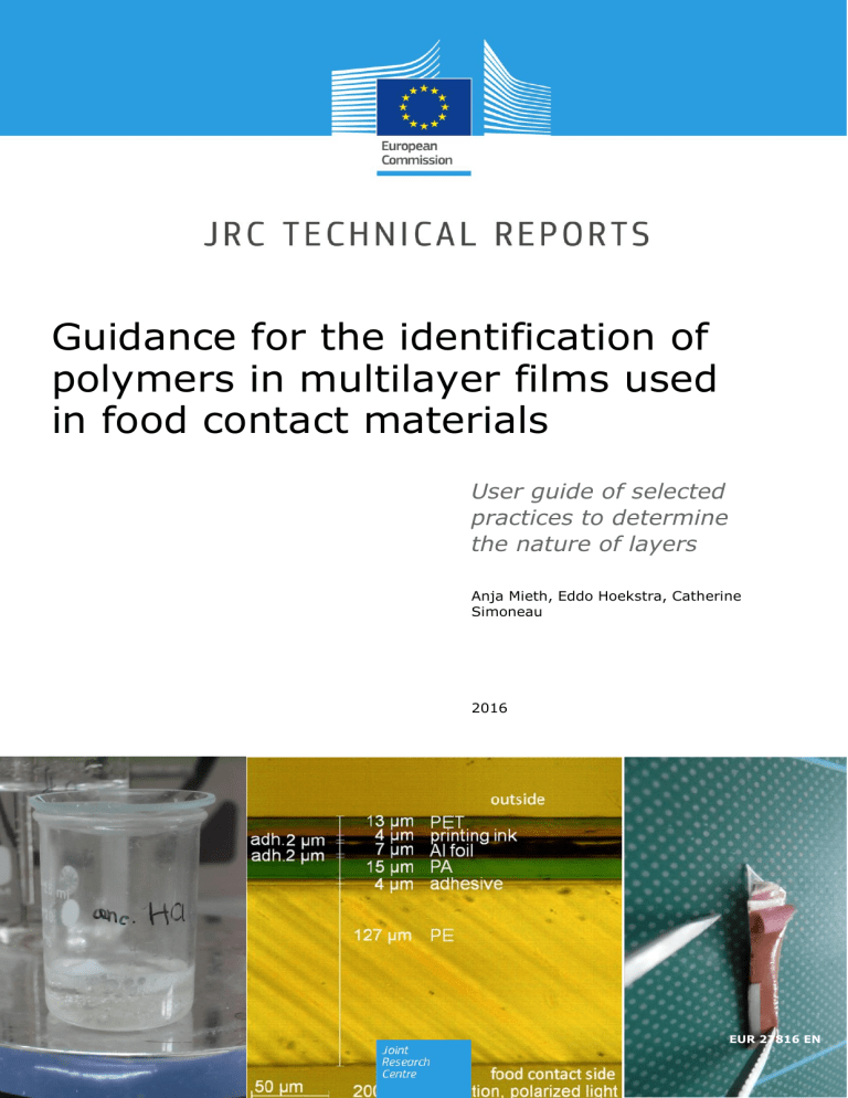
Guidance for the identification of polymers in multilayer films used in food contact materials User guide of selected practices to determine the nature of layers Anja Mieth, Eddo Hoekstra, Catherine Simoneau 2016 EUR 27816 EN 1 This publication is a Technical report by the Joint Research Centre, the European Commission’s in-house science service. It aims to provide evidence-based scientific support to the European policy-making process. The scientific output expressed does not imply a policy position of the European Commission. Neither the European Commission nor any person acting on behalf of the Commission is responsible for the use which might be made of this publication. Contact information Catherine Simoneau Address: Joint Research Centre, TP 260 E-mail: [email protected] JRC Science Hub https://ec.europa.eu/jrc JRC100835 EUR 27816 EN ISBN 978-92-79-57561-7 (PDF) ISSN 1831-9424 (online) doi:10.2788/10593 (online) © European Union, 2016 Reproduction is authorised provided the source is acknowledged. All images © European Union 2016 How to cite: Anja Mieth, Eddo Hoekstra, Catherine Simoneau - Guidance for the identification of polymers in multilayer films used in food contact materials: User guide of selected practices to determine the nature of layers; EUR 27816 EN; doi:10.2788/10593 2 Table of contents Abstract ............................................................................................................................. 4 1. Introduction ................................................................................................................ 4 2. Terms and definitions ................................................................................................... 5 3 Analysing the composition of a multilayer plastic film: Separation and identification of layers . 5 3.1 Scope .................................................................................................................... 5 3.2 Materials and reagents ............................................................................................. 6 3.2.1 Organic solvents (all of reagent grade)................................................................. 6 3.2.2 Organic and inorganic acids ................................................................................ 6 3.2.3 Reagents and materials for analytical tests ........................................................... 6 3.2.4 Other materials ................................................................................................. 6 3.3 Equipment .............................................................................................................. 6 3.3.1 Microtome and polarization microscope ................................................................ 6 3.3.2 ATR-FTIR system............................................................................................... 6 3.3.3 DSC system ...................................................................................................... 6 3.3.4 Glassware and other laboratory equipment ........................................................... 6 3.4 General composition of multilayer packaging films ....................................................... 7 3.4.1 Plastic polymers ................................................................................................ 7 3.4.2 Aluminium foil ................................................................................................... 8 3.4.3 Bonding of the different layers ............................................................................ 8 3.4.4 Coatings/lacquers ............................................................................................ 10 3.4.5 Non-food contact printings ............................................................................... 11 3.4.6 Direct food contact printings ............................................................................. 12 3.5 Analysis of the composition of a Multilayer film ......................................................... 12 3.5.1 Microtome sectioning and microscopy ................................................................ 12 3.5.2 Identification of food contact layer and outside layer by ATR-FTIR ......................... 13 3.5.3 Separation of layers ......................................................................................... 13 3.5.3.1 Selection of solvents .................................................................................. 13 3.5.3.2 Immersing the sample in solvent ................................................................. 15 3.5.4 Identification of the layers peeled off and laid open (ATR-FTIR spectroscopy) ........ 16 3.5.5 Specification of layers with DSC ........................................................................ 19 3.5.6 Special analytical tests for the identification of printing inks and coatings ............... 20 3.6 Examples ............................................................................................................. 21 References ....................................................................................................................... 34 Annex 1: Example of protocol for operating a rotary microtome .............................................. 37 Annex 2: Example of protocol for microscopy of microtome cross-sections ............................... 50 Annex 3: Example of protocol for handling/maintenance of an ATR-FTIR spectrometer .............. 67 3 Abstract This guidance describes how to characterize the composition of a multilayer plastic film for food packaging, with respect to the consecutive order of the layers and their identity. It provides background information on the general composition of multilayer plastic packaging and it illustrates in detail the separation of layers for some examples. It also provides in annexes additional information related to the use of a microtome and of optical microscopy using one common instrument for illustrative purposes. 1. Introduction Being able to identify the composition of a packaging film is essential to confirm the correctness of specifications given in the Declaration of Compliance that accompanies all food contact materials made of plastic (see EU 10/2011 Art. 15 and Annex IV (3) [1]). Furthermore, each type of polymer, printing, coating and adhesive contains typical additives and monomers or oligomers with a potential for migration, while at the same time certain layers can act as functional barriers for particular migrants. Therefore, knowing the composition of a multilayer film enables laboratories to predict which migrants can be expected from the packaging film and thus also helps to check the plausibility of obtained migration results. A PT was organised in 2013 which for the first time aimed at estimating performance of qualitative analysis. The aim of the exercise was to test the laboratories' ability to identify unknown plastics materials via quick screening tools. The results demonstrated a spread and some level of difficulty for laboratories to identifying particular multilayer materials. A follow up was organised in 2014 specifically for multilayers, in order to ensure improved laboratory performance. Considering there are no reference methods and the novelty to tackle assessments of qualitative nature, EU-RL FCM produced a number of methods descriptions to assist NRLs in doing this type of analysis. The descriptions have been integrated in the present guidelines. 4 2. Terms and definitions AlOx ATR BOPP CN DMSO DSC EAA EB EMA EVA EVOH FTIR HDPE ionomers LDPE LLDPE PA PA6 PA6/66 PA PA6/MXD6 PA66 PAMXD6 PC PE PEN PET PET-G PMMA PP PS PTFE PUR PVC PVDC SiOx UV aluminium oxide attenuated total reflection biaxially-oriented polypropylene cellulose nitrate (nitrocellulose) dimethyl sulfoxide differential scanning calorimetry ethylene acrylic acid copolymer electron beam ethylene methyl acrylate copolymer ethylene vinyl acetate copolymer ethylene vinyl alcohol copolymer Fourier Transform Infrared high density polyethylene EAA or EMA neutralised with cations (e.g. Na+, Zn2+, Li+) low density polyethylene linear low density polyethylene polyamide PA homopolymer made from caprolactam copolymer made from caprolactam, hexamethylenediamine and adipinic acid PA copolymer made from caprolactam, m-xylylenediamine and adipinic acid PA homopolymer made from hexamethylenediamine and adipinic acid PA homopolymer made from caprolactam, m-xylylenediamine and adipinic acid polycarbonate polyethylene polyethylene naphthalate polyethylene terephthalate glycol-modified polyethylene terephthalate polymethyl methacrylate polypropylene polystyrene polytetrafluorethylene polyurethane polyvinylchloride polyvinylidene chloride silicon oxide ultraviolet. 3 Analysing the composition of a multilayer plastic film: Separation and identification of layers 3.1 Scope This procedure describes how to characterize the composition of a multilayer plastic film for food packaging, meaning the consecutive order of the layers and their identity. It provides background information on the general composition of multilayer plastic packaging and it illustrates in detail the separation of layers for some examples. 5 3.2 Materials and reagents 3.2.1 Organic solvents (all of reagent grade) Acetone, butyl acetate, carbon disulfide, chloroform, cyclohexanone, cycloheptanone, din-butyl sulfoxide, N,N-dimethylacetamide, N,N-dimethylformamide, dimethyl sulfoxide, ethyl acetate, methylene chloride, methyl ethyl ketone, tetrahydrofuran, toluene, pxylene 3.2.2 Organic and inorganic acids Formic acid, reagent grade, ≥95% Hydrochloric acid, reagent grade, 37% Sulfuric acid, reagent grade, 95-98 % 3.2.3 Reagents and materials for analytical tests Sodium hydroxide, reagent grade, ≥98% Diphenylamine Copper wire 3.2.4 Other materials Cotton sticks Paper towels Glass beads 3.3 Equipment 3.3.1 Microtome and polarization microscope Boeckeler S300 rotary microtome Zeiss Axioskop 2 MAT, equipped with AxioCam MRc 3.3.2 ATR-FTIR system Perkin Elmer Spectrum One FT-IR Spectrometer Perkin Elmer Universal ATR Sampling Accessory Software: Spectrum Version 6.3.1, Perkin Elmer 3.3.3 DSC system TA Instruments DSC Q100 TA Instruments DSC Refrigerated Cooling System Software: Advantage for Q Series Version 2.5, Thermal Advantage Release 4.7.3 3.3.4 Glassware and other laboratory equipment 25 mL and 50 mL glass beakers Watch glasses, diameter 4-5 cm Glass test tubes Tweezers Scissors Heating plate Bunsen burner 6 3.4 General composition of multilayer packaging films 3.4.1 Plastic polymers Several plastic polymers, copolymers and blends are used for the production of multilayers. The most important polymers are polyethylene (LDPE, HDPE, LLDPE), polypropylene, polyamide (PA6, PA6/66, PA6/MXD6), polyethylene terephthalate, polystyrene, ethylene vinyl alcohol, polyvinylidene chloride, ethylene vinyl acetate and polycarbonate ([2]-[14]). Polyvinyl chloride may also be used as a plastic layer in multilayer food packaging ([1]). Table 1 lists the most common polymers in multilayer packaging materials, their functions in the packaging and some applications. Table 1 Plastic polymers used in multilayer food packaging ([1], [4], [5], [6], [8], [9], [18], [26], [27], [28], [35]) Plastic polymer Functions in multilayers Applications Polyethylene (PE) heat-sealable food contact layer moisture barrier can be combined with gas/aroma barriers (e.g. PA, EVOH) breathable packaging for fresh produce (LDPE, HDPE) carton liners (LLDPE) Polypropylene (PP) moisture barrier to provide mechanical strength can be coated with heat seal coatings (PVDC, acrylate) can be combined with gas/aroma barriers (e.g. PVDC coatings, PA, EVOH) modified atmosphere packaging thermoformed containers for microwavable packaging, hotfilled packaging Polyamide (PA) gas/aroma barrier to provide mechanical strength heat resistance can be used as outside layer of a heat seal film → film will not stick to the sealing bar surface boil-in-bag packaging thermoformed packaging Polyethylene terephthalate (PET) gas/aroma barrier moisture barrier to provide mechanical strength heat resistance plastic bottles for carbonated softdrinks, meat and cheese packaging, snack food wrapper boil-in-bag, sterilisable pouches, ovenware containers Polystyrene (PS) gas permeability printability can be combined with gas/aroma barriers (coextruded or laminated) → commercially available structures: e.g. PS/PVDC/PS, PS/PVDC/PE, PS/EVOH/PE, PS/EVOH/PP breathable packaging for fresh produce (e.g. fresh-meat packaging) printable outside layers Ethylene vinyl alcohol (EVOH) oxygen barrier needs to be protected from moisture → often sandwiched (coextruded) between PE or PP, in some applications also sandwiched between PET, PA or PS modified atmosphere packaging packing of oxygen-sensitive food Polyvinylidene chloride (PVDC) gas/aroma and/or moisture barrier to protect the surface from scratches and abrasion heat-sealable food contact layer often copolymers of vinylidene chloride and ester-type monomers (e.g. ethyl acrylate) modified atmosphere packaging applied as coating or coextruded film 7 Plastic polymer Functions in multilayers Applications Ethylene vinyl acetate (EVA) moisture barrier adhesion layer (tie layer) for co-extrusion of polar (e.g. PA, PET-G) and non-polar (e.g. PE) polymers heat-sealable food contact layer; heatsealable extrusion coatings on PET or BOPP films modified atmosphere packaging applied as coating or coextruded film Polycarbonate (PC) heat resistance mechanical strength moisture barrier microwavable packaging, hotfilled packaging modified atmosphere packaging barriers for fruit juice cartons Polyvinylchloride (PVC) gas/aroma barrier mechanical strength fresh food packaging (e.g. PVC/PE films) modified atmosphere packaging (e.g. PVC/EVOH/PE films) Polyethylene naphthalate (PEN) gas/aroma and moisture barrier heat resistance Glycol modified polyethylene terephthalate (PET-G) heat-sealable food contact layer Ethylene acrylic acid (EAA) extrusion coating tie layer between aluminium foil and other polymers heat-sealable food contact layer for hot refills, rewashing, reuse beverage bottles (e.g. beer) 3.4.2 Aluminium foil Many packaging materials, especially for high value foods (e.g. dried soups, herbs, spices), contain a layer of aluminium foil as it is an effective barrier against moisture, air, odours and UV light [26]. 3.4.3 Bonding of the different layers Multilayer films can be prepared by co-extrusion or by lamination. Co-extrusion can be performed as blown film extrusion or cast extrusion [14]. In both cases, granules of the different plastics are melted separately and the melts are brought together in the extruder. The combined plastics then are extruded as a single product in which the coextruded layers bond directly to each other. In order to achieve bonding of two molten polymer layers during co-extrusion, it can be necessary to apply a third polymer ("extrudable adhesive") in between. For example, anhydride-modified polyethylene or anhydride-modified EVA can act as an extrudable adhesive in the co-extrusion of PE and PA [9]. In contrast to co-extrusion, lamination combines two or more plastic or non-plastic materials (e.g. aluminium foil) in web-form by applying a certain type of adhesive between them. Three main principles of lamination can be distinguished. These are extrusion lamination, adhesive lamination and wax/hot melt lamination. In extrusion lamination, a molten polymer (e.g. LDPE) is applied to one of the webs and a second web is fed from the top. In adhesive lamination, an adhesive is applied instead. These can be solvent based adhesives ("dry bond lamination"), one- or two-component water 8 based adhesives ("wet-bond adhesion"), two-component solid adhesives ("solventless lamination"), UV-curing or electron beam-curing adhesives ([14], [25]). In wax/hot melt lamination, a layer of molten wax or hot melt is applied between two webs and then cooled to bond them together. The wax remains heat sensitive. Therefore this technique is not suitable if the packaging will be heat-sealed afterwards or filled very hot ([9], [14]). Table 2 lists some of the most important adhesives for the different types of lamination and their applications. Table 2 Adhesives and waxes/hot melts used for lamination ([9], [14], [39], [40]) Type of lamination extrusion lamination Adhesive Examples of selected applications LDPE multilayer-multimaterial packaging (combination of plastic or aluminium foil with paper or paperboard → e.g. fruit juice cartons) EVA bonding PE to PVC acid-containing adhesives: EAA or EMA ionomers terpolymers of ethylene, acid and acrylate (e.g. methyl acrylate or isobutyl acrylate) used to bond with aluminium foil anhydride modified polyolefins: terpolymers of ethylene, maleic anhydride and acrylates (e.g. ethyl acrylate, butyl acrylate) dry bond lamination flexible packaging → lamination of plastic films (bags, pouches, wraps for snack food, meat and cheese packs, boil-in-bag food pouches) solvent based PUR adhesives poly(vinyl acetate) emulsion (or coploymers of vinyl acetate and ethylene or acrylic esters) crosslinking acrylic-vinyl acetate copolymer emulsions snack packages (e.g. potato chip bags) acrylic emulsion pressure-sensitive adhesives pressure-sensitive labels polyurethane dispersions lamination of plastic films in mediumperformance flexible packaging where some chemical resistance is required UV-curing acrylics and acrylates, epoxies, polyurethanes, polyesters, silicones, and vinyl and vinyl esters flexible plastic packaging (at least one of the webs must be transparent to allow UV penetration) EB-curing see UV-curable adhesives solventless lamination polyurethanes wet-bond adhesion hot-melt lamination EVA ethylene-butyl acrylate 9 Type of lamination Adhesive Examples of selected applications LDPE for paper bonding constructions, case seaming, bag seaming and sealing block copolymers of styrene, butadiene or isoprene hot-melt pressure-sensitive adhesives for tapes and labels, attachment of PEbase cups to polyester soft-drink bottles, sealing of film-laminated frozen-food cartons moisture curing PUR hot melts 3.4.4 Coatings/lacquers Coatings or lacquers can be applied to the inside (food contact layer), outside (non-food contact layer) or in between layers to enhance the appearance or alter the physical properties of the packaging [17]. An overview of the most common types of coatings is given in Table 3. Table 3 Coatings used in multilayer plastic food packaging ([8], [14], [16], [17], [26], [31]). *Ionomers: EAA or EMA neutralised with cations (e.g. Na+, Zn2+, Li+) Type of coating Purpose Substances in use protective coatings to protect the surface from mechanical damage nitrocellulose coatings, epoxy resins, reactive polyurethane systems, ionomers heat-seal coatings to allow heat sealability for non-sealable materials (e.g. flexible aluminium lids for yoghurt containers, polypropylene) EAA, EVA, PVDC, PMMA, ionomers* primers to improve the bond between a substrate and an otherwise incompatible coating polyethyleneimines, EAA dispersions, reactive polyethylene amines cold seal coatings coatings that can be sealed self-to-self using just pressure (e.g. for flow-wrapping heat-sensitive products like chocolate or ice cream) blends of acrylic resins and latex; synthetic rubber release lacquers applied to the opposite surface of a plastic film that is coated with coldseal to prevent sticking and to promote easy unwinding from the reel PA-based coatings antimists to prevent formation of condensate droplets/fogging on the food contact side (e.g. in fresh salad packaging) PVDC coatings gloss/matt coatings to alter the visual perception of parts of the design PVDC, PMMA, nitrocellulose coatings gas and moisture barrier barrier to gases/odours/flavours and moisture PVDC, PMMA, SiOx, AlOx, aluminium vapour coating UV light barrier barrier to UV light aluminium vapour coating 10 3.4.5 Non-food contact printings Parts of the sample may also be covered with printings. A printing can be applied as a surface printing on the outside (i.e. non-food contact side) of the multilayer packaging or as a reverse printing where it is trapped between two layers of the multilayer packaging ([25], [29]). In reverse printing, the printing is usually applied to the inside surface of the outermost plastic layer of the packaging ([25]) (see Figure 1). This way, the printing is best protected against mechanical damage (scratching, rubbing) and moisture, as well as set-off (i.e. transfer of printing ink components to the food contact layer when the packaging film is stored in staples or rolled) ([29]). Figure 1 Laminate with reverse printing (PA/print//PURPE) after boiling in ethyl acetate for 2 min To protect surface printings (e.g. improve rub resistance) and to provide gloss or matt effects, overprint coatings can be applied. They have the same composition as the corresponding inks but do not contain pigments [15]. An overview on the main compositions of the most commonly used ink systems is provided in Table 4. Table 4 Printing ink systems for non-food contact applications ([16], [19], [20], [21], [22]) Printing ink system Principle composition surface printing inks solventbased inks pigments binders: nitrocellulose, maleic resin, polyvinyl butyral, polyamide, polyurethane solvents: alcohols (ethanol, isopropanol), esters (ethyl acetate, isopropyl acetate), ethoxypropanol additives: plasticisers, slip additives (lubricants), adhesion promoters UV-curing inks (free radical curing) Pigments vehicles: oligomers: epoxy acrylates, polyester acrylates, polyether acrylates, urethane acrylates; monomers/reactive diluents: di-, tri-, tetrafunctional acrylates photoinitiators additives: waxes (PE/PTFE waxes), silicone oils, stabilisers cationic UV-curing inks pigments cationic photoinitiators: triarylsulphonium salts, diaryliodonium salts vehicles: diepoxide compounds and vinyl ethers additives electron beamcuring inks pigments vehicles: oligomers: epoxy acrylates, polyester acrylates, polyether acrylates, urethane acrylates 11 Printing ink system Principle composition monomers/reactive diluents: di-, tri-, tetrafunctional acrylates additives waterbased inks pigments binders: styrene-acrylic co-polymers, acrylic co-polymers, maleic resins solvents: water, isopropanol, glycol ether, propylene glycol additives: amines, biocides, defoamers, wetting agents, waxes (PE, PTFE), slip agents Reverse printing inks solventbased inks pigments binders: vinyl resins, polyurethane, nitrocellulose, polyamide solvents: alcohols (ethanol, isopropanol), esters (ethyl acetate, isopropyl acetate), ethoxypropanol additives waterbased inks pigments binder: acrylic resin solvents: water additives 3.4.6 Direct food contact printings For promotional reasons (e.g. special campaigns), sometimes also parts of the food contact layer may be printed (see Figure 2). For this purpose, special "direct food contact printing inks" are used. These are usually pigmented coatings based on vinyl resins or modified cellulose ([16], [23], [24]). Figure 2 Chocolate packaging with printing on the food contact layer 3.5 Analysis of the composition of a Multilayer film 3.5.1 Microtome sectioning and microscopy It is recommended to prepare a microtome section (usually of 5 µm thickness) of the sample first and investigate it using a transmitted light microscope with polarized light. This will allow you to see of how many layers the sample consists of and whether they are co-extruded or laminated. Adhesives in laminated samples are visible in the microtome section as thin layers of about 1-3 µm thickness between the polymer films. The coloration of the different layers in polarized light also gives a first idea about their identity. Transparent polyamide layers may turn green in polarized light and polyethylene terephthalate layers may appear in rainbow colours, while polyethylene or polypropylene remain colourless (see Figure 3). 12 Figure 3 Microtome sections of multilayer plastic laminates in polarised light 3.5.2 Identification of food contact layer and outside layer by ATR-FTIR To identify the food contact layer (inside) and the outermost non-food contact (outside) layer of the sample, ATR-FTIR spectra of the inside and outside of the multilayer film are recorded. ATR-FTIR settings: spectral range: 4000-600 cm-1 number of scans: 4 background: air (4 scans, 4000-600 cm-1) resolution: 4 cm-1 The spectra are identified by comparison to the spectra database, other available standard spectra and the information provided in section 0 Table 8. 3.5.3 Separation of layers The separation of the different layers can be achieved by taking advantage of their different solubility properties. Having identified the food contact layer and the outermost layer (coating, printing ink or polymer film), a proper solvent or acid can be selected to dissolve and remove these outer layers. Alternatively, it is possible to split laminated layers by dissolving the adhesive in between and to split samples that contain an aluminium layer by dissolving the aluminium. 3.5.3.1 Selection of solvents Solvents for plastic polymers An overview about the solubility of the most common plastic polymers used for the production of flexible packaging is given in Table 5. Table 5 Solvents and acids suitable to dissolve plastic polymers ([32], [33], [34]) Polymer/coating Soluble/decomposed in Polyethylene (PE) boiling p-xylene, boiling toluene, boiling dimethyl sulfoxide Polypropylene (PP) boiling toluene, boiling dimethyl sulfoxide Ethylene vinyl alcohol (EVOH) boiling N,N-dimethylformamide, hot concentrated sulfuric acid (turns black) 13 Polymer/coating Soluble/decomposed in Polyamide (PA) hot concentrated formic acid, 37% hydrochloric acid, hot concentrated sulfuric acid (96 w%), boiling N,N-dimethylformamide Polyethylene terephthalate (PET) boiling N,N-dimethylformamide (swells), hot concentrated sulfuric acid Polystyrene (PS) toluene, chloroform, cyclohexanone, butyl acetate, carbon disulfide, acetone, methylene chloride, ethyl acetate, boiling N,Ndimethylformamide*, hot concentrated sulfuric acid (96 w%) (turns black) Polyvinylchloride (PVC) tetrahydrofuran, cyclohexanone, N,N-dimethylformamide, methyl ethyl ketone Polyvinylidene chloride (PVDC) boiling xylene, boiling N,N-dimethylacetamide, boiling cycloheptanone, di-n-butyl sulfoxide Polycarbonate (PC) methylene chloride, boiling N,N-dimethylformamide*, boiling dimethylsulfoxide*, hot concentrated sulfuric acid (96 w%) (turns black), acetone (swells), boiling toluene (swells) Ethylene vinyl acetate (EVA) toluene *requires strong heating and long time Solvents for adhesives in laminated samples (PUR adhesives, Polyester acrylates) Most commonly used adhesives in the manufacturing of flexible food packaging are polyurethane (PUR) and acrylic or acrylate based adhesives. PUR adhesives can be easily dissolved in formic acid. Note that this procedure might also dissolve possible polyamide layers and others soluble in formic acid. An alternative can be to use ethyl acetate or other suitable solvents instead (see Table 6). Note that all samples which contain an aluminium foil or an aluminium vapour coating as well as reverse printed samples are somehow laminated to bond the aluminium layer or reverse printed film, respectively, to the other plastic layers. In conclusion, one can always try to immerse any sample containing an aluminium layer or any reverse printed sample in formic acid or ethyl acetate (or other suitable solvents) to dissolve the adhesive layers (see Figure 4). Figure 4 Sample (PET/print//PURAl foil//PURPE) before and after boiling in HCOOH for 5 min. Resulting 3 separated layers: PET/print, Al foil, PE (top and bottom view shown) Table 6 Solvents and acids suitable to dissolve adhesive ([32], [33]) Polymer/coating Soluble/decomposed in Polyurethane (PUR) formic acid, methylene chloride (swells), acetone (swells), ethyl acetate (swells) Polyacrylates boiling xylene, toluene, methylene chloride, acetone, ethyl acetate 14 Dissolving aluminium foil or aluminium vapour coatings An aluminium foil or an aluminium vapour coating (i.e. a thin aluminium layer applied by thermal evaporation) itself can be dissolved by immersion in concentrated hydrochloric acid (37 % w/w). If the complete sample is immersed, the sample will split in two parts, the one above and the one below the aluminium layer (see Figure 5). Figure 5 Sample with an inner aluminium foil before and after boiling in conc. HCl for 4 min (Al is partly dissolved) and 5 min (Al is completely dissolved) Removal of printings and coatings Ink films and coatings often can be wiped off with a paper towel or cotton stick soaked in a suitable solvent. For example, acrylate and nitrocellulose coatings usually can be wiped off with ethyl acetate. If this is not successful, the sample can be immersed in solvent and, if necessary, heated until the coating is completely dissolved (NOTE: sometimes additional rubbing with a paper towel is required). An overview about suitable solvents for the most common coatings and printing ink films is provided in Table 7. Table 7 Solvents and acids suitable to dissolve binders of printing inks and coatings ([32], [33], [34]) Polymer/coating Soluble/decomposed in Polyvinylidene chloride (PVDC) boiling xylene, boiling N,N-dimethylacetamide, boiling cycloheptanone, di-n-butyl sulfoxide Ethylene vinyl acetate (EVA) toluene Nitrocellulose (CN) ethyl acetate, acetone Polyacrylates boiling xylene, toluene, methylene chloride, acetone, ethyl acetate Polymethyl methacrylate (PMMA) toluene, methylene chloride, acetone, ethyl acetate Polyurethane (PUR) formic acid, methylene chloride (swells), acetone (swells), ethyl acetate (swells) Aluminium metallisation 37% hydrochloric acid 3.5.3.2 Immersing the sample in solvent Cut a sample piece of about 1 cm x 3 cm. It should be of asymmetric shape (e.g. trapezoid) to distinguish between the inner and outer side of the multilayer (and later on the layers peeled off) (see Figure 6). 15 Inner side (food conact) Outer side (non-food contact) Figure 6 Sample preparation Approximately 5 to 10 mL of solvent or acid (selected according to 0) are filled into a 25 or 50 mL glass beaker. To prevent boiling retardation later on, add 2-3 glass beads. The sample piece is added (use tweezers!) and the beaker is covered with a watch glass. Never put more than 1 sample piece into the beaker at the same time. Otherwise there is a risk to lose track of which sample eventually split off layers belong. The beaker is placed on a hot plate and heated carefully to make the solvent boil. Depending on the type of plastic, it may take up to 10-15 min until the respective sample layer is dissolved completely in the boiling solvent. However, often it is useful to immerse the sample only for a short time until parts of the respective layer are dissolved and then to tear or peel off the upper layer. Therefore, check every 1-2 minutes whether parts of the sample are already swollen or dissolved (especially at the edges of the sample piece). If so, take out the sample with a pair of tweezers, rinse it with a nontoxic solvent (e.g. ethanol) or distilled water to remove adhering solvent and acid, respectively, dry it with a paper towel and then try to peel off the upper layer using some tweezers. Do not forget to remove the beaker from the hot plate when you are finished. Heating can also be performed in glass test tubes using a Bunsen burner. This process is faster but more difficult to control. Some coatings and adhesives as well as aluminium foil or aluminium vapour coatings are dissolvable already at room temperature but the process may take 1-24 h. By additional heating (hot plate or Bunsen burner), the process can be accelerated and shortened to a couple of minutes. 3.5.4 Identification of the layers peeled off and laid open (ATR-FTIR spectroscopy) Once a layer is peeled off, record an ATR-FTIR spectrum of the surface laid open. It is advisable to record also ATR-FTIR spectra of top and bottom of the layer peeled off. This will help to make sure that the layer peeled off was a monolayer. If the respective layers were laminated, the adhesive was laid open. It may stick to both sides, i.e. the upper layer peeled off and also to the remaining multilayer (see Figure 7). PUR adhesive Figure 7 PUR adhesive sticks to the aluminium foil and partly also to the PA layer after separation 16 ATR-FTIR settings: spectral range: 4000-600 cm-1 number of scans: 4 background: air (4 scans, 4000-600 cm-1) resolution: 4 cm-1 The spectra are identified by comparison to the spectra database or other available standard spectra. An overview on characteristic bands in FTIR spectra of some plastic polymers, binders for printing inks and adhesives is also provided in Table 8. Table 8 Characteristic bands in FTIR spectra ([32], [37], [41], [42]). Polymer ATR-FTIR spectrum 100 90 Polyethylene νaCH2 and νsCH2 δCH2 Wave number** [cm-1] 3000-2840 1463 ρCH2 725 νaCH2 and νsCH2 νaCH3 and νsCH3 δCH2 and δaCH3 δsCH3 ρCH3 νC-C 3000-2840 3000-2840 1459 1376 1167 998, 973 ρCH2 840 ν=CH νaCH2 and νsCH2 νC=O νPh νC(=O)O and δ=CH νO-C and δ=CH δ=CH γ=CH 3150-3000 3000-2840 1718 1600-1325 1260 1100 1018 971, 872 γPh 726 ν=CH νaCH2 and νsCH2 overtones νPh γ=CH 3150-3000 3000-2840 2000-1660 1600-1375 1180, 1154, 1069, 1028 906, 842, 758 γPh 700, 540 Vibration* [%T] 80 70 60 CH2 CH2 n 50 40 30 4000 3500 3000 2500 2000 1500 1000 500 [cm-1] 100 Polypropylene 90 [%] 80 CH3 70 CH CH2 n 60 50 4000 3500 3000 2500 2000 1500 1000 500 [cm-1] 105 95 85 Polyethylene terephthalate 75 [%T] 65 O O 55 O O CH2 CH2 45 n 35 25 15 4000 3500 3000 2500 2000 1500 1000 500 [cm-1] 100 Polystyrene 90 80 [%] 70 60 δ=CH 50 40 CH CH2 30 n 20 10 4000 3500 3000 2500 2000 1500 1000 [cm-1] 17 500 Polymer ATR-FTIR spectrum Vibration* ν=CH νaCH3 and νsCH3 νC=O νPh νaC-O-C 105 100 95 90 Polycarbonate [%] 85 80 O CH3 75 O O CH3 n δ=CH 70 65 60 4000 3500 3000 2500 2000 1500 1000 500 [cm-1] Polyamide 6 100 90 O 80 NH n [%T] 70 60 50 40 30 20 4000 Ethylene acetate vinyl 3500 3000 2500 [cm-1] 2000 1500 1000 500 100 90 CH2 CH2 CH2 80 [%T] O n O 70 CH3 m 60 50 40 4000 3500 3000 2500 2000 1500 1000 Wave number** [cm-1] 3150-3000 3000-2840 1773 1506 1228 1192, 1162, 1126, 1015 γ=CH 831 νNH νaCH2 νsCH2 νC=O δNH and νCN δCH2 ωCH2 and τCH2 3303 2935 2860 1635 1539, 1276 1465 1200 γNH and γC=O 690 νaCH2 and νsCH2 νaCH3 and νsCH3 νC=O δCH2 and δaCH3 δsCH3 νC(=O)O νO-C and ρCH3 ρCH2 3000-2840 3000-2840 1740 1469 1371 1241 1020 720 νOH...O νaCH2 and νsCH2 νaNO2 νsNO2 νC-O ρCH2 νC(=O)O νO-C νN-C ρCH2 νaCH2 and νsCH2 δCH2 γCH2 νsC-C νCCl2 3400 3000-2840 1654 1279 1063 841 1223 1111 1000 809 3000-2840 1405 1064 1044 600, 655 500 [cm-1] Nitrocellulose (cellulose nitrate) 100 90 80 O 2N NO 2 O H O O O H [%] NO 2 O H HH O O H O H O H O NO 2 H O H NO 2 O 2N 70 60 50 n 40 30 4000 3500 3000 2500 2000 1500 1000 500 2000 1500 1000 500 [cm-1] Polyvinylidene chloride 105 100 95 90 Cl 85 Cl [%T] CH2 C 80 n 75 70 65 60 55 4000 3500 3000 2500 [cm-1] 18 Polymer ATR-FTIR spectrum Polyester acrylate Vibration* 105 95 85 R 75 [%T] O O 65 55 CH2 n R: aromatic polyester 45 35 25 4000 3500 3000 2500 2000 1500 1000 500 [cm-1] ν=CH (aryl) νaCH2 and νsCH2 νC=O νC=C (vinyl ester) νPh δCH2 δC=C (vinyl ester) νC(=O)O νasC(=O)-O-C (acrylate polymer) γC=C (vinyl ester) γPh Wave number** [cm-1] 3150-3000 3000-2840 1728 1636***, 1610*** 1600-1325 1453 1408*** 1242 1163 810*** 726 *: νa: antisymmetric stretching vibration, νs: symmetric stretching vibration, δ: in-plane deformation, γ: out-of-plane, ρ: rocking vibration, ω: wagging vibration, τ: twisting vibration **: The wave numbers given refer to transmission spectra and differ slightly from those obtained by ATR measurement.) ***: The characteristic vinyl ester moieties CH2=CH-C(=O)-O~ of UV/EB curable acrylate printings or adhesives react upon curing and form the acrylate polymer backbone. As a consequence, the respective characteristic vinyl ester bands at 1636 (νC=C), 1610 (νC=C), 1408 (δC=C) and 810 (γC=C) cm -1 decrease and are no longer present/barely-there if the printing ink or adhesive was well cured ([41], [42], [43], [44]). 3.5.5 Specification of layers with DSC The separated polymer layers can be further characterised by their melting points with differential scanning calorimetry (DSC) to confirm the results obtained by ATR-FTIR and to specify the findings. DSC will enable to distinguish between LDPE, LLDPE and HDPE as well as PA 6, PA 66 and PA MXD6 and blends of different polymers [30]. You can also perform a DSC of the complete laminate. This will give an idea about the types of plastic polymers that are contained in the multilayer sample (see Figure 8). 2 ML2_complete ML2_PE ML2_PET ML2_PA 0 Heat Flow [W/g] -2 -4 219.78°C -6 -8 (PA6) 112.78°C -10 252.87°C (PET) (LDPE) -12 -14 253.24°C -16 220.88°C -18 (PA6) 109.84°C 117.19°C -20 0 50 100 150 200 250 (PET) 300 Temperature [°C] Figure 8 DSC diagrams of a laminate (PET/acrylate printing//PURAl foil//PURPA//PURPE) and its single polymer layers (PET, PA, PE) after separation 19 DSC settings: Start temperature: 50°C End temperature: 300°C Heating rate: 20°C/min Reference: air Amount of sample: for separated monolayers: 0.5-2.5 mg for multilayer samples: 10 mg An overview about melting temperatures of some common plastic polymers in flexible packaging is provided in Table 9. Table 9 Glass transition (Tg) and crystalline melting temperatures (Tm) of some common plastic polymers ([30], [36], [38]) Polymer Tg [°C] Polyethylene Tm [°C] 105-113 (LDPE) -20 120 (LLDPE) 135 (HDPE) Polypropylene 5 160 Polyvinylchloride 80 - Polyvinylidene chloride -18 190 Atactic polystyrene 95 - Isotactic polystyrene 100 230 Polyethylene terephthalate 67 256 Polycarbonate (made of Bisphenol A) 150 225 Polyamide 6 50 215 Polyamide 66 60 266 Ethylene vinyl acetate 80-110 (depends on EA content) 3.5.6 Special analytical tests for the identification of printing inks and coatings Besides identification with ATR-FTIR spectroscopy, some of the printing inks and coatings can also be identified by simple chemical tests. Nitrocellulose (cellulose nitrate, CN) - Diphenylamine test To prepare the reagent, 0.1 g diphenylamine are suspended in 30 mL water and then 100 mL concentrated sulfuric acid are added carefully. A drop of the freshly prepared solution is applied to the sample surface. The reaction is positive for nitrocellulose if the reagent turns dark blue. [33] Polyvinylidene chloride (PVDC) – Reaction with pyridine and Beilstein test A drop of pyridine is applied to the sample surface. After 2-5 min, a drop of 5% w/v methanolic sodium hydroxide solution is added (1 g sodium hydroxide dissolved in 20 mL methanol). The reaction is positive for PVDC if the surface turns brown-black 20 immediately. PVC instead would show a red-brown colour first, and after approx. 5 min the colour would turn into black-brown. [33]. In addition, the Beilstein test can be performed to proof the presence of chlorine. A copper wire is heated in a Bunsen flame until the flame is colourless. The wire is allowed to cool down. Then a piece of the sample is placed on it (the chlorine containing layer should face the wire) and the copper wire plus sample are heated at the edge of the colourless part of the Bunsen flame. A green or blue-green flame will indicate the presence of chlorine when the sample burns. [33] 3.6 Examples Laminate 1 (PET/polyester acrylate printing//PURAl foil//PURPA//PURPE) Sample: food contact side outside Step I: General observations The sample is printed (not yet known whether it is a surface or reverse printing) The sample contains an aluminium layer (not yet known whether it is an aluminium foil or an aluminium vapour coating). Step II: Microtome section (5 µm thickness) adh. adh. polymer film 1 printing ink aluminium foil polymer film 2 adhesive polymer film 3 The microtome section displays that: The sample consists of 3 different polymer films and an aluminium foil. Polymer film 1 appears in rainbow colours in polarized light and therefore is likely to consist of PET. Polymer film 2 appears green, so it might be a polyamide layer. The printing was applied as a reverse print on the bottom side of polymer film 1. 21 There are no co-extruded layers, all layers are laminated with adhesives. schematic composition: polymer 1 print adhesive aluminium foil adhesive polymer 2 adhesive polymer 3 Step III: ATR-FTIR of the inside (food contact layer) and outside (non-food contact) ATR-FTIR: complete laminate(outside) = PET ATR-FTIR: PET polymer 1 = PET print adhesive Al foil adhesive polymer 2 adhesive ATR-FTIR: complete laminate(inside) = PE ATR-FTIR: polymer 3 = PE PE Results: The outside layer consists of PET. The food contact layer consists of PE. Step IV: Separation of layers From the information gained in step I-III, the following options arise to start the separation of the layers: 1) The food contact layer consists of PE. → Try to dissolve/swell the PE layer in DMSO, toluene or xylene and remove it. 2) The sample contains an aluminium foil. → Try to dissolve the aluminium in conc. HCl to split the sample. 3+4) The sample contains an aluminium foil. The plastic films on top of and below the aluminium foil must be somehow adhered to it. → Try to dissolve the adhesive on top of and below the aluminium foil in conc. HCOOH or ethyl acetate to split the sample. 22 Step IV: Separation of layers – option 1 – start with DMSO 1 complete laminate Cut a piece of the sample (complete laminate) (approx. 1 cm x 3 cm) and boil it for 1-2 min in DMSO to partly dissolve/swell the PE layer. 2 tear here! The edges of the PE layer are swollen and the adhesive that bonds polymer 2 to the aluminium foil is partly dissolved. → Part B (adhesive/polymer 2/adhesive/PE) can be peeled off from the bottom side of the Al foil. Part A (PET/print/adhesive/Al) cannot be further separated yet. 3 part A ATR-FTIR: - (Al) ATR-FTIR: PUR part B polymer 1 = PET ATR-FTIR: part A (top) = PET, part A (bottom) = no polymer/coating (aluminium), part B (top) = polyester urethane (adhesive), part B (bottom) = PE ATR-FTIR: PA tear here! 5 tear here! adhesive = PUR polymer 2 adhesive Dip part B in cold HCOOH and take it out immediately(!) to dissolve the PUR adhesive. → Rub its top side with a paper towel to remove the remaining PUR adhesive. → ATR-FTIR: part B (top, PUR adh. removed) = PA NOTE: If you immerse part B for >5 s in cold HCOOH, the PA will be dissolved. If you immerse it in hot HCOOH, the PA will be dissolved immediately. Separate the PA and PE layer of part B: Go back to step 3 → Having a closer look to the edges of part B, you can see that the PA layer (part B1) can be peeled off. 6 part B1 part A Al foil part B polymer 3 = PE Remove the PUR adhesive on top of part B to check what is below: 4 print adhesive part B2 peel off part B1 23 polymer 1 = PET print adhesive Al foil adhesive = PUR polymer 2 = PA adhesive polymer 3 = PE part B 7 part B1 ATR-FTIR: part B1 (top) = polyester urethane (adhesive), part B1 (bottom) = PA, part B2 part A part B2 (top) = polyester urethane (adhesive), part B2 (bottom) = PE polymer 1 = PET print adhesive Al foil adhesive = PUR polymer 2 = PA part B1 adhesive = PUR polymer 3 = PE part B2 Separate the PA and PE layer of part B: 8 part B Go back to step 3 → If you peel off the PA layer (part B1) 5-7, you can boil part B for acetate → the PUR adhesive PE swells/is partly dissolved were not able to as shown in step 2-3 min in ethyl between PA and 9 peel off the PA layer (part B1) tear here! polymer 1 = PET print 10 ATR-FTIR: part B2 part B1 adhesive Al foil part B1 (top) = polyester urethane (adhesive), part B1 (bottom) = PA, adhesive = PUR part B2 (top) = polyester urethane (adhesive), part B2 (bottom) = PE adhesive = PUR polymer 2 = PA polymer 3 = PE 11 ATR-FTIR: 12 PE part A Remove the PUR adhesive on top of part B2 to check what is below: polymer 1 = PET boil part B2 for at least 15 min in HCOOH and/or acetone, rub its top side with a paper towel to remove the remaining PUR adhesive → ATRFTIR: part B2 (top) = PE adhesive part B1 part B2 print Al foil adhesive = PUR polymer 2 = PA adhesive = PUR polymer 3 = PE Characterisation of the adhesive that bonds the reverse printed PET-film to the Al foil: Go back to step 3 → boil part A for 30 s in conc. HCl part B2 polymer 1 = PET print adhesive = PUR part A Al foil 13 adhesive = PUR ATR-FTIR: The Al foil on the bottom of part A is completely dissolved. → ATR-FTIR: part A (bottom, Al foil removed) = polyester urethane (adhesive) PUR 14 ATR-FTIR: PET To confirm that the “white layer” on the bottom side of part A is a reverse printing and to check what is below, try to rub it off with a paper towel soaked in ethyl acetate (requires some force!). → PUR adhesive and the printing are removed. → ATR-FTIR: part A (bottom, print removed) = PET → same like outside → PET-monolayer polymer 2 = PA adhesive = PUR polymer 3 = PE polymer 1 = PET print adhesive = PUR part A Al foil adhesive = PUR polymer 2 = PA adhesive = PUR polymer 3 = PE Characterisation of the printing: 15 ATR-FTIR: PET ATR-FTIR: Go back to step 3 → boil part A for 3-5 min DMSO → PET layer can be removed, printing remains on the aluminium foil → ATR-FTIR: aluminium foil (top) = polyester acrylate (printing) polymer 1 = PET print = acrylate adhesive = PUR Al foil adhesive = PUR polymer 2 = PA adhesive = PUR polyester acrylate polymer 3 = PE 24 part A Step IV: Separation of layers – option 2 – start with conc. HCl 1 complete laminate polymer 1 = PET Cut a piece of the sample (complete laminate) (approx. 1 cm x 3 cm) and boil it in conc. HCl to partly dissolve the aluminium foil print adhesive = PUR part A Al foil adhesive = PUR polymer 2 = PA adhesive = PUR polymer 3 = PE 2 ATR-FTIR: PET part A ATR-FTIR: PUR part B after 30 s the adhesive that laminates part A (PET/print) to the aluminium foil is (partly) dissolved, while the aluminium foil is only dissolved at its edges → peel off part A → ATR-FTIR: part A (top) = PET, part A (bottom) = polyester urethane (adhesive) polymer 1 = PET print adhesive = PUR Al foil adhesive polymer 2 adhesive ATR-FTIR: PUR 3 part B* immerse part B again for 10 s in boiling conc. HCl → the aluminium layer is dissolved immediately → part B* (adhesive/polymer 2/adhesive/PE) remains → ATR-FTIR: part B* (top) = polyester urethane (adhesive), part B*(bottom) = PE part A part B polymer 3 = PE polymer 1 = PET print adhesive = PUR 4 Separate polymer 2 and the PE layer: part B* 5 boil part B* for 2 min in ethyl acetate to partly dissolve/swell the adhesive between polymer 2 and the PE layer Al foil adhesive = PUR polymer 2 adhesive part B* polymer 3 = PE scratch/tear polymer 2 (part B1) became brittle already while boiling in conc. HCl → now it can be scratched off with a spatula 6 ATR-FTIR: part B1 (top) = polyester urethane (adhesive), part B1 (bottom) = PA, ATR-FTIR: PUR part B2 part B2 (top) = polyester urethane (adhesive), part B2 (bottom) = PE part B1 7 ATR-FTIR: PA part A ATR-FTIR: PET Go back to step 2 – To confirm that the “white layer” on the bottom side of part A is a reverse printing and to check what is below, try to rub it off with a paper towel soaked in ethyl acetate (requires some force!). → PUR adhesive and the printing are removed. → ATR-FTIR: part A (bottom, print removed) = PET → same like outside → PET-monolayer 25 polymer 1 = PET print adhesive = PUR Al foil adhesive = PUR polymer 2 = PA adhesive = PUR polymer 3 = PE part A Step IV: Separation of layers – option 3 – start with conc. HCOOH 1 complete laminate Cut a piece of the sample (complete laminate) (approx. 1 cm x 3 cm) and boil it for 1 min in conc. HCOOH to partly dissolve the adhesives above and below the aluminium foil. (Attention: If you boil the sample for >5min, polymer 2 (PA) is dissolved as well.) tear here! 2 Part A (PET/print/adhesive) can be peeled off from the top of the aluminium foil... tear here! ...and part B (adhesive/polymer 2/adhesive/PE) can be peeled off from the bottom of the aluminium foil. 3 ATR-FTIR: ATR-FTIR: PUR PUR part A part B → ATR-FTIR: polymer 1 = PET part A (top) = PET, part A (bottom) = polyester urethane (adhesive), adhesive = PUR part B (top) = polyester urethane (adhesive), part B (bottom) = PE adhesive = PUR print part A Al foil polymer 2 adhesive 4 Separate polymer 2 and the PE layer: part B part B polymer 3 = PE boil part B for 2 min in ethyl acetate to partly dissolve/swell the adhesive between polymer 2 and the PE layer 5 polymer 1 = PET print tear here! part B1 ATR-FTIR: adhesive = PUR part B1 (top) = polyester urethane (adhesive), part B1 (bottom) = PA, adhesive = PUR part B2 (top) = polyester urethane (adhesive), part B2 (bottom) = PE adhesive = PUR Al foil polymer 2 = PA polymer 3 = PE 6 ATR-FTIR: PA part B1 part B2 part B2 ATR-FTIR: PUR Go back to step 3 – Remove the PUR adhesive and the acrylate printing on the bottom side of part A to check what is below and to confirm that the "white layer" is a reverse printing as described in option 2 step 7 part A ATR-FTIR: PET polymer 1 = PET print adhesive = PUR Al foil adhesive = PUR polymer 2 = PA adhesive = PUR polymer 3 = PE 26 part A Step IV: Separation of layers – option 4 – start with ethyl acetate 1 complete laminate Cut a piece of the sample (complete laminate) (approx. 1 cm x 3 cm) and boil it for 1 min in ethyl acetate to partly dissolve/swell the adhesives on top of and below the aluminium foil. ATR-FTIR: 2 ATR-FTIR: - (Al) PUR part B part A The adhesive on the bottom of the aluminium foil is partly dissolved. → part B (adhesive/polymer 2/adhesive/PE) can be peeled off from the bottom side of the Al foil. → ATR-FTIR: part A (top) = PET, part A (bottom) = no polymer/coating (aluminium foil), part B (top) = polyester urethane (adhesive), part B (bottom) = PE polymer 1 = PET print adhesive part A Al foil adhesive = PUR polymer 2 adhesive 3 Separate polymer 2 and the PE layer: part B 4 part B polymer 3 = PE If necessary: Boil part B again for 1 min in ethyl acetate to partly dissolve/swell the adhesive between polymer 2 and the PE layer. polymer 2 (part B1) can be peeled off tear here! part B2 polymer 1 = PET part B1 print adhesive ATR-FTIR: part B1 (top) = polyester urethane (adhesive), part B1 (bottom) = PA, 5 part B1 ATR-FTIR: PA part B2 part B2 (top) = polyester urethane (adhesive), part B2 (bottom) = PE ATR-FTIR: Al foil adhesive = PUR polymer 2 = PA part B1 adhesive = PUR polymer 3 = PE part B2 polymer 1 = PET print PUR adhesive Al foil adhesive = PUR 6 Remove the PUR adhesive on top of part B2 as described in option 1 step 11 polymer 2 = PA adhesive = PUR polymer 3 = PE ATR-FTIR: PE 7 ATR-FTIR: PA Go back to step 2 to remove the PUR adhesive on top of the PA layer and confirm that there is no more layer on top of the PA → follow step 4 described in option 1 NOTE: If you try to immerse the isolated PA layer (part B1) in HCOOH (cold or hot) to remove the PUR adhesive on its top, the whole PA layer will be dissolved immediately! You have to go back to step 2 first and immerse the PA/PE laminate (part B) instead! part B2 polymer 1 = PET print adhesive Al foil adhesive = PUR polymer 2 = PA adhesive part B polymer 3 = PE polymer 1 = PET 8 ATR-FTIR: PUR Characterisation of the adhesive that bonds the reverse printed PET-film to the Al foil: Go back to step 2 and follow step 12-13 described in option 1 print adhesive = PUR Al foil adhesive = PUR polymer 2 = PA adhesive = PUR polymer 3 = PE 27 part A 9 ATR-FTIR: PET To confirm that the “white layer” on the bottom side of part A is a reverse printing and to check what is below, follow the descriptions in option 1 step 14 polymer 1 = PET 10 Characterisation of the printing: ATR-FTIR: ATR-FTIR: PET Go back to step 2 and follow step 15 described in option 1 polyester acrylate print = acrylate adhesive = PUR Al foil adhesive = PUR polymer 2 = PA adhesive = PUR polymer 3 = PE SUMMARY OF RESULTS: Identified composition of layers: PET/polyester acrylate printing//PURAl foil//PURPA//PURPE outside polymer 1 = PET print = polyester acrylate adhesive = polyester urethane aluminium foil adhesive = polyester urethane polymer 2 = PA adhesive = polyester urethane polymer 3 = PE food contact side 28 part A Laminate 2 (PA/polyester urethane printing//PURPE) Sample: outside food contact side Step I: General observations The sample is partially printed (surface or reverse printing → not yet known). The sample does not contain an aluminium layer. Step II: Microtome section - not available Step III: ATR-FTIR of the inside (food contact layer) and outside (non-food contact) ATR-FTIR: complete laminate (outside) = PA ATR-FTIR: complete laminate (inside) = PE Results: The outside layer consists of PA. The food contact layer consists of PE. Step IV: Separation of layers From the information gained in step I-III, the following options arise to start the separation of the layers: 1) The food contact layer consists of PE. → Try to dissolve/swell the PE layer in DMSO, toluene or xylene and remove it. 2) The outside layer consists of PA. → Try to completely dissolve the PA layer in conc. HCOOH to check what is below. 29 Step IV: Separation of layers – option 1 – start with DMSO 1 complete laminate Cut a piece of the sample (complete laminate) (approx. 1 cm x 3 cm) and boil it in DMSO to swell the PE layer on the bottom of the sample. 2 After 1-2 min, the upper layer starts to separate (see edges) but cannot yet be peeled off. Therefore, boil the sample for another 2 min in DMSO. 3 Part A can be peeled off from the top of the sample. The printing is laid open, it sticks to the bottom of part A and the top of part B. part A part B 4 part B ATR-FTIR: part A (bottom, unprinted) = PA, part A (bottom, green printing) = polyester urethane (printing), part A part B (top, unprinted) = polyester urethane (adhesive), part B (top, green printing) = polyester urethane (printing) 5 part B part A 6 Remove the printing from the bottom of part A and from the top of part B to check what is below: The printing can be rubbed off with a paper towel soaked in ethyl acetate. (If necessary, you can also boil the sample in ethyl acetate.) → ATR-FTIR: part A (bottom, print removed) = PA, part B (top, print+adhesive removed) = PE part A Check whether part A consists only of PA by dissolving it completely in cold HCOOH: Dip part A into cold HCOOH. 30 7 Part A is dissolved immediately. Only the green printing ink film remains in the beaker. → Part A seems to consist only of PA. 8 part B Check whether part B is really a monolayer or whether it consists of coextruded layers, e.g. two PE layers with an EVOH layer in between: boil part B for 5 min in conc. H2SO4 → if there was an EVOH layer in between, it would be decomposed (turn black) and you would see a thin black-brown line at the edges of the sample 9 Residues of the green printing on top of part B were decomposed (turned black). → Can be rubbed off with a paper towel. 10 Having a closer look to the edges of part B after treatment with H2SO4, you can see that there is no black line visible. → There is no EVOH layer sandwiched between two PE layers. → The PE layer is probably a monolayer. 31 Step IV: Separation of layers – option 2 – start with HCOOH 1 complete sample Cut a piece of the sample (complete sample) (approx. 1 cm x 3 cm) and boil it for 15 min in HCOOH to completely dissolve the PA layer on top of the sample 2 ATR-FTIR: top (PA removed, green printed parts) = polyester urethane (printing), top top (PA removed, unprinted/clear parts) = polyester urethane (adhesive), bottom bottom = PE Remove the printing and adhesive to check what is below: 3 The printing and adhesive can be rubbed off with a paper towel soaked in ethyl acetate. (If necessary, you can also boil the sample in ethyl acetate.) → ATR-FTIR: top (PA, print and adhesive removed) = PE 4 Now that you know, that the sample is reverse printed, you should check that you did not dissolved anything else during step 1 apart from the PA layer. Therefore, try to separate the PA layer from the complete sample and check its bottom side: Take a piece of the complete sample and boil it for 2 min in ethyl acetate to partly dissolve/swell the adhesive that bonds the reverse printed PA film to the rest of the sample. 5 part A The PA layer (part A) can be peeled off. The printing sticks to both sides (the bottom of part A and the top of part B). part B 6 part A part B Remove the printing from the bottom of part A and from the top of part B to check what is below: part B part A 7 part B The printing can be rubbed off with a paper towel soaked in ethyl acetate. (If necessary, you can also boil the sample in ethyl acetate.) → ATR-FTIR: part A (bottom, print removed) = PA, part B (top, print+adhesive removed) = PE Check whether part B is really a monolayer or whether it consists of coextruded layers, e.g. two PE layers with an EVOH layer in between: → follow the descriptions in option 1 step 8-10 32 8 Check whether part A consists only of PA by dissolving it completely in cold HCOOH: part A Go back to step 6 and follow the descriptions in option 1 step 67 SUMMARY OF RESULTS: Identified composition of layers: PA/polyester urethane printing//PURPE outside PA print = polyester urethane adhesive = polyester urethane PE food contact side 33 References [1] Alfatherm SpA (2014). Packaging Films. Retrieved http://www.alfatherm.it/en/applications/packaging-films/ April 24, 2014, from [2] Sidwell JA (1992). Food Contact Polymeric Materials. Rapra Review Reports. Vol. 6 (1), Report 61 [3] Anon. (1996). Understanding Plastic Film: Its Uses, Benefits and Waste Management Options. Retrieved April 24, 2014, from http://plastics.americanchemistry.com/Understanding-Plastic-Film [4] Krohn JV, Todd WG, Culter JD (n.d.). Optimizing barrier performance of multi-layer polyethylene films. Retrieved April 24, 2014, from http://www.lyondellbasell.com/techlit/techlit/Brochures/Barrier%20Performance%20 Multi-Layer%20Film%209366.pdf [5] Haux V (2006). Multilayer films: a solution for demanding food packaging applications. Food, 3, p. 19-22. Retrieved April 24, 2014, from http://www.kpfilms.com/de/news/pdfs/case%20studies/Multilayer%20films%20a%2 0solution%20for%20demanding%20food%20packaging%20applications%20English. pdf [6] Goetz W (2003). Polyamide for Flexible Packaging Film. 2003 PLACE Conference, 1214 May 2003, Rome, Italy. Retrieved April 24, 2014, from http://www.tappi.org/content/enewsletters/eplace/2004/10-2goetz.pdf [7] PolyOne Corporation (2013). Spartech Packaging Sheet Products. Retrieved April 24, 2014, from http://www.spartech.com/packaging/products.html#productSummaryChart [8] Kenneth J. Valentas, Enrique Rotstein, R. Paul Singh (1997). Handbook of Food Engineering Practice. CRC Press LLC, p. 305 et seqq. [9] Kit L. Yam (ed.) (2009). The Wiley Encyclopedia of Packaging Technology. 3rd edition, John Wiley & Sons, p. 974 [10] ILSI Europe Packaging Material Task Force (2000). Packaging Materials: 1. Polyethylene Terephthalate for Food Packaging Applications. ILSI Europe Report Series. Brussels. Retrieved April 24, 2014, from http://www.ilsi.org/Europe/Publications/R2000Pac_Mat1.pdf [11] ILSI Europe Packaging Material Task Force (2002). Packaging Materials: 2. Polystyrene for Food Packaging Applications. ILSI Europe Report Series. Brussels. Retrieved April 24, 2014, from http://www.ilsi.org/Europe/Publications/R2002Pac_Mat2.pdf [12] Tice P (2002). Packaging Materials: 3. Polypropylene as a Packaging Material for Foods and Beverages. ILSI Europe Report Series. Brussels. Retrieved April 24, 2014, from http://www.ilsi.org/Europe/Publications/R2002Pac_Mat3.pdf [13] Tice P (2003). Packaging Materials: 4. Polyethylene for Food Packaging Applications. ILSI Europe Report Series. Brussels. Retrieved April 24, 2014, from http://www.ilsi.org/Europe/Publications/R2003Pac_Mat.pdf [14] Dixon J (2011). Packaging Materials: 9. Multilayer Packaging for Food and Beverages. ILSI Europe Report Series. Brussels. Retrieved April 24, 2014, from http://www.ilsi.org/Europe/Publications/ILSI-11-011%209%20pack%2003.pdf [15] Sutter J, Dudler V, Meuwly R (2011). Packaging Materials: 8. Printing Inks for Food Packaging. Composition and Properties of Printing Inks. ILSI Europe Report Series. Brussels. Retrieved April 24, 2014, from http://www.ilsi.org/Europe/Documents/ILSI11-015%20printing%20inks_finallr.pdf 34 [16] CEPE (2009). Code of Practice for Coated Articles where the Food Contact Layer is a Coating. Working Document. Edition 4. 2nd February 2009 (available at http://www.cepe.org/EPUB/easnet.dll/ExecReq/Page?eas:template_im=100087&eas: dat_im=05043D) [17] Americk Primopost (2014). Coatings. http://www.primopost.com/coatings.asp Retrieved April 24, 2014, from [18] Paisley K (2007). PVDC – New Developments, New Opportunities. 2007 TAPPI PLACE Conference, 19 September 2007, St. Louis, USA. Retrieved April 24, 2014, from http://www.tappi.org/content/events/07place/papers/paisley.pdf [19] Packflex Flexible Packaging Inks Division (2012). Rotogravure Printing Inks. Retrieved April 24, 2014, from http://www.flexiblepackaginginks.com/rotogravureprinting-inks.htm [20] Packflex Flexible Packaging Inks Division (2012). Flexographic Printing Inks. Retrieved April 24, 2014, from http://www.flexiblepackaginginks.com/flexographicprinting-inks.htm [21] Xiamen Green spring environmental Greenway Technology Materials Co. (2013). Rotogravure reverse printing water based ink for laminating plastic film. Retrieved April 24, 2014, from http://www.greenspring-ink.com/en/pd.jsp?id=653&_pp=2_351 [22] DIC India Limited (n.d.). Gravure Ink. Retrieved http://www.dicindialtd.co/pns-gravure-ink.html April 24, 2014, from [23] Colorcon (2012). No-Tox Products. Technical Data. No-Tox® Gravure Vinyl Inks. NT14 (02/12). Retrieved April 24, 2014, from http://www.colorcon.com/notox/literature/sales/NT14-GravureOffset%20Gravure%20Vinyl_2.pdf [24] Colorcon (2012). No-Tox Products. Technical Data. No-Tox® Flexo/Gravure Inks. NT04 (02/12). Retrieved April 24, 2014, from http://www.colorcon.com/notox/literature/sales/NT04-Flexo-Gravure_2.pdf [25] Lapin SC (2006). Ink Bonding Properties of Electron Beam Cured Adhesive Laminates for Flexible Packaging. 2006 PLACE Conference 17-21 September 2006, Cinninati, Ohio, USA. Retrieved April 24, 2014, from http://www.tappi.org/content/enewsletters/eplace/2007/06PLA45.pdf [26] Marsh K and Bugusu B (2007). Food Packaging – Roles, Materials, and Environmental Issues. Journal of Food Science. 72(3), pp. R39-R55 [27] Richter L, Biedermann-Brem S, Simat TJ, Grob K (2014). Internal bags with barrier layers for foods packed in recycled paperboard: recent progress. Eur Food Res Technol. DOI 10.1007/s00217-014-2208-x [28] Schut JH (2000). Polystyrene blown film starts to get some respect. Plastics Technology, November 2000. Retrieved April 24, 2014, from http://www.ptonline.com/articles/polystyrene-blown-film-starts-to-get-some-respect [29] U.S. Environmental Protection Agency (2002). Flexographic Ink Options: A Cleaner Technologies Substitutes Assessment. Volume 1. Chapter 4: Performance. EPA 744R-02-001A. Retrieved April 24, 2014, from http://www.epa.gov/dfe/pubs/flexo/ctsa/index.html [30] Forrest JM (2002). Analysis of Plastics. Rapra Review Reports. Vol. 13 (5), Report 149 [31] Dow Wolff Cellulosics GmbH (n.d.). Nitrocellulose. Retrieved April 25, 2014, from http://msdssearch.dow.com/PublishedLiteratureDOWCOM/dh_08a6/0901b803808a6 80d.pdf?filepath=/822-00007.pdf&fromPage=GetDoc 35 [32] Verleye GAL, Roeges NPG, De Moor MO (2001). Easy Identification of Plastics and Rubbers. Shawbury, Shrewsbury, Shropshire (UK): Rapra Technology Limited. [33] Braun D (1996). Simple Methods for Identification of Plastics. With the Plastics Identification Table by Hansjürgen Saechtling (translated by Edmund Immergut). 3 rd, revised edition. Cincinnati, Ohio : Hanser/Gardner Publications; Munich: Hanser [34] Dow Chemical Company (2000). Vinylidene Chloride Monomer and Polymers. A Technical Report on VDC and PVDC. (Reprinted from Kirk-Othmer: Encyclopedia of Chemical Technology, 4th edition, vol. 24. New York: John Wiley & Sons, Inc. 1997, pp. 882-923.) Retrieved May 3, 2014, from http://msdssearch.dow.com/PublishedLiteratureDOWCOM/dh_005d/0901b8038005d 707.pdf?filepath=plastics_ap/pdfs/noreg/190-00347.pdf&fromPage=GetDoc [35] Scheirs J (2000). Compositional and Failure Analysis of Polymers. A practical approach. Chichester: John Wiley & Sons, Ltd., p. 102 [36] Vasile C, Pascu M (2005). Practical Guide to Polyethylene. Shrewsbury: Rapra Technology Limited [37] Bower DI, Maddams WF (1989). The vibrational spectroscopy of polymers. Cambridge Solid State Science Series. Cambridge: Cambridge University Press [38] Arsac A, Carrot C, Guillet J (2000). Determination of primary relaxation temperatures and melting points of ethylene vinyl acetate copolymers. J. Therm. Anal. Cal., 61(3): 681-685 [39] Jopko L (2004). Flexible Packaging Adhesives – The Basics. Retrieved May 08, 2014, from http://www.tappi.org/content/enewsletters/eplace/2004/14-3Jopko.pdf [40] Rolando T.E. (1998). Solvent-Free Adhesives. Rapra Review Reports. Vol. 9 (5), Report 101 [41] Bauer F, Decker U, Czihal K, Mehnert R, Riedel C, Riemschneider M, Schubert R, Buchmeister MR (2009). UV curing and matting of acrylate nanocomposite coatings by 172 nm excimer irradiation. Prog. Org. Coat., 64: 474-481 [42] Photoinitiator-free UV curing and matting of acrylate-based nanocomposite coatings: Part 3. Prog.Org. Coat., 77: 1085-1094 [43] Park Y-J, Lim D-H, Kim H-J, Park D-S, Sung I-K (2009). UV- and thermal-curing behaviors of dua-curable adhesives based on epoxy acrylate oligomers. International Journal of Adhesion and Adhesives, 29: 710-717 [44] Lee B-H, Choi J-H, Kim H-J (2006). Coating performance and characteristics for UV-curable aliphatic urethane acrylate coatings containing Norrish Type I photoinitiators. JCT Research, 3(3): 221-229 36 Annex 1: Example of protocol for operating a rotary microtome Note: the example given is the one used for JRC FCM activities. It is intended as an illustrative example only and in no way constitutes a recommendation nor any commercial endorsement. Scope and field of application This procedure describes the activities to be carried out in order to properly handle and maintain the rotary microtome SLEE medical CUT 4062 accessible in the FIT-EURL group of the CAT Unit, Bld. 26, Lab 017. The rotary microtome is used to perform crosssections of multilayer plastic films. Strict respect of this SOP is required to all operators within FIT-EURL laboratories. Materials and reagents Materials needed for cross-sectioning foldable plastic plates in which the multilayer plastic film is sandwiched during sectioning (e.g. 40 x 10 x 1.5 mm, polystyrene, white, purchasable from MicroKern (www.micro-kern.de), Art.no. MK99997); sectioning tape (highly transparent, with low birefringence) (e.g. 21 x 14.8 cm, Avery GRAPHICS, purchasable from MicroKern (www.micro-kern.de), Art.no. MK99990_100B). Materials needed for embedding of cross-sections microscope slides (76 x 26 mm); cover slips (24 x 24 mm); mounting medium for coverslipping of slides (e.g. LEICA CV Ultra, Art.no. 0709 36261); paper towels. Equipment Rotary microtome rotary microtome: SLEE medical CUT 4062 (SN: A 14 0017) specimen holder: standard object clamp, orientable (SLEE medical, Art.no. 10090006); blade holder; disposable blades (stainless steel, low profile, e.g. SLEE medical, Art.no. 28407000). Other laboratory equipment scissors (1 pair of ordinary scissors, 1 pair of dissecting scissors); paint brush; tweezers; dissecting needle; permanent marker; Allen key (size 3 x 100 mm, 4 x 150 mm, 6 x 100 mm). 37 Use of the equipment (Microtome sectioning) To set up the microtome and to perform the microtome sectioning, carry out the following instructions in the given order. Please fill in the instrument table after use (see Annex 1). Install the orientable standard object clamp A The microtome should be equipped with the object orientation (A). If not, install it and fasten all 4 screws using an Allen key (size 4 x 150 mm). B NOTE: To access the screws of the object orientation, it may be necessary to move the specimen holder towards you by turning the trimming hand wheel (B) on the left counterclockwise. Now take the standard object clamp and unscrew it. C Install the corpus of the standard object clamp (C) at the object orientation. Use an Allen key (size 3 x 100 mm) to fasten the two screws. 38 Reassemble the remaining parts of the standard object clamp. Check the position of the specimen holder. If it sticks out a lot, move it back inwards by turning the trimming hand wheel (B) clockwise. Otherwise it might be difficult later on to position the blade holder correctly and the limit for moving the specimen holder outwards might be reached during trimming or sectioning. An acoustic noise informs you that the limit to move the specimen holder inwards/outwards was reached. Stop turning the trimming hand wheel (B) immediately when you hear it. B Inserting a sample of a multilayer plastic film into the standard object clamp Cut a strip of approx. 0.8 cm x 1.5 cm, but no bigger than 1 cm x 2 cm, from the multilayer plastic film sample. Use the dissecting scissors to do so. With an ordinary pair of scissor, you might squeeze the edges of the sample strip. Insert the sample strip in between one of the foldable plastic plates. Pay attention whether the non-food contact side (outside) of the sample faces upwards or downwards. To avoid confusion, it should always face upwards. 39 NOTE: If later on problems appear and the sections are not cut off completely from the specimen, the right edge (which will be the upper edge when inserted into the standard object clamp) of the foldable plate with the sample strip in between can be cut off with a razor blade. F D Insert the folded plastic plate into the standard object clamp. Again the nonfood contact side (outside) of the sample should face upwards. outside E If necessary, adjust the angle of the object orientation as described in the operation manual (see Error! eference source not found. p. 15 section 7.3). Adjusting the screws on top (D) and on the left (E) of object orientation, the sample should be oriented parallel to the x-z-plane. This is necessary to avoid problems in setting the blade angle in a later stage. Then loosen the metal arm (F) on the right to change the orientation in the x-z-plane. The sample should be oriented neither parallel nor perpendicular to the blade (see Error! Reference source not ound.). Best is an oblique angle of about 15°-45° in the x-z-plane. 30° z y x Install the blade holder and adjust the blade angle G After having inserted the specimen and adjusted the object orientation, take out the blade holder from the plastic box. Be careful when handling the blade holder. The blade is very sharp and you can easily injure yourself when touching it. The red metal bar ("finger protection guard") (G) should always be folded up to shield the blade and to prevent accidently touching it. 40 Put the blade holder on the metal rail and fasten it by pulling tight the metal arm (H) on the lower left. H Set the blade angle by loosening the screw on the lower right (I) with an Allen key (size 6 x 100 mm). As all blades are wedge shaped, the correct blade angle should always be set in the following way (see Error! Reference ource not found.): The lower bevel face has to be positioned parallel to the specimen block and the plane of motion. Then the angle should be slightly raised by another 1/2 degree. This prevents the lower bevel face from sliding over the specimen block and possibly producing friction damage. Using the low profile disposable blades made of stainless steel (SLEE medical Art.no. 28407000), an angle of about 2° should be suitable for use. 2° I Position the blade holder for trimming Before trimming, check again the position of the blade holder. Loosen the hand wheel stop (J) on the right and slowly turn the hand wheel (K) on the right to move the specimen holder down. J K 41 The blade holder should be positioned close to the specimen but the blade should not yet touch the specimen. 0.51 mm of distance between blade and specimen are enough. Otherwise trimming will take much longer. If necessary, reposition the blade holder by loosening the metal arm (H) on the lower left. (NOTE: Refasten the hand wheel stop (J) before you readjust the blade holder position.) Select a virgin part of the blade for cutting by shifting the blade holder horizontally. To do so, loosen the metal arm (L) on the lower right of the blade holder block. L Those parts of the blade that were used already for sectioning are marked with a small dot. Starting from the last mark on the blade, move the blade holder about 1 cm further left to arrive at a "virgin" part of the blade. If you have reached the end of the blade already, change the blade and use a new one (see 0) Mark the new position where the specimen will touch the blade with a small dot on the blade using a permanent marker. 42 Perform trimming Set the trimming thickness at the upper grey knob (M) on the left front side of the microtome, labelled with "TRIM". It can be varied from 10 to 40 µm. Usually a trimming thickness of 30 µm is ok. M To set the cutting thickness for the actual microtome sections, turn the lower, black/grey knob (N). The current setting for the section thickness is shown in the small window next to "CUT" (O). For the analysis of plastic multilayers, sections of 5 µm are generally well suited. For very fragile samples, also thicker sections of 10 µm could be performed. O N M G J To start trimming, fold down the finger protection guard (red metal bar) (G), loosen the hand wheel stop (J) and slowly turn the hand wheel (K) on the right clockwise while pressing the grey "TRIM" knob (M) on the left. With every turn of the hand wheel, the specimen holder will move 30 µm towards you as set before on the "TRIM" knob (M). NOTE: If you turn the hand wheel (K) anticlockwise, the specimen holder will move 30 µm towards you as well. It will not move backwards. After some turns of the hand wheel (K), the specimen will touch the blade and sections of 30 µm thickness will be cut from the specimen. Continue like this until a plane and clear specimen surface is obtained (incl. the white plastic plates in between which the specimen is sandwiched). Do not perform the trimming sections too fast to avoid damage of the specimen surface. 43 Then perform another 5-10 sections of 5 µm without pressing the "TRIM" knob (M) to really obtain a smooth specimen surface. When finished, fasten the hand wheel stop (J). Remove section material from the blade and the specimen holder with a dry paint brush. Be careful not to cut yourself. The blade and specimen block should be completely clean before you proceed with the next steps. After cleaning, fold up again the finger protection guard (G). G 44 Perform sectioning Cut a piece (approx. 1 cm x 1.5 cm) from the sectioning tape. You can use an ordinary pair of scissors to do so. Do not use the dissecting scissors, otherwise they may become blunt. Remove the paper from the backside of the sectioning tape and stick the sectioning tape to the surface of the specimen. Carefully press it to the specimen surface, e.g. with the wooden backside of the paint brush. Do not touch the tape with your fingers, use tweezers instead. To prepare the microtome section, fold down the finger protection guard (G) and loosen the hand wheel stop (J). J G Turn clockwise the hand wheel (K) on the right with your right hand without stopping until the specimen completely passed the blade, while at the same time you keep holding the sectioning tape with some tweezers in your left hand. (NOTE: If you turn the hand wheel (K) anticlockwise, the specimen will also move 5 µm towards you, not backwards.) 45 The moment the blade cuts off the section of the specimen, tear off the sectioning tape with the tweezers in your left hand. To obtain a good section, the cutting has to be performed at a continuous, average speed – neither too fast, nor too slow. It will need some training to find an optimum cutting speed and to coordinate all steps. Fasten the hand wheel stop (J) and fold up the finger protection guard (G) after the section. J G The section is acceptable from a first glance when the white strips cut off from the sandwich plastic plate are straight, homogeneous and eventual knife marks (periodic waves) are only barely visible. Whether the section is really fine, can only be seen afterwards during the microscopy. Prepare the microtome section for microscopy If an acceptable section was obtained, take a microscope slide and apply some mounting medium with the dissecting needle. Spread the mounting medium and put the section tape on it. The sticky surface of the tape should face the microscope slide. Gently press with the wooden shaft of the paint brush to remove eventually present air bubbles. 46 Apply some more mounting medium on top of the sectioning tape and cover everything with a cover slip. Again gently press with the wooden shaft of the paint brush to remove eventually present air bubbles. Clean the dissecting needle with a paper towel. Label the microscope slide with the sample name, date, thickness of the section and the initials of the operator and draw an arrow on the cover slip pointing to the outside of the multilayer sample. Change the blade For changing the blade, refer to the user manual (see Error! Reference source ot found., chapter 7.5, p. 17). Fold down the finger protection guard (G) and loosen the metal arm on the left (P). P G Q Push the button (Q) on the left. The blade will be pushed about 1 cm out of the slot on the right side. Then pull it out completely. Be careful not to injure yourself when handling the blade! There is a compartment for used blades in the bottom of the blade dispenser. Insert the used blade there to store it safely. NOTE: When all blades of the dispenser are finished (and the "used blades" compartment is full), throw away the entire dispenser (incl. the used blades) into a container for contaminated solid 47 waste without opening blades" compartment. the "used Take out a new blade from the upper part of the blade dispenser. Insert the blade in the blade holder. Use the wooden shaft of the paint brush to push it in completely. Q G When finished, refasten the metal arm (Q) on the left and fold up the finger protection guard (G). Clean the microtome after use Clean the blade holder with a dry paint brush and put it back into the plastic box for storage. Be careful again not to injure yourself while handling the block. Remove the specimen and clean the specimen holder with a dry paint brush. Take a paper towel and remove the section waste from the waste tray (R). R 48 Troubleshooting The following table lists some errors and possible sources of error Characteristics of the error periodic waves, different thicknesses along the section changing thicknesses along the section strongly compressed section Possible origin damaged or blunt knife edge improperly or loosely placed knife elastic sample insufficiently fixed sample or sample holder sample is too soft inhomogeneities (i.e. hard and soft phases) in the sectioning area wrong trimming wrong knife angle wrongly adjusted free angle (x-z-plane) sample material is too soft In case of a malfunctioning of the instrument, open a non-conformity in LAD and if necessary require a technical assistance by an external company and register the intervention in LAD. Maintenance A check of the instrument functions, lubricating of movable parts, a check of the driving system and a complete cleaning should be performed by an authorised service technician when needed. Safety Blades for microtomes are extremely sharp and need to be handled with care to prevent any kind of injury. Please respect the following rules to prevent any injury of yourself or other persons in the laboratory when using the rotary microtome: Before installing the blade holder, FIRST install the proper sample holder and insert your sample. During use, ALWAYS remove the blade holder BEFORE changing the sample holder or the sample. AFTER having finished your work, remove the blade holder and put it back into the plastic box for storage. References [1] Microtome User Manual (SLEE medical GmbH (2014). Operating Instructions – CUT 4062 / CUT 5062 / CUT 6062) [2] Charles W. Scouten, Ph.D., Leica Microsystems (2011). Tips & Tricks in Sample Preparation. Knife Angle in Microtomy. Retrieved January 19, 2015 from http://www.leica-microsystems.com/uploads/media/Knife_Angle_in_Microtomy.pdf [3] Michler GH (2008). Electron microscopy of polymers. Berlin/Heidelberg: SpringerVerlag, p. 213 [4] Sawyer LC, Grubb DT, Meyers GF (2008). Polymer microscopy. 3rd edition. Springer 49 Annex 2: Example of protocol for microscopy of microtome crosssections The protocol shown is for polarised light microscopy and digital imaging Note: the example given is the one used for JRC FCM activities. It is intended as an illustrative example only and in no way constitutes a recommendation nor any commercial endorsement. Scope and field of application This procedure describes how to analyse microtome cross-sections of multilayer plastic laminates in plane and crossed polarised light using transmitted polarised light microscopy (incident light microscope Zeiss Axioskop 2 MAT available in the "PRODUCT SAFETY & INNOVATION" group of the CAT Unit, Bld. 26, Lab 023), how to perform digital imaging and how to determine the thickness of individual layers. Strict respect of this SOP is required to all operators within FIT-EURL laboratories. Equipment Incident light microscope incident light microscope: Zeiss Axioskop 2 MAT (SN: 005 007 249), equipped with Moticam 10 (10.0 MP) personal computer: Dell Optiplex 740MT software: Motic Images Plus 2.0 Other laboratory equipment microscope slide with calibration and scale cross for calibration Microscopy Set the microscope for transmitted-light brightfield according to KÖHLER. Refer to the instrument manual (0 section 4.3.6 (3) p. 89-90). Switch on the microscope. Place a high-contrast specimen on the specimen holder (e.g. the calibration slide → circle with 0.15 mm diameter). NOTE: The calibration slide can be found in one of the drawers labelled as "Moticam accessories". 50 Switch the turret disk of the condenser to position "H" for brightfield. Swing in the 10x object lens and adjust the focus so that the object is clearly visible. Close the luminous-field diaphragm until its border becomes visible (even if not in focus) in the field of view. Use drive to lower the condenser until the edge of the luminous-field diaphragm is in focus. Use both centering screws of the universal condenser to centre the luminous-field diaphragm and then open the diaphragm until its edge just disappears from the field of view. 51 To set the aperture diaphragm (contrast), remove one eyepiece from the tube (it can be pulled out) and look into the tube with your naked eye. Use the sliding knob to set the aperture diaphragm to approx. 2/3 or 4/5 of the diameter of the objective exit pupils as described in the instrument manual 0. Insert the eyepiece in the tube again when ready. Calibration of the digital camera Switch on the computer. Take the microscope slide with calibration circles and scale cross and place it on the specimen holder. NOTE: The calibration slide can be found in one of the drawers labelled as "Moticam accessories". Select the object lens that needs to be calibrated and adjust the microscope focus to view the appropriate calibration circle or the scale cross (see Table 10). NOTE: The calibration circles are more suitable if round shaped items need to be measured. If cross-sections of multilayer plastic films shall be analysed, it is advised to perform the calibration using the scale cross. 52 Table 10 Object lenses and the appropriate calibration circle or scale cross for calibration Object lens Calibration circle or scale cross 10x/0.25 Ph1 scale cross 0.01 mm OR circle Ø 0.15 mm (150 µm) 20x/0.45 Ph2 scale cross 0.01 mm OR circle Ø 0.15 mm (150 µm) or 0.07 mm (70 µm) 40x/0.65 Ph2 scale cross 0.01 mm OR circle Ø 0.07 mm (70 µm) Open the software "Motic Images Plus 2.0". Go to menu "File" and select "Capture Window". A new window ("Motic Live Imaging Module") will open. Pull out the metal bar in the upper part of the microscope. Now the live picture of the camera (Moticam 10) can be seen on the screen (in the "Motic Live Imaging Module" window). 53 Readjust the focus again if necessary and adjust the position of the microscope slide to see the entire calibration circle or the origin of the scale cross. The calibration circle or the origin of the scale cross, respectively, should be in the centre of the picture. To properly align the scale cross, it may be necessary to slightly turn the camera. If necessary, change the settings for the contrast and sharpness. NOTE: For calibrations using a calibration circle, a sufficient contrast between the circle and the background is needed. Otherwise the automatic calibration cannot be carried out. The settings shown here in the print screens should be sufficient. In addition, the power of the light source on the microscope should be no higher than "5". Then go to the tab "Video Capture" (see camera symbol) and click "Capture" to take a snap shot of the current camera view. 54 Each snap shot is saved automatically and can be seen on the right in the window "Motic Images Plus 2.0 ML". The files are all named as "Capture_xx.sfc" with numbers in chronological order. They can be found in the folder C:\Motic\Motic Images Plus 2.0\Capture Folder. NOTE: Do not change the position of the camera after having taken the snap shot for calibration. Otherwise a new snap shot should be taken to carry out the calibration. Calibration with calibration circle Go to window "Motic Images Plus 2.0 ML". Open the menu "Measure". Select "Calibration Wizard". The window "Calibration Wizard" will open. Go to tab "Calibrate with calibration circle" and click the button "Load Image". 55 A dialog box opens. Select the snap shot file of the calibration circle that you want to use for calibration and click "Open". NOTE: All snap shots taken before can be found in the folder C:\Motic\Motic Images Plus 2.0\Capture Folder. Enter the diameter [µm] of the selected calibration circle and specify the object lens (e.g. 10x, 20x or 40x) with which the snap shot was taken. Then click the button "Calibration" and the calibration is done automatically. A new window "Save Sign" will open. Select a "Sign Name" from the column on the left (e.g. "40X (object lens)") or define a new one. Then click the button "Save". The calibration is finished now and you can close the "Calibration Wizard" window by clicking the button "Close". 56 Calibration with scale cross Go to window "Motic Images Plus 2.0 ML". Open the menu "Measure". Select "Calibration Wizard". The window "Calibration Wizard" will open. Go to tab "Calibrate with scale cross" and click the button "Load Image". A dialog box opens. Select the snap shot file of the scale cross that you want to use for calibration and click "Open". NOTE: All snap shots taken before can be found in the folder C:\Motic\Motic Images Plus 2.0\Capture Folder. The selected photo will be opened in the "Calibration Wizard" window. Use the scroll bars on the right and on the bottom to move to the centre of the photo. There you will find a white circle with two red dots and a dialog box that shows the current dimension of the white circle. 57 The circle must be positioned in the very centre (origin) of the scale cross. If necessary, reposition it. To do so, move the cursor above the white circle. The cursor will transform into a cross. Now click on the circle with the left mouse button and drag and drop it. B A B Move the cursor to the upper red dot (B). The cursor will transform into a hand. Now click on the red dot (B), drag it along the y-axis of the scale cross and drop it exactly at a certain scale division/bar (e.g. 5 bars = 0.05 mm = 50 µm away from the origin). The circle will deform into an ellipse. Then move the cursor to the right red dot (A). Again the cursor will transform into a hand. Click on the red dot (A), drag it along the x-axis of the scale cross and drop it exactly at a certain scale division/bar. Use the same distance as before at the y-axis (e.g. 5 bars = 0.05 mm = 50 µm away from the origin). A If you positioned each of the red dots (A, B) with the same distance to the origin of the scale cross, the pixel number for the x- and y-dimension ("Major/Minor half axis") of the circle must be the same. If not, readjust the positions of the red dots. Sometimes it can be necessary to reposition/recentre the entire circle. Specify the object lens (e.g. 10x, 20x or 40x) with which the snap shot was taken and enter the x,y-dimensions of the circle. "Width" and "Height" refer 58 ̅̅̅̅ and 𝑂𝐵 ̅̅̅̅ , to the distance 𝑂𝐴 respectively, with "O" being the origin of the circle. In the present example, the dots were positioned 5 bars (50 µm) away from the origin (O). ̅̅̅̅) and "Height" (𝑂𝐵 ̅̅̅̅) of the "Width" (𝑂𝐴 circle are then 50 µm each. Then click the button "Calibration". A new window "Save Sign" will open. Select a "Sign Name" from the column on the left (e.g. "40X (object lens)") or define a new one. Then click the button "Save". The calibration is finished now and you can close the "Calibration Wizard" window by clicking the button "Close". Set the microscope for transmitted-light polarisation Refer to the instrument manual (0 section 4.3.9.1 (3) p. 96). Take the microscope slide with the microtome section of the sample and place it on the specimen holder. 59 Push back in the metal bar to be able to view the sample in the eyepiece of the microscope. Adjust the focus so that the sample is clearly visible. For sufficient magnification use the 40x object lens. To view the object in plane polarised light, swing the polariser in the beam path. To view the object in crossed polarised light, swing in the analyser module (filter No. 2 "Epi Pol") on the reflector turret in addition. sample in crossed polarised light: The field of view will appear dark now because the polarisers are crossed. Only birefringent (i.e. anisotropic) objects (e.g. polymer films where polymer chains have an orientation, e.g. due to shear stress in the die or due to stretching during extrusion) will display a colour now (white or brightly coloured) (see 0, 0). NOTE: Anisotropic materials may also remain dark between crossed polars if the sample is oriented so that it is being 60 viewed down an optic axis and therefore appears isotropic or the sample is in one of 4 possible extinction positions (see 0). If this happens, microscopy should be carried out in plane-polarised light. It is not possible to make the specimen visible by rotating the specimen itself or by changing the direction of the polarised by rotating the polariser because the microscope is equipped with a non-rotatable mechanical stage and a non-rotatable polariser/analyser. sample in plane-polarised light: sample in crossed polarised light: Carefully "scan" the entire microtome section from head to tail. It may happen that certain parts are damaged and one or more of the sample layers can be delaminated. Try to find an intact part of the section to continue. Otherwise you have to prepare a new section. sample in plane-polarised light: Take a digital image of the sample Pull out the metal bar in the upper part of the microscope. Now the live picture of the camera (Moticam 10) can be seen on the screen (in the "Motic Live Imaging Module" window). 61 sample in crossed polarised light: Readjust the focus again if necessary and adjust the position of the microscope slide to see all the layers of the sample in the live picture on the screen. Go to the tab "Video Capture" (see camera symbol) and click "Capture" to take a snap shot of the current camera view. sample in plane-polarised light: Each snap shot is saved automatically and can be seen on the right in the window "Motic Images Plus 2.0 ML". The files are all named as "Capture_xx.sfc" with numbers in chronological order. They can be found in the folder C:\Motic\Motic Images Plus 2.0\Capture Folder. Measure the sample dimensions in the digital image 62 Go to the window "Motic Images Plus 2.0 ML" and load an image. You can either select one of the photos taken before from the right side of the window or load an image from a file. IN the latter case, open the menu "File" and select "Open". A new window will open. Select a file and click the button "Open" to open it. Select the correct object lens that was used to take the image. Open the tab "Measure" on the lower left of the window and select an object lens from the drop-down menu. NOTE: The object lens must have been calibrated before as described in section 0. Select the object lens name from the list that was used in the calibration (here "40X (object lens)"). 63 To measure the thickness of the layers in the picture, open the menu "Measure" on the upper left of the window and select "Line" from the drop-down menu. You can draw click and hold while dragging the line will be small text box. a line now. To do so, the left mouse button the line. The length of shown immediately in a NOTE: If necessary, move the scroll bars on the right and the bottom to select the area of the image where you want to carry out the measurements. Start and end point of the line can be readjusted. Simply click on the start or end point with the left mouse button and drag and drop the point. Like this you can also elongate a line if the measurement area is bigger than the actual area shown on the screen. Also the entire line as well as the text box can be moved via drag and drop with the left mouse button. The measurement should be carried out perpendicular to the layers. ● 64 If you want to add more lines, just click and hold the left mouse button and draw the line as described before. There is no need to activate the "Line" function again in the "Measure" menu. A table that lists the dimensions of all lines drawn can be accessed via the button "Measure Table" in the lower left corner of the screen, tab "Measure". A new window ("Measure") opens. The tab "Measure Tables" shows the length of each drawn line. In the tab "Options" the line style and the format of the text boxes can be changed. When you are finished, save the file. To do so, open the drop-down menu "File" in the upper left corner of the screen and select "Save As". 65 A new window ("Save Image") will open. It is advised to save the file as a "sfc"-project which can be reworked if necessary. If you want to export it, save it also as a "tif"-file which provides a sufficient resolution for publication in reports or scientific journals. Never overwrite the original file "Capture_xx.sfc". Always select a new file name. You can also create a new folder for your project and save the files in the project folder. Reference documents [1] Carl Zeiss Light Microscopy (2002). Operating Manual. Axioskop 2 MAT/2 MAT mot. Incident Light Microscope. B 46-0012 e 10/02 [2] Zhang XM, Elkoun S, Ajji A, Huneault MA (2004). Oriented structure and anisotropy properties of polymer blown films: HDPE, LLDPE and LDPE. Polymer 45: 217-229 [3] Sawyer LC, Grubb DT, Meyers GF (2008). Polymer microscopy. 3rd edition. Springer 66 Annex 3: Example of protocol for handling/maintenance of an ATR-FTIR spectrometer Note: the example given is the one used for JRC FCM activities. It is intended as an illustrative example only and in no way constitutes a recommendation nor any commercial endorsement. Scope and field of application This procedure describes how to perform ATR-FTIR spectroscopy to characterise liquid or solid substances (e.g. polymer films). Terms and definitions ATR attenuated total reflection -- FTIR Fourier Transform Infrared Materials and reagents organic solvent (e.g. acetone) to clean the ATR diamond crystal paper towels Equipment ATR-FTIR system Perkin Elmer Spectrum One FT-IR Spectrometer equipped with Perkin Elmer Universal ATR Sampling Accessory personal computer: Dell Optiplex GX270 software: Spectrum Version 6.3.1, Perkin Elmer Other laboratory equipment tweezers Pasteur pipettes glass beaker (25-50 mL) spatula ATR-FTIR Spectroscopy Start up Switch on the computer and monitor. The ATR-FTIR spectrometer should be already turned on as it is kept ON at any time. Open the software "Spectrum" (Version 6.3.1) and log in Check the intensity of the laser beam as described in the user's guide for the "Spectrum" software. If the criteria are not met, refer to the troubleshooting section of the instrument manual 0. Confirm that the ATR crystal is clean and collect a background spectrum for air without any sample. To do so, proceed as indicated in the "Spectrum" software user's guide 0. Collecting sample spectra 67 Collect sample spectra according to the "Spectrum" software user's guide 0. Avoid any scratching of the ATR crystal when installing a test specimen. Lift the plunger well up before moving it to avoid scratching the ATR crystal and the surrounding metal plate. When installing the specimen, never fasten the plunger too tight. Otherwise the ATR crystal may break. Data processing Perform data processing, such as baseline correction, smoothing, subtraction or labelling of spectra, as needed. Refer to the "Spectrum" software user's guide and proceed as indicated in there 0. Files of all recorded spectra are saved automatically in the folder C:\pel_data\spectra. You can create an additional folder for your project and move all files there. Indicate the data path in the instrument table (Annex 1). Save also your processed files if wanted under this data path. Shut down Fill in the instrument table Shut down the computer and switch off the monitor. DO NOT SWITCH OFF THE ATR-FTIR SPECTROMETER. The instrument should remain ON at all times. This keeps it stable and prevents excessive accumulation of water which might damage the beam splitter. Troubleshooting For troubleshooting refer to the instrument manual 0 or the software user's guide 0. Cleaning and maintenance Clean the ATR crystal and the plunger with a paper towel soaked in an organic solvent (e.g. acetone) after use. DO NOT SCRATCH THE CRYSTAL SURFACE! In case of errors, problems or general maintenance consult the instrument manual 0. The desiccant inside the instrument has to be removed and dried once every year. To do so, proceed as indicated in the instrument manual 0. For extraordinary maintenance, contact an authorised service technician. Register all errors, problems and maintenance activities in the instrument table Safety The instrument contains a low power, visible (red) laser. Do not stare into the laser beam to prevent damage of your eye. Reference document [1] Perkin Elmer. Spectrum One. Instrument Manual [2] Perkin Elmer. IR Spectroscopy Software User's Guide 68 Europe Direct is a service to help you find answers to your questions about the European Union Free phone number (*): 00 800 6 7 8 9 10 11 (*) Certain mobile telephone operators do not allow access to 00 800 numbers or these calls may be billed. A great deal of additional information on the European Union is available on the Internet. It can be accessed through the Europa server http://europa.eu How to obtain EU publications Our publications are available from EU Bookshop (http://bookshop.europa.eu), where you can place an order with the sales agent of your choice. The Publications Office has a worldwide network of sales agents. You can obtain their contact details by sending a fax to (352) 29 29-42758. 69 LB-NA-27816-EN-N JRC Mission As the Commission’s in-house science service, the Joint Research Centre’s mission is to provide EU policies with independent, evidence-based scientific and technical support throughout the whole policy cycle. Working in close cooperation with policy Directorates-General, the JRC addresses key societal challenges while stimulating innovation through developing new methods, tools and standards, and sharing its know-how with the Member States, the scientific community and international partners. Serving society Stimulating innovation Supporting legislation doi:10.2788/10593 ISBN 978-92-79-57561-7 70
