Glyphosate Formulations Induce Apoptosis and
Anuncio
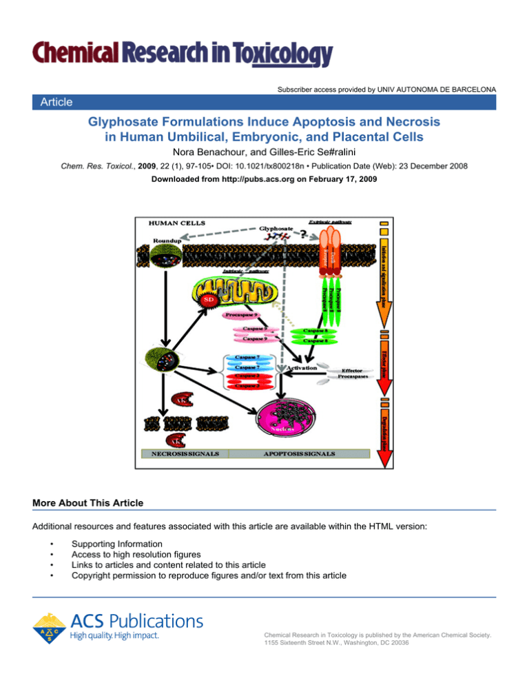
Subscriber access provided by UNIV AUTONOMA DE BARCELONA Article Glyphosate Formulations Induce Apoptosis and Necrosis in Human Umbilical, Embryonic, and Placental Cells Nora Benachour, and Gilles-Eric Se#ralini Chem. Res. Toxicol., 2009, 22 (1), 97-105• DOI: 10.1021/tx800218n • Publication Date (Web): 23 December 2008 Downloaded from http://pubs.acs.org on February 17, 2009 More About This Article Additional resources and features associated with this article are available within the HTML version: • • • • Supporting Information Access to high resolution figures Links to articles and content related to this article Copyright permission to reproduce figures and/or text from this article Chemical Research in Toxicology is published by the American Chemical Society. 1155 Sixteenth Street N.W., Washington, DC 20036 Subscriber access provided by UNIV AUTONOMA DE BARCELONA Chemical Research in Toxicology is published by the American Chemical Society. 1155 Sixteenth Street N.W., Washington, DC 20036 Chem. Res. Toxicol. 2009, 22, 97–105 97 Glyphosate Formulations Induce Apoptosis and Necrosis in Human Umbilical, Embryonic, and Placental Cells Nora Benachour and Gilles-Eric Séralini* UniVersity of Caen, Laboratory Estrogens and Reproduction, UPRES EA 2608, Institute of Biology, Caen 14032, France ReceiVed June 16, 2008 We have evaluated the toxicity of four glyphosate (G)-based herbicides in Roundup (R) formulations, from 105 times dilutions, on three different human cell types. This dilution level is far below agricultural recommendations and corresponds to low levels of residues in food or feed. The formulations have been compared to G alone and with its main metabolite AMPA or with one known adjuvant of R formulations, POEA. HUVEC primary neonate umbilical cord vein cells have been tested with 293 embryonic kidney and JEG3 placental cell lines. All R formulations cause total cell death within 24 h, through an inhibition of the mitochondrial succinate dehydrogenase activity, and necrosis, by release of cytosolic adenylate kinase measuring membrane damage. They also induce apoptosis via activation of enzymatic caspases 3/7 activity. This is confirmed by characteristic DNA fragmentation, nuclear shrinkage (pyknosis), and nuclear fragmentation (karyorrhexis), which is demonstrated by DAPI in apoptotic round cells. G provokes only apoptosis, and HUVEC are 100 times more sensitive overall at this level. The deleterious effects are not proportional to G concentrations but rather depend on the nature of the adjuvants. AMPA and POEA separately and synergistically damage cell membranes like R but at different concentrations. Their mixtures are generally even more harmful with G. In conclusion, the R adjuvants like POEA change human cell permeability and amplify toxicity induced already by G, through apoptosis and necrosis. The real threshold of G toxicity must take into account the presence of adjuvants but also G metabolism and time-amplified effects or bioaccumulation. This should be discussed when analyzing the in vivo toxic actions of R. This work clearly confirms that the adjuvants in Roundup formulations are not inert. Moreover, the proprietary mixtures available on the market could cause cell damage and even death around residual levels to be expected, especially in food and feed derived from R formulation-treated crops. Introduction Humans are exposed daily to a great number of xenobiotics and their metabolites, present as pollutants (1). They act as mixtures having compensatory, multiplicative, or synergistic effects, as we have shown (2) with others (3, 4). The main glyphosate (G) formulations, commercialized as Roundup (R) from the Monsanto Company, are themselves already mixtures of G and various adjuvants at different concentrations. We have studied these products, which are the major nonselective herbicides worldwide (5); moreover, their use and presence in the food chain (6) are increasing since more than 75% of genetically modified edible plants have been designed to tolerate high levels of these compounds (7). G and its major metabolite aminomethylphosphonic acid (AMPA) were classified among the first contaminants in rivers (8). The adjuvants, less measured in the environment, are usually considered as inert and are protected as a “trade secret” in manufacturing (9). However, among them, the predominant one appears to be the polyethoxylated tallowamine or POEA (10, 11), which has itself some toxicity (12), such as causing ocular burns, redness, swellings and blisters, short-term nausea, and diarrhea. In combination with G, the mixture becomes more active (13). These products, like detergents, could allow facilitated G penetration through plasmatic membranes, potentialization of its action, increased stability, and bioaccumulation (14, 15). * To whom correspondence should be addressed. Tel: 33(0)2-31-56-5684. Fax: 33(0)2-31-56-53-20. E-mail: [email protected]. The dose- and time-dependent cytotoxicity of R Bioforce (360 g/L of G, R360) on human placental and embryonic cells (15) could explain at least in part some reproductive problems (16). Among the two lines, 293 embryonic cells have proven to be very suitable for estimating the hormonal activity for xenobiotics (17), and JEG3 cells are also considered a useful model for examining placental toxicity (18). These lines may be equally or even less sensitive to xenobiotics than primary cultures (19). In the present study, we also tested the mechanism by which R mixtures affect human primary cells of the umbilical vein cord endothelial cells (HUVEC) for comparative purposes. The endothelial lining of blood vessels constitutes a permeable barrier between the blood and the underlying tissues. The endothelium also plays an important role in various physiological processes, such as metabolism of vasoactive substances and maintenance of antithrombotic factors on the vessel wall (20). The endothelial cells are exposed directly to chemicals circulating in the blood of the umbilical cord and pass through the placenta (21). It is known that HUVEC cells may be a target for adverse effects of xenobiotics activated into reactive metabolites (22, 23). Other somatic cell types have been used to study pesticide toxicity and apoptosis such as HeLa (24) and Jurkat (25), but none was before treated by glyphosate. In human cells, we have demonstrated that G mixed with adjuvants in R360 was cytotoxic through alteration of succinate dehydrogenase SD (14, 15). With isolated rat liver mitochondria, it is demonstrated that R depresses the mitochondrial complexes 10.1021/tx800218n CCC: $40.75 2009 American Chemical Society Published on Web 12/23/2008 98 Chem. Res. Toxicol., Vol. 22, No. 1, 2009 Benachour and Séralini Figure 1. Cytotoxic effects of four Roundup formulations (R) on three human cell types. The R (from 10 to 2 × 10⁴ ppm) contain different glyphosate (G) concentrations (7.2, 360, 400, or 450 g/L) and adjuvants. G alone was used as control at equivalent quantities to R360 and at similar pH 5.8. The cells were either primary from neonate umbilical cord (HUVEC) or lines from embryo (293) or placenta (JEG3). The actions on the mitochondrial succinate dehydrogenase (SD) activity (cellular viability in %, A) and on the release of cytoplasmic adenylate kinase (AK) activity [cell death in relative luminescence units (RLU), B] were compared in serum-free medium after 24 h of exposure. The 50% lethal dose (LD50) is indicated by a dashed line. SEs are shown in all instances (n ) 12). II (SD) and III (26). In sea urchin eggs, R deteriorated cell cycle checkpoints, and G with its adjuvants inhibited hatching enzyme transcription synergistically (27, 28). Recently, it was shown in this model to activate the DNA damage checkpoint CDK1/ cycline B of the first cell cycle of development (29, 30) for commitment to cell death by apoptosis in the case of failure of DNA repair. This work focuses on the cell death mechanism in human cells induced by four different G formulations with a large number of agricultural applications. We have chosen Roundup Express (R7.2), Roundup Bioforce or Extra 360 (R360), Roundup Grand Travaux (R400), and Roundup Grand Travaux Plus (R450) at subagricultural dilutions. We tested them on three important enzymatic biomarkers. First, at the membrane level, we measured adenylate kinase (AK) activity after its release in the medium (31), revealing cytoplasmic membrane rupture, corresponding to a necrosis and/or a secondary necrosis at the end of apoptosis (32). Second, at the mitochondrial respiration level, we measured succinate dehydrogenase (SD) activity (33). Third, we tested the cytosolic level with caspase 3 and 7 activities to determine the apoptosis pathway (34-36) and in situ DNA fragmentation (DAPI). Necrosis is evinced by cytoplasmic swelling, rupture of the plasma membrane, swelling of cytoplasmic organelles (particularly mitochondria), and some condensation of nuclear chromatin, whereas apoptosis is manifested by cytoplasmic and nuclear condensation (pyknosis), nuclear fragmentation (karyorrhexis), normal morphological appearance of cytoplasmic organelles, and an intact plasma membrane; following nuclear fragmentation, the cell disaggregates into a number of membrane-bound apoptotic bodies (37, 32). By contrast, cell death is now known to be perpetrated through a variety of mechanisms. It can be classified into four different types, based upon morphological characteristics: apoptosis (type 1), autophagy (type 2), necrosis (oncosis, type 3), and mitotic catastrophe (37). The three human cell types allowed us to establish not only the differential sensitivity of these models but also the general human cell pathways of G-based pesticides actions from 1 ppm (0.0001%); these were produced by G itself, its major metabolite AMPA, and the main adjuvant POEA, singly or in combination. Materials and Methods Chemicals. N-Phosphonomethyl glycine (glyphosate, G, PM 169.07) and its major metabolite AMPA (PM 111.04) were purchased from Sigma-Aldrich (Saint Quentin Fallavier, France). Herbicide Roundup formulations (Monsanto, Anvers, Belgium) were available on the market: Roundup Express 7.2 g/L of G, homologation 2010321 (R7.2); Bioforce or Extra 360 at 360 g/L of G, homologation 9800036 (R360); Grands Travaux 400 g/L of G, homologation 8800425 (R400); and Grands Travaux plus 450 g/L of G, homologation 2020448 (R450). A 2% solution of Roundup (1 or 2% is recommended by the company for agricultural use) and an equivalent solution of glyphosate to Roundup Bioforce were prepared in serum-free medium and Glyphosate Formulations Toxicity in Human Cells Chem. Res. Toxicol., Vol. 22, No. 1, 2009 99 Figure 2. Nonlinear dose effects of R formulations. The LD50 (%) measured by SD are compared for the 4 R (see the Figure 1A legend) and G for the three cell types in similar conditions. Figure 3. Cytotoxicity of R adjuvant (POEA) and glyphosate (G) metabolite (AMPA) on three human cell types. G and R360 were used as controls in similar conditions as in Figure 1 (see legend), in comparison to R adjuvant POEA and G metabolite AMPA (1-105 ppm). The 50% lethal dose (LD50) is indicated by a dashed line. SEs are shown in all instances (n ) 12). adjusted to pH 5.8 of the 2% Roundup Bioforce solution. The major adjuvant of Roundup, polyethoxylated tallowamine (POEA at 785 g/L), was a gift from Pr. Robert Bellé (UMR 7150 CNRS/ UPMC, Station Biologique de Roscoff, France). Successive dilutions were then obtained with serum-free medium. 4′,6′Diamidino-2-phenylindole, dihydrochloride (DAPI) nucleic acid stain powder was obtained from Lonza (Saint Beauzire, France). 3-(4,5-Dimethylthiazol-2-yl)-2,5-diphenyl tetrazolium bromide (MTT) and all other compounds, otherwise precised, were obtained from Sigma-Aldrich. MTT was prepared as a 5 mg/ mL stock solution in phosphate-buffered saline, filtered through a 0.22 µm filter before use, and diluted to 1 mg/mL in a serumfree medium. Cell Cultures. Human Primary Cells. The human primary cells used in this work were HUVEC (C2519A) provided by Lonza. Cells (passage 5 or 6) were grown according to the supplier, in specific endothelial growth medium EGM-2 SingleQuots (CC-4176) containing hEGF, hydrocortisone, GA-1000 (Gentamicin, Amphoteri- 100 Chem. Res. Toxicol., Vol. 22, No. 1, 2009 Benachour and Séralini Figure 4. Combined effects of G, AMPA, and POEA on three human cell types. The cells were incubated in serum-free medium for 24 h, and the products were tested by pairs to a final concentration, where they are nontoxic alone on succinate dehydrogenase, of 0.05 (HUVEC) and 0.5% (293, JEG3). Results of cellular death are evaluated through AK activity in relative units in comparison to nontreated cells (control ) 1), and values are blank-subtracted (blank ) no AK); see the Materials and Methods. R360 and G are used as controls. SEs are shown in all instances (n ) 16; **p < 0.01). cin-B), FBS (fetal bovine serum), VEGF, hFGF-B, R3-IGF-1, ascorbic acid, and heparin. Fifty thousand cells per well were grown at 37 °C (5% CO2, 95% air) over a 24 h period to 80% confluence in 48 well plates and were washed with serum-free EGM-2. Human Cell Lines. The human embryonic kidney 293 cell line (ECACC 85120602) and the human choriocarcinoma-derived placental JEG3 cell line (ECACC 92120308) were provided by CERDIC (Sophia-Antipolis, France). Cells were grown in phenol red-free Eagle’s modified minimum essential medium (EMEM; Abcys, Paris, France) containing 2 mM glutamine, 1% nonessential amino acid, 100 U/mL antibiotics (a mix of penicillin, streptomycin, and fungizone; Lonza), 10 mg/mL of liquid kanamycin (Dominique Dutscher, Brumath, France), and 10% FBS (PAA, les Mureaux, France). The JEG3 cell line was supplemented with 1 mM sodium pyruvate. Fifty thousand cells per well were grown at 37 °C (5% CO2, 95% air) over a 48 h period to 80% confluence in 48 well plates and were washed with serum-free EMEM. Cell Treatments. Cells were exposed for 24 h in serum-free medium to various dilutions of the different treatments including the four Roundup formulations (R7.2, R360, R400, and R450), G, AMPA, or POEA (14 concentrations from 10 ppm to 2%) and, particularly for POEA, were tested at the very low concentrations of 1 and 5 ppm; for AMPA, we tested in addition 4, 6, 8, and 10%. In another case, cells were incubated with G, AMPA, and POEA mixtures by pairs at the final nontoxic dilution on SD of 0.5% on the human cell lines (293 or JEG3) and 0.05% on the human primary cells (HUVEC) in comparison to R360. For the details, in each cell type, three combinations were studied. For the two cell lines, the first mixture was the combination of G (0.4999%) with POEA (0.0001%); the second was the combination of G (0.4%) with AMPA (0.1%), and the third was AMPA (0.4999%) plus POEA (0.0001%). For the primary HUVEC cells, the first mixture was G (0.04999%) with POEA (0.0001%); the second was G (0.04%) with AMPA (0.01%), and the third was AMPA (0.04999%) plus POEA (0.0001%). Cell Death Measurements. Mitochondrial Activity Measurement. This measure was based on the cleavage of MTT into a blue-colored product (formazan) by the mitochondrial enzyme succinate dehydrogenase (38, 39, 33); it was used to evaluate human cell viability. After cell treatments, the supernatants were recovered for the ToxiLight bioassay, and adherent cells were washed with serum-free medium and incubated with 200 µL MTT per well after each treatment. The 48 well plates were incubated for 3 h at 37 °C, and 200 µL of 0.04 N hydrochloric acid-containing isopropanol solution was added to each well. The plates were then vigorously shaken to solubilize the blue formazan crystals formed. The optical density was measured at 570 nm using a luminometer Mithras LB 940 (Berthold, Thoiry, France). Cell Membrane Damage Assay. The bioluminescent ToxiLight bioassay (Lonza) was a nondestructive cytotoxicity highly sensitive assay designed to measure toxicity in mammalian cells and cell lines in culture. It quantitatively measured the release of cytosolic AK from the membranes of damaged cells (40, 31). AK is a robust protein present in all eukaryotic cells, which is released into the culture medium when cells die, described as an important necrosis marker. The enzyme actively phosphorylated ADP, and the resultant ATP was then measured using the bioluminescent firefly luciferase reaction with the ToxiLight reagent. After 24 h of different treatments, 50 µL of cell supernatants was deposited in 96 well black plates. Then, 50 µL of the AK detection reagent (AKDR) was added by well. Plates were then placed under agitation for 15 min safe from the light, and then, luminescence was measured using the luminometer Mithras LB 940 (Berthold) at 565 nm. The serumfree medium was the negative control, and a positive control was the active reagent AKDR mixed with cells treated in the serumfree medium to determine the basal activity. Glyphosate Formulations Toxicity in Human Cells Chem. Res. Toxicol., Vol. 22, No. 1, 2009 101 Figure 5. Time-dependent apoptosis through caspases 3/7 induction by R and G in three human cell types. R360 and G, at similar concentrations and pH (as in Figure 1), were incubated for 6, 12, 18, or 24 h. The apoptotic pathway was tested by the Caspase-Glo 3/7 assay, and results are presented in relative units to nontreated cells (control ) 1). SEs are shown in all instances (n ) 8). Apoptotic Cell Death Measurements. The Caspase-Glo 3/7 assay (Promega, Paris, France) was a luminescent kit designed for automated high-throughput screening of caspases activity or apoptosis. It can measure caspase 3 and 7 activities in purified enzyme preparations or cultures of adherent or suspension cells (41, 42, 36). The assay provided a pro-luminescent caspase 3/7 substrate, which contains the tetrapeptide sequence DEVD active group. This substrate was cleaved to release amino-luciferin, a substrate of luciferase used in the production of light. The Caspase-Glo 3/7 reagent was optimized for caspase activity, luciferase activity, and cell lysis. The addition of the single Caspase-Glo 3/7 reagent, in an “add-mix-measure” format, resulted in cell lysis followed by caspase cleavage of the substrate and generation of a “glow type” luminescent signal. The Caspase-Glo 3/7 bioassay was carried out in 96 well white plates. After cell cultures and their treatments by 50 µL of various dilutions, an equal volume of the reagent was added to each well. Plates were then agitated for 15 min safe from the light, to stabilize the light signal before measuring luminescence. Again, the negative control was the serum-free medium, and the positive control was the active reagent mixed with cells treated in the serum-free medium to determine the basal activity of the caspases 3/7. Luminescence was measured using the luminometer Mithras LB 940 (Berthold) at 565 nm. Cell Microscopy. At the end of the 24 h cell treatment, the serum-free medium was removed, and cells were fixed in absolute ethanol-chloroform-acetic acid (6:3:1, v/v/v) for 1 day at -20 °C. Each well was washed with PBS (pH 7.4) and incubated with 1 µg/mL DAPI solution (43). Staining of DNA with DAPI was examined with a microscope using a fluorescent mode (model Leïca LMD 6000, Rueil Malmaison, France). Labeled DNA of viable cells was scattered throughout the nucleus, and bright condensation of chromatin revealed apoptotic cells (magnification, 400×). At the end of the cell treatment, the microphotographs (magnification, 100×; blue filter) of cells without coloration were also obtained with the Leïca Microscopy Systems (model Leïca DC 100, Germany). Statistical Analysis. The experiments were repeated at least three times during different weeks on three independent cultures each time. All data were presented as the means ( standard errors (SEs). Statistical differences were determined by a Student’s t test using significant levels of 0.01 (**). Results We have studied for the first time the mechanism of cellular action of different R on human cells, from placenta, embryonic kidney, and neonate. The first surprising results show that the four R herbicides and G cause cellular death for all types of human cells, with comparable toxicity for each one but at different concentrations. For instance, 20 ppm for R400 at 24 h, 102 Chem. Res. Toxicol., Vol. 22, No. 1, 2009 Figure 6. Microphotographs of R-treated human cells. The cell types were without coloration (magnification, 100×; blue filter), HUVEC (A, B), 293 (C, D), and JEG3 (E, F), and were incubated with 0.005% of R400 or not (controls) in serum-free medium for 24 h. Microphotographs were obtained with the Leïca Microscopy Systems (model Leïca DC 100). the most toxic, corresponds approximately to 47 µM G (8 ppm) with adjuvants (Figure 1). However, 4-10 ppm G alone is nontoxic; its toxicity begins around 1%. The mechanism is constant for all R: There is a release of AK, indicative of cell membrane damage, and an inhibition of the mitochondrial SD (Figure 1). For all R, the membrane damage (AK) is 1.5-2 times more sensitive than mitochondrial activity (SD) for 293 and JEG3 or equally sensitive for HUVEC. By contrast, G induces mitochondrial toxicity without cell membrane damage. Unexpectedly, R400 is more toxic than another formulation containing more G, such as R450; the latter is in turn more harmful than R360, R7.2, and G in last, but all of them are detrimental nonproportionally to the G concentration that they contain. This is illustrated in Figure 2. The mitochondrial SD inhibition measures cell asphyxia. It is obvious from Figure 2 that 7.2 or 360 g/L G with adjuvants in R formulations has closely comparable actions on cell death, while 400 or 450 g/L gives inversely proportional effects in another range. This is not an artifact since the embryonic and placental cell lines behave remarkably similarly in that regard and the primary umbilical cord cells have sensitivity for all R and G just analogous to these cell lines (Figure 2). The mortality in all cases is not linearly linked to G. The hypothesis that other substances are implicated has thus to be investigated in the formulation of the product. Consequently, the major G metabolite, AMPA, and the surfactant POEA, the main claimed adjuvant by the manufacturer (the exact composition is a secret of formulation), have been tested separately in a first approach, in comparison to G and R360 as controls, and in similar conditions as in Figure 1, Benachour and Séralini from very low subagricultural dilutions (10-6 if used pure like claimed by some farmers and 10-4 if diluted as recommended at 1%). G is claimed by the manufacturer to be the active ingredient, and it is claimed to be not toxic for human cells but toxic for vegetable ones when mixed with inert components. Our study demonstrates for the first time that all products including AMPA and POEA provoke SD and AK effects in human cells, and thus mortality (Figure 3), but at different concentrations. Astonishingly, the supposed inert product POEA is the most potent one. From 1 ppm, it begins to alter SD in HUVEC and AK in 293 and JEG3. The mixture R is then more poisonous than G or AMPA. The metabolite AMPA itself destroys the cell membrane (AK release), whatever the cell type. This is not observed with G, which is, however, 3-8 times more inhibitory on SD than AMPA, with some differences between cells. However, because the cell membrane damage is generally more sensitive, the metabolite AMPA is finally more toxic than G on human cells. POEA is the most toxic; if it was the only adjuvant of R360, its maximal concentration would be around 1-24 ‰, according to the cells. Thus, POEA could be considered as the active ingredient on human cell death and more damaging than G. As R is more viscous than 1‰ POEA plus G, it is obvious that other compounds are in the mixture. Thus, it was necessary to study the combined effects on cell membrane integrity (by AK release). We have tested the compounds by pairs at maximal levels where alone they do not influence SD (Figure 4). This was to assess the respective role of each one, knowing that R contains all tested compounds when metabolized. In contrast to previous results, the cells reacted differently. The mixtures were more disrupting on embryonic and umbilical cells, respectively, while placental carcinoma cells appeared to be more membrane-resistant but to mixtures only. It is very clear that if G, POEA, or AMPA has a small toxic effect on embryonic cells alone at low levels, the combination of two of them at the same final concentration is significantly deleterious (Figure 4). We have thus elucidated that R- and G-induced cell death can be due, at least in part, to apoptosis via caspases 3/7 induction (Figure 5). The caspases are activated from 6 h with a maximum at 12 h in all cases, but umbilical primary cells are 60-160 times more sensitive than lines (293 and JEG3, respectively) at this level. Moreover, G and R360 enhance exactly at the same concentration caspases, from 50 ppm (HUVEC). The adjuvants do not appear to be necessary to render G as a death inducer at this level. Even G alone is 30% more potent on this pathway than R. Surprisingly, G acted very rapidly at concentrations 500-1000 times lower than agricultural use on human cell apoptosis. This apoptotic pathway was also activated at levels 200 times lower for G on caspases than its action on SD for umbilical cells, and for R at levels 60 times lower, in a four times shorter period (6-24 h). After 24 h of treatment, the caspases returned to basal level when SD and AK react significantly. These data are consistent with a gradual loss of caspases 3/7 activity in apoptotic cells that undergo secondary necrosis in vitro (44). Our results are confirmed by the morphology of the cells after treatment by R (for instance R400, Figure 6B,D,E) in comparison with the normal cell types (A, C, F). Indeed, the very weak R concentration of 0.005% causes a very important cell death, lack of adhesion, shrinking, and fragmentation in apoptotic bodies. This is confirmed in Figure 7 with the DNA fluorescent labeling with DAPI, for example, with R360 at 0.5% over 24 h. The characteristic fluorescence of apoptotic cells evidencing Glyphosate Formulations Toxicity in Human Cells Chem. Res. Toxicol., Vol. 22, No. 1, 2009 103 Figure 7. Increase of DNA condensation (DAPI test) in R360- or G-treated human cells. The cell types HUVEC (A-C), 293 (D-F), and JEG3 (G-I) were incubated for 24 h with or without 0.5% R360 or G at equivalent concentrations. Staining of DNA with DAPI was examined with a microscope model Leïca LMD 6000, using a fluorescent mode. Labeled DNA of viable cells was scattered throughout the nucleus, and bright condensation of chromatin revealed apoptotic cells (magnification, 400×). DNA condensation is more visible with the herbicide than in controls (A, D, G) and more after R treatment (C, F, I) than with G alone (B, E, H), for cell lines. The primary cells are similarly sensitive to G than to R, as for caspases activation in Figure 5. Discussion We had previously demonstrated (14) that G-based formulations were able to affect human placental cell viability at subagricultural doses (0.1% in 18 h) and sexual steroid biosynthesis at lower nontoxic doses (0.01%) and that this was due at least in part to G, but its action was highly amplified by adjuvants, the so-called inert ingredients of R formulations, kept confidential by the companies (9). However, the question of a specific cell line action or a time reversible effect remained open. Benachour et al. (15) demonstrated that in embryonic cells as well as in normal human placenta and equine testis, there was a similar G-dependent endocrine disruption, through aromatase inhibition, at nontoxic levels. The embryonic cells were even more sensitive: It was discovered that the cell mitochondrial activity was also reached in time- and dose-dependent manners by the G formulation R360. The cytotoxicity was amplified around 14 times between 24 and 72 h (15), suggesting either a bioaccumulation or a time-delayed effect and suggesting a cumulative impact, after endocrine disruption, of very low doses around G acceptable daily intake (ADI: 0.3 mg/kg/j), according to the nature of the adjuvants. To understand in vivo effects through the interpretation of the in cell impacts described above, it is necessary to have knowledge of the dilution and of the processes leading to an elimination of the product in the body. This must be taken into account in regard to its bioaccumultation potential and timedelayed effects. This is why we have measured the caspases activities at different times and G or R concentrations, after having previously demonstrated their effects amplified with time within 3 days, on SD in embryonic and placental cells (15). Moreover, the metabolism of the herbicide has to be considered, and the tests in this study of all the above-cited products approach this question. All cell types, including primary cultures, react similarly at the membrane and mitochondrial level, justifying the hypothesis that the cell lines used provide excellent models to study human cell toxicity, for instance in placental cells (18). We show for the first time that embryonic and umbilical cells also have comparable sensitivity. The most reactive level reached appears to be the cell membrane level for the different formulations, but not for G. The supposed “inert ingredients” play obviously and differently the role of cell membrane disruptors, independently to G, as we have previously proposed (14), and this was suggested in fish, amphibians, and microorganisms (27, 45) or in plants (46). We now demonstrate that in human cells. The second level is the mitochondrial membrane and the enzymatic reaction in it, SD, localized in the internal membrane in complex II of the respiratory chain (47). It is altered in a comparable way, not proportional to G but relatively to the nature and the quantity of the adjuvants that we have previously listed (15). This means that the toxicity of G clearly varies with formulations that must imperatively now be used in in vivo tests to study any toxicity (45); this also means that the ADI of G must take into account its formulation, since 7.2 or 360 g/L of G may have comparable effects, considerably different to 400 g/L. It would even be more correct to use precisely an ADI of R instead of G. It may also be time-dependent. These ideas are not taken into account yet for regulatory legislation. The necessity to study combined effects also appears from our results. In fact, the body is always exposed to mixtures and not to single compounds. We have previously demonstrated that mixtures could amplify toxicity for other widely spread pollutants (2). For embryonic or neonatal cells, POEA, the major adjuvant, has the highest toxicity, either by itself or amplified 2-5 times in combination with G or AMPA. It has already been shown that POEA is highly toxic for sea urchin embryos, impinging on transcription (28). It is also known that in an 104 Chem. Res. Toxicol., Vol. 22, No. 1, 2009 aquatic environment, POEA has higher effects than R and G on bacteria, microalgae, protozoa, and crustaceans (12). In addition, the known metabolism of G in the soil or plants is supposed to detoxify it in AMPA (11); however, here, we demonstrate that AMPA is more toxic than G in human cells, especially on cell membrane. AMPA is also more stable in soil (48), in plants, and in food or feed residues (49), and more present in wastewater (2-35 ppm) than G [0.1-3 ppm; (50)]. It is not toxic alone at these concentrations in our experiments, but it amplifies G or POEA toxicity in combination. The synergic toxicity of all of these compounds is now more obvious. The induced resistance of placental cancer cells (51) could explain a specific difference for JEG3 cells at these levels. The placental cells could form an efficient barrier to mixtures before their death, since the membranes are more resistant, and this could be due to the fact that there are carcinoma-derived cells that have acquired a capacity to excrete xenobiotics. The caspases 3/7 inducing apoptosis were in fact activated first within 6 h, and then, they decreased with cell mortality. This corroborates the timing observed for another compound (36). The caspases induction by G alone is observed at doses that do not provoke cell or mitochondrial membrane damages, indicating a clear G-apoptotic pathway always at subagricultural doses. Mixed with adjuvants, G in R formulations reached the other end points. This suggests that the adjuvants could also play a role in total cell death, through necrosis characterized by organelle alterations with mitochondrial and cell membranes swelling and ruptures (52). The most sensitive are umbilical HUVEC cells, for which apoptosis has been described (53-55), but very rarely induced by a pesticide, for example, in the case of diallyl trisulfide (56). Surprisingly, this phenomenon was observed for G and R at similar and low concentrations, as if a cell membrane death receptor was activated (57, 32), with no G penetration necessary. The modification of a dependency receptor is another pathway that could be studied (58). The apoptotic cell appearance was microscopically confirmed. Then, our next step could be to study the necrotic/apoptotic ratio within short times. In conclusion, mixtures called “formulations” change cell permeability, toxicity, and pathways of xenobiotics: In all cases, cell death is induced more by R than by AMPA or G, and the latter provokes apoptosis (from 50 ppm in HUVEC cells) without membrane damage. By contrast, G mixed with adjuvants in R formulations disrupts cell and mitochondrial membranes and promotes necrosis. It becomes obvious that the “threshold” level of action of the herbicide should take into account the period and length of exposure, the presence of adjuvants, in particular POEA, metabolism, and bioaccumulation or timedelayed effects. All of the above effects are demonstrated below the recommended herbicide agricultural dilutions (from 104 ppm). This clearly confirms that the adjuvants in Roundup formulations are not inert. Moreover, the proprietary mixtures available on the market could cause cell damage and even death around residual levels to be expected, especially in food and feed derived from R formulation-treated crops. Acknowledgment. We thank CRIIGEN, Regional Council of Basse-Normandie, and Human Earth Foundation. This work was also supported by a grant from Fondation Denis Guichard under the aegis of the Fondation de France. We declare that we have no competing financial interest. We thank Dr. Carine Travert for scientific revision of the manuscript. Benachour and Séralini References (1) Feron, V. J., Cassee, F. R., Groten, J. P., van Vliet, P. W., and van Zorge, J. A. (2002) International issues on human health effects of exposure to chemical mixtures. EnViron. Health Perspect. 110, 893– 899. (2) Benachour, N., Moslemi, S., Sipahutar, H., and Séralini, G. E. (2007) Cytotoxic effects and aromatase inhibition by xenobiotic endocrine distrupters alone and in combination. Toxicol. Appl. Pharmacol. 222, 129–140. (3) Tichy, M., Borek-Dohalsky, V., Rucki, M., Reitmajer, J., and Feltl, L. (2002) Risk assessment of mixtures: Possibility of prediction of interaction between chemicals. Int. Arch. Occup. EnViron. Health 75, S133-S136. (4) Monosson, E. (2005) Chemical mixtures: Considering the evolution of toxicology and chemical assessment. EnViron. Health Perspect. 113, 383–390. (5) Acquavella, J. F., Bruce, H., Alexander, B. H., Mandel, J. S., Gustin, C., Baker, B., Champan, P., and Bleeke, M. (2004) Glyphosate biomonitoring for farmers and their families: Results from the farm family exposure study. EnViron. Health Perspect. 112, 321–326. (6) Takahashi, M., Horie, M., and Aoba, N. (2001) Analysis of glyphosate and its metabolite, aminomethylphosphonic acid, in agricultural products by HPLC. Shokuhin Eiseigaku Zasshi 42, 304–308. (7) Clive, J. (2007) The global status of the commercialized biotechnological/genetically modified crops: 2006. Tsitol. Genet. 41, 10–12. (8) IFEN (2006) Report on pesticides in waters: Data 2003-2004. Institut Français de l’Environnement, Orleans, France. Dossier 5, 15–20. (9) Cox, C. (2004) Glyphosate. J. Pest. Reform. 24, 10–15. (10) Acquavella, J. F., Weber, J. A., Cullen, M. R., Cruz, O. A., Martens, M. A., Holden, L. R., Riordan, S., Thompson, M., and Farmer, D. (1999) Human ocular effects from self-reported exposures to Roundup herbicides. Hum. Exp. Toxicol. 18, 479–86. (11) Williams, G. M., Kroes, R., and Munro, I. C. (2000) Safety evaluation and risk assessment of the herbicide Roundup and its active ingredient, glyphosate, for human. Regul. Toxicol. Pharmacol. 31, 117–65. (12) Tsui, M. T., and Chu, L. M. (2003) Aquatic toxicity of glyphosatebased formulations: comparison between different organisms and the effects of environmental factors. Chemosphere 52, 1189–1197. (13) Cox, C. (1998) Glyphosate (Roundup). J. Pest. Reform. 18, 3–17. (14) Richard, S., Moslemi, S., Sipahutar, H., Benachour, N., and Séralini, G. E. (2005) Differential effects of glyphosate and Roundup on human placental cells and aromatase. EnViron. Health Perspect. 113, 716– 720. (15) Benachour, N., Sipahutar, H., Moslemi, S., Gasnier, C., Travert, C., and Séralini, G. E. (2007) Time and dose-dependent effects of Roundup on human embryonic and placental cells and aromatase inhibition. Arch. EnViron. Contam. Toxicol. 53, 126–133. (16) Savitz, D., Arbuckle, T., Kaczor, D. M., and Curtis, K. (1997) Male pesticide exposure and pregnancy outcome. Am. J. Epidemiol. 146, 1025–1036. (17) Kuiper, G. G., Lemmen, J. G., Carlsson, B., Corton, J. C., Safe, S. H., Van der Saag, P. T., Van der Burg, B., and Gustafsson, J. A. (1998) Interaction of estrogenic chemicals and phytoestrogens with estrogen receptor β. Endocrinology 139, 4252–4263. (18) Letcher, R. J., Van Holstein, I., Drenth, H. J., Norstrom, R. J., Bergman, A., Safe, S., Pieters, R., and van den Berg, M. (1999) Cytotoxicity and aromatase (CYP19) activity modulation by organochlorines in human placental JEG-3 and JAR choriocarcinoma cells. Toxicol. Appl. Pharmacol. 160, 10–20. (19) L’Azou, B., Fernandez, P., Bareille, R., Beneteau, M., Bourget, C., Cambar, J., and Bordenave, L. (2005) In vitro endothelial cell susceptibility to xenobiotics: Comparison of three cell types. Cell Biol. Toxicol. 21, 127–137. (20) Thorin, E., and Shreeve, S. M. (1998) Heterogeneity of vascular endothelial cells in normal and disease states. Pharmacol. Ther. 78, 155–166. (21) Peters, R. J. B. (2005) Man-Made Chemicals in Maternal and Cord Blood. TNO report B&O-A R 2005/129. (22) Annas, A., Granberg, A. L., and Brittebo, E. B. (2000) Differential response of cultured human umbilical vein and artery endothelial cells to Ah receptor agonist treatment: CYP-dependent activation of food and environmental mutagens. Toxicol. Appl. Pharmacol. 169, 94–101. (23) Choi, W., Eum, S. Y., Lee, Y. W., Hennig, B., Robertson, L. W., and Toborek, M. (2003) PCB 104-induced proinflammatory reactions in human vascular endothelial cells: Relationship to cancer metastasis and atherogenesis. Toxicol. Sci. 75, 47–56. (24) Rathinasamy, K., and Panda, D. (2006) Suppression of microtubule dynamics by benomyl decreases tension across kinetochore pairs and induces apoptosis in cancer cells. FEBS J. 273, 4114–4128. (25) Kannan, K., Holcombe, R. F., Jain, S. K., Alvarez-Hernandez, X., Chervenak, R., Wolf, R. E., and Glass, J. (2000) Evidence for the Glyphosate Formulations Toxicity in Human Cells (26) (27) (28) (29) (30) (31) (32) (33) (34) (35) (36) (37) (38) (39) (40) (41) induction of apoptosis by endosulfan in a human T-cell leukemic line. Mol. Cell. Biochem. 205, 53–66. Peixoto, F. (2005) Comparative effects of the Roundup and glyphosate on mitochondrial oxidative phosphorylation. Chemosphere 61, 1115– 1122. Marc, J., Mulner-Lorillon, O., Boulben, S., Hureau, D., Durand, G., and Bellé, R. (2002) Pesticide Roundup provokes cell division dysfunction at the level of CDK1/cyclin B activation. Chem. Res. Toxicol. 15, 326–331. Marc, J., Le Breton, M., Cormier, P., Morales, J., Bellé, R., and Mulner-Lorillon, O. (2005) A glyphosate-based pesticide impinges on transcription. Toxicol. Appl. Pharmacol. 203, 1–8. Bellé, R., Le Bouffant, R., Morales, J., Cosson, B., Cormier, P., and Mulner-Lorillon, O. (2007) Sea urchin embryo, DNA-damaged cell cycle checkpoint and the mechanisms initiating cancer development. J. Soc. Biol. 201, 317–27. Le Bouffant, R., Cormier, P., Cueff, A., Bellé, R., and Mulner-Lorillon, O. (2007) Sea urchin embryo as a model for analysis of the signaling pathways linking DNA damage checkpoint, DNA repair and apoptosis. Cell. Mol. Life Sci. 64, 1723–1734. Squirrell, D., and Murphy, J. (1997) Rapid detection of very low numbers of micro-organisms using adenylate kinase as a cell marker. A Practical Guide to Industrial Uses of ATP Luminescence in Rapid Microbiology, pp 107-113 Cara Technology Ltd., Lingfield, Surrey, United Kingdom. Taatjes, D. J., Sobel, B. E., and Budd, R. C. (2008) Morphological and cytochemical determination of cell death by apoptosis. Histochem. Cell. Biol. 129, 33–43. Scatena, R., Messana, I., Martorana, G. E., Gozzo, M. L., Lippa, S., Maccaglia, A., Bottoni, P., Vincenzoni, F., Nocca, G., Castagnola, M., and Giardina, B. (2004) Mitochondrial damage and metabolic compensatory mechanisms induced by hyperoxia in the U-937 cell line. J. Biochem. Mol. Biol. 37, 454–459. Los, M., Wesselborg, S., and Schulze-Osthoff, K. (1999) The role of caspases in development, immunity, and apoptotic signal transduction: Lessons from knockout mice. Immunity 10, 629–639. Omezzine, A., Chater, S., Mauduit, C., Florin, A., Tabone, E., Chuzel, F., Bars, R., and Benahmed, M. (2003) Long-term apoptotic cell death process with increased expression and activation of caspase-3 and -6 in adult rat germ cells exposed in utero to flutamide. Endocrinology 144, 648–661. Liu, J. J., Wang, W., Dicker, D. T., and El-Deiry, W. S. (2005) Bioluminescent imaging of TRAIL-induced apoptosis through detection of caspase activation following cleavage of DEVD-aminoluciferin. Cancer Biol. Ther. 4, 885–892. Galluzzi, L., Maiuri, M. C., Vitale, I., Zischka, H., Castedo, M., Zitvogel, L., and Kroemer, G. (2007) Cell death modalities: Classifications and pathophysiological implications. Cell Death Differ. 14, 1237–43. Mosmann, T. (1983) Rapid colorimetric assay for cellular growth and survival: Application to proliferation and cytotoxicity assays. J. Immunol. Methods 65, 55–63. Denizot, F., and Lang, R. (1986) Rapid colorimetric assay for cell growth and survival modifications to the tetrazolium dye procedure giving improved sensitivity and reliability. J. Immunol. Methods 89, 271–277. Crouch, S. P. M., Kozlowski, R., Slater, K. J., and Fletcher, J. (1993) The use of ATP bioluminescence as a measure of cell proliferation and cytotoxicity. J. Immunol. Methods 160, 81–88. O’Brien, M., Moravec, R., Riss, T., and Promega Corporation (2003) Caspase-Glo 3/7 assay: Use fewer cells and spend less time with this homogeneous assay. Cell Notes 6, 13-15. Chem. Res. Toxicol., Vol. 22, No. 1, 2009 105 (42) Ren, Y. G., Wagner, K. W., Knee, D. A., Aza-Blanc, P., Nasoff, M., and Deveraux, Q. L. (2004) Differential regulation of the TRAIL death receptors DR4 and DR5 by the signal recognition particle. Mol. Biol. Cell 15, 5064–5074. (43) Travert, C., Carreau, S., and Le Goff, D. (2006) Induction of apoptosis by 25-hydroxycholesterol in adult rat Leydig cells: Protective effect of 17beta-estradiol. Reprod. Toxicol. 22, 564–570. (44) Riss, T. L., and Moravec, R. A. (2004) Use of multiple assay endpoints to investigate the effects of incubation time, dose of toxin, and plating density in cell-based cytotoxicity assays. Assay Drug DeV. Technol. 2, 51–62. (45) Cox, C., and Surgan, M. (2006) Unidentified inert ingredients in pesticides: Implications for human and environmental health. EnViron. Health Perspect. 114, 1803–806. (46) Haefs, R., Schmitz-Eiberger, M., Mainx, H. G., Mittelstaedt, W., and Noga, G. (2002) Studies on a new group of biodegradable surfactants for glyphosate. Pest. Manage. Sci. 58, 825–833. (47) Thomas, P. K., Cooper, J. M., King, R. H., Workman, J. M., Schapira, A. H., Goss-Sampson, M. A., and Muller, D. P. (1993) Myopathy in vitamin E deficient rats: Muscle fibre necrosis associated with disturbances of mitochondrial function. J. Anat. 183, 451–461. (48) Gard, J. K., Feng, P. C., and Hutton, W. C. (1997) Nuclear magnetic resonance timecourse studies of glyphosate metabolism by microbial soil isolates. Xenobiotica 27, 633–644. (49) Scalla, R. (1997) Sécurité alimentaire biotransformation des herbicides par les plantes transgéniques résistantes. Oléagineux, Corps Gras, Lipides. Dossier: Génie Génét. Oléagineux 4, 113–119. (50) Ghanem, A., Bados, P., Kerhoas, L., Dubroca, J., and Einhorn, J. (2007) Glyphosate and AMPA analysis in sewage sludge by LC-ESIMS/MS after FMOC derivatization on strong anion-exchange resin as solid support. Anal. Chem. 79, 3794–801. (51) Xu, J., Peng, H., and Zhang, J. T. (2007) Human multidrug transporter ABCG2, a target for sensitizing drug resistance in cancer chemotherapy. Curr. Med. Chem. 14, 689–701. (52) Okada, H., and Mak, T. W. (2004) Pathways of apoptotic and nonapoptotic death in tumour cells. Nat. ReV. Cancer 4, 592–603. (53) Solovyan, V. T., and Keski-Oja, J. (2005) Apoptosis of human endothelial cells is accompanied by proteolytic processing of latent TGF-beta binding proteins and activation of TGF-beta. Cell Death Differ. 12, 815–826. (54) Song, Y., Shen, K., and He, C. X. (2007) Construction of autocatalytic caspase-3 driven by amplified human telomerase reverse transcriptase promoter and its enhanced efficacy of inducing apoptosis in human ovarian carcinoma. Zhonghua Fu Chan Ke Za Zhi 42, 617–622. (55) Qiu, J., Gao, H. Q., Li, B. Y., and Shen, L. (2008) Proteomics investigation of protein expression changes in ouabain induced apoptosis in human umbilical vein endothelial cells. J. Cell. Biochem. 104, 1054–1064. (56) Xiao, D., Li, M., Herman-Antosiewicz, A., Antosiewicz, J., Xiao, H., Lew, K. L., Zeng, Y., Marynowski, S. W., and Singh, S. V. (2006) Diallyl trisulfide inhibits angiogenic features of human umbilical vein endothelial cells by causing Akt inactivation and down-regulation of VEGF and VEGF-R2. Nutr. Cancer 55, 94–107. (57) Ryu, H. Y., Emberley, J. K., Schlezinger, J. J., Allan, L. L., Na, S., and Sherr, D. H. (2005) Environmental chemical-induced bone marrow B cell apoptosis: Death receptor-independent activation of a caspase-3 to caspase-8 pathway. Mol. Pharmacol. 68, 1087–1096. (58) Kroemer, G., Galluzzi, L., and Brenner, C. (2007) Mitochondrial membrane permeabilization in cell death. Physiol. ReV. 87, 99–163. TX800218N
