Plaque de calcanéus Placa de Calcâneo Пластина пяточной кости
Anuncio
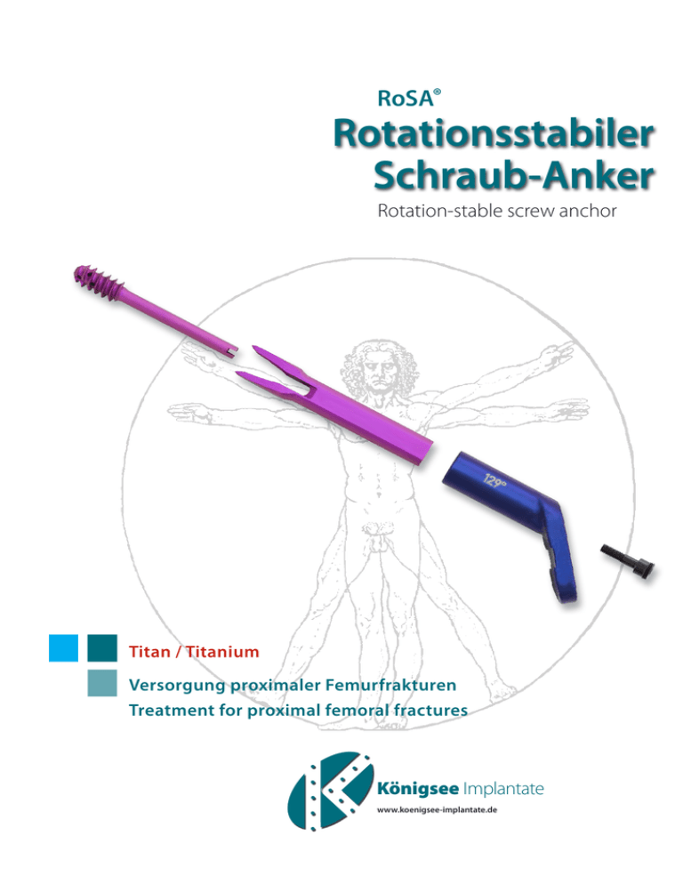
RoSA® Rotationsstabiler Schraub-Anker Rotation-stable screw anchor Titan / Titanium Versorgung proximaler Femurfrakturen Treatment for proximal femoral fractures Königsee Implantate www.koenigsee-implantate.de 2 Introduction Die OP-Anleitung und die Gebrauchsanweisung für das Instrumentarium erheben bei der Komplexität der Anwendung keinen Anspruch auf Vollständigkeit. Eine zusätzliche Einweisung in die Handhabung dieses Instrumentariums und die OP Methode durch einen in dieser Methode erfahrenen Chirurgen vor der ersten OP wird dringend empfohlen. The surgical technique and the operating instructions for the instrumentation do not claim to be exhaustive due to the complexity of their use. Prior to the first surgery, additional training in the use of this instrumentation by an experienced surgeon in these surgical techniques is strongly recommended. Indikationen Indications Einleitung Die rotationsstabile Fixation des Hüftkopfes/ des Kopf-Hals-Fragmentes bei Schenkelhalsfrakturen bzw. hüftnahen Oberschenkelfrakturen ist das Ziel der Osteosynthese mit RoSA®, dem rotationsstabilen Schraub-Anker. RoSA® ermöglicht erstmalig die Kombination von Gleitprinzip, Kompressionsmöglichkeit und Rotationssicherung mit einem kompakten, aus mehreren Teilen bestehenden Kraftträger. Die Vorteile der „Klinge“ (hohe Rotationsstabilität, insbesondere auch im osteoporotischen Knochen, hohe Belastbarkeit...) werden mit den Vorteilen der „Schraube“ (Ausreißfestigkeit, Kompressionsmöglichkeit...) in einem einzigen Kraftträger kombiniert und erreicht. Als extramedulläres Implantat wird der Schraub-Anker in einer Gleithülsenplatte aufgenommen. Die Möglichkeit des dynamischen Gleitens und die rotationsstabile Verankerung des Schraub-Ankers im KopfHals-Fragment verhindern das Eindringen des Implantates in das Gelenk („Cut Out“ bzw. „Cut Through“). The aim of osteosynthesis with RoSA®, the rotation-stable screw anchor, is to achieve rotationally stable fixation of the femoral head/head-neck fragment in cases of femoral neck fractures or femoral fractures in proximity to the hip. For the first time, RoSA® allows for the combination of the sliding principle, compression option and stability against rotation with a compact, multi-component load-bearing device. The benefits of the “blade” (high rotational stability, including in particular in osteoporotic bones, high load-bearing capacity, etc.) are combined with the benefits of the “screw” (pull-out resistance, compression option, etc.) in a single load-bearing device. As an extramedullary implant, the screw anchor is integrated in a sliding sleeve plate. The possibility of dynamic sliding and the rotationally stable anchorage of the screw anchor in the head-neck fragment prevent the implant from penetrating the joint (“cut-out” or “cut-through”). RoSA®, P1 (1 Schaftloch) - Alle medialen und lateralen Schenkelhalsfrakturen - Garden I-IV - Pauwels I-III - Frakturtypen B1-B3 nach AO RoSA®, P1 (1 shaft hole) - All medial and lateral femoral neck fractures - Garden I-IV - Pauwels I-III - Fracture types B1-B3 according to AO RoSA®, P3 und P5 (3 bzw. 5 Schaftlöcher) - Pertrochantäre Frakturen Typ A1.1-1.3 und A2.1 ggf. auch A2.2 nach AO entsprechend Evans und Jensen Typ I-III RoSA®, P3 and P5 (3 or 5 shaft holes) - Pertrochanteric fractures type A1.1-1.3 and A2.1 and possibly also A2.2 according to AO and corresponding to Evans and Jensen types I-III 3 Das extramedulläre Implantat besteht aus: The extra-medullary implant consists of: Einer Tragschraube, die über einen Führungsdraht mit Trokar Ø 3,0 mm in den Hüftkopf eingebracht wird. A support screw, which is inserted into the femoral head via a guide wire with trocar Ø 3.0mm. Einer Klinge, die über die Schraube eingeschlagen wird. A blade which is driven in over the screw. Einer Gleithülsenplatte. A sliding sleeve plate. Vorteile: Advantages: Relativ kleiner Zugang bei kopferhaltender Osteosynthese der Schenkelhalsfraktur unter weitgehender Schonung der Muskulatur (kleines Implantat, anwenderfreundliches Instrumentarium). Relatively small access for osteosynthesis of a femoral neck fracture with preservation of the head and with extensive protection for the musculature (small implant, user-friendly instrumentarium). Gute Abstützung durch kompakten und knochensparenden Kraftträger (Klinge und Trageschraube). Good support thanks to the compact and bone-conserving load-bearing device (blade and support screw). Einer Verbindungsschraube für Schraube und Klinge. Connecting screw for screw and blade. Rotationsstabilität zwischen Knochen und Implantat Rotational stability between the bone and the implant Um alle CCD-Winkel (vom Varus bis zum Valgus) versorgen zu können, wird die Gleithülsenplatte mit Winkeln in 7°-Schritten (122°, 129°, 136° und 143°) angeboten. Rotationsstabilität zwischen den Implantatkomponenten The sliding sleeve plate is available with angles at intervals of 7° (122°, 129°, 136° and 143°), in order to allow for treatment of all CCD angles (from varus to valgus). Rotational stability between the implant components RoSA® gibt es in 3 Längen (Schraube und Klinge paarweise). Im Zusammenhang mit der Gleithülsenplatte wird ein Längenbereich von 75 bis 130 mm abgedeckt. Extrem hohe Ausreißfestigkeit auch bei Osteoporose RoSA® is available in 3 lengths (screw and blade as a pair). In conjunction with the sliding sleeve plate, this covers a length range of 75 to 130 mm. Extremely high pull-out resistance, even in cases of osteoporosis Die Gleithülsenplatte gibt es in 3 Schaftlängen, mit 1, 3 und 5 Löchern. Die Schaftlöcher sind Kombi-Kompressionslöcher mit folgenden Optionen zur Besetzung mit Kortikalisschrauben Ø 4,5 mm (Großfragment): - Standardschrauben (variable Richtung) ohne Kompression - Standardschrauben (etwa senkrecht zum Schaft) mit Kompression - Winkelstabile Kopfgewindeschrauben (senkrecht zur Platte) ohne Kompression - Winkelstabile Kopfgewindeschrauben (senkrecht zur Platte) mit Kompression Intraoperative kontrollierte Reposition und Kompression The sliding sleeve plate is available in three shaft lengths, with 1, 3 and 5 holes. The shaft holes are combicompression holes with the following options for the use of cortical screws Ø 4.5 mm (large fragment): - Standard screws (variable direction) without compression - Standard screws (approximately perpendicular to the shaft) with compression - Angle-stable screws with a threaded head (perpendicular to the plate) without compression - Angle-stable screws with a threaded head (perpendicular to the plate) with compression Intraoperative controlled reduction and compression Geringe Abhängigkeit von der Knochenqualität Low dependency on bone quality Kein Risiko für intraoperativen Repositionsverlust beim Einbringen des Implantats No risk of intraoperative loss of reduction during the insertion of the implant 4 Introduction Einleitung RoSA® Operationsschritte im Überblick RoSA® surgical steps at a glance Seite 7 Lagerung und Reposition Positioning and reduction Page 7 Seite 8 Zugang Approach Page 8 Seite 8 Bestimmung des ap und des Antetorsionswinkels Determining the ap angle and the antetorsion angle Page 8 Seite10 Setzen der Drähte Positioning of the wires Page 10 Seite16 Längenmessung und Bohren Length measurement and drilling Page 16 Seite17 Eindrehen der Tragschraube Screwing in the support screw Page 17 Seite19 Austausch Führungsdraht gegen Gewindedraht Replacing the guide wire with a threaded wire Page 19 Seite20 Einbringen der Klinge und Gleithülsenplatte Inserting the blade with the sliding sleeve plate Page 20 Seite23 Kompression der Fraktur Compression of the fracture Page 23 Seite24 Einbringen der Verbindungsschraube Inserting the connecting screw Page 24 Seite25 Einbringen der Schaftschrauben Inserting the shaft screws Page 25 Seite25 Abschluss Completion Page 25 5 RoSA® Die korrekte Positionierung der Drähte zu Beginn der Operation ist Grundvoraussetzung für die ideale Lage der Implantate. The correct setting of the wires in the beginning of surgery forms the basis for an ideal positioning of the implants. 6 OP-Anleitung Vorüberlegungen Preliminary considerations Die Operationstechnik wird im folgenden am Beispiel einer Schenkelhalsfraktur dargestellt. The surgical technique is explained on the following pages using the example of a femoral neck fracture. OP-Instruction Voraussetzung für eine stabile und sichere Osteosynthese ist die exakte Lage des Implantats. Der Schraub-Anker (=Schraubengewinde und Klinge) muss möglichst optimal im Hüftkopfzentrum platziert werden, geringe Abweichungen vom Zentrum dürfen allenfalls nach kaudal (im ap Röntgenbild) oder nach dorsal (im axialen Röntgenbild) toleriert werden. Eine Fehlplatzierung kranial oder ventral im Hüftkopf erhöht das Risiko für eine Implantatlockerung bis hin zum Cut-Out. Auch im Schenkelhals ist eine zentrale Lage des Schraub-Ankers (=Schaftanteil der Klinge) anzustreben, im ap Röntgenbild darf der Schaftanteil der Klinge nahe am Adam´schen Bogen zu liegen kommen, da hier am Calcar eine gute knöcherne Abstützung erreicht wird. An dieser Stelle ist zu beachten, dass es erhebliche interindividuelle Unterschiede in der Größe (=Breite im ap Röntgenbild) des Schenkelhalses gibt. Bei sehr schmalen Schenkelhälsen muss der Schraub-Anker mit seiner kaudalen Begrenzung auf jeden Fall sehr nahe am Adam´schen Bogen platziert werden, während sehr breite Schenkelhälse an dieser Stelle naturgemäß mehr Spielraum bieten. Eine exzentrisch kraniale Lage im ap Röntgenbild oder eine exzentrische Lage im axialen Röntgenbild muss vermieden werden. Bei der Auswahl des Winkels der Gleithülsenplatte spielen der individuelle CCDWinkel, die Reposition der Fraktur und der Frakturverlauf eine Rolle. Mit den zur Verfügung stehenden Winkelmaßen der Gleithülsenplatte (122°, 129°, 136° und 143°) werden alle individuellen CCD-Winkel, von der Varushüfte bis zur Valgushüfte, abgedeckt. Der Winkel der Gleithülsenplatte gibt die Richtung des postoperativen Gleitweges des mit dem Schraub-Anker fixierten Kopf-HalsFragmentes vor. Daher sollte bei sehr steilem Frakturverlauf (z.B. Frakturtyp Pauwels 3) eine Gleithülse mit eher stumpfem („varischem“) Winkel ausgewählt werden, um ein Abgleiten des Kopf-Hals-Fragmentes am Calcar in der postoperativen Phase (=Belastung) zu vermeiden. Der postoperative Gleitweg sollte gerade in dieser Situation möglichst senkrecht zur Fraktur, d.h. in vermehrt horizontaler Richtung verlaufen, um ein varisches Fehlgleiten des Kopf-Hals-Fragmentes bzw. ein Abgleiten mit dem Resultat eines Versatzes ad latus im Rahmen der postoperativen Belastung zu vermeiden. Man sollte sich vor Augen halten, dass die Richtung und das Ausmaß des postoperativen Gleitens („Sintern“ des Hüftkopfes auf den Schenkelhals in der Phase der Frakturheilung) immer eine Resultierende des vorgegebenen Winkels der ausgewählten Gleithülsenplatte, des Frakturwinkels (Frakturtyp Pauwels 1–3) und der Frakturform (glatter Bruch, dorsomedial ausgesprengtes Schenkelhalsfragment, Trümmerzone…) ist. Diese Überlegungen gelten sinngemäß natürlich auch für pertrochantäre Frakturen. A prerequisite for stable and secure osteosynthesis is the precise positioning of the implant. The screw anchor (=screw thread and blade) must be positioned as optimally as possible in the center of the femoral head, although small deviations from the center in either a caudal direction (in the ap X-ray) or a dorsal direction (in the axial X-ray) can be tolerated if necessary. Incorrect positioning either cranially or ventrally in the femoral head increases the risk of the implant loosening and may even result in cut-out. It is also important to strive for a central position of the screw anchor (= shaft of the blade) in the femoral neck; in the ap X-ray the shaft of the blade may lie close to Shenton’s line, since good osseous support is achieved here at the calcar. At this juncture, it should be noted that there are considerable differences in the size (=width in the ap X-ray) of the femoral neck between individuals. If the femoral neck is very narrow, the screw anchor with its caudal margin must always be positioned very close to Shenton’s line, whereas very broad femoral necks naturally offer greater leeway here. An eccentric cranial position in the ap X-ray or an eccentric position in the axial X-ray should be avoided. When selecting the angle of the sliding sleeve plate, the individual CCD angle, the reduction of the fracture and the fracture line must all be considered. Since the sliding sleeve plate is available in various angular dimensions (122°, 129°, 136° and 143°), all individual CCD angles are covered, from the varus hip to the valgus hip. The angle of the sliding sleeve plate determines the direction of the post-operative slide path of the headneck fragment fixed with the screw anchor. Consequently, in the case of a very steep fracture line (e.g. fracture type Pauwels 3), a sliding sleeve with a more obtuse (“varus”) angle should be selected in order to prevent the headneck fragment at the calcar from sliding away during the post-operative phase (=loading). It is precisely in this situation that the post-operative slide path should be as perpendicular as possible to the fracture, i.e. it should take a more horizontal course of direction in order to prevent incorrect varus sliding of the head-neck fragment or a sliding-away, resulting in lateral offset during post-operative loading. It should be kept in mind that the direction and extent of the postoperative sliding (the “sintering” of the femoral head onto the femoral neck during the fracture healing phase) are always a result of the defined angle of the selected sliding sleeve plate, the fracture angle (fracture type Pauwels 1–3) and the fracture shape (clean fracture, femoral neck fragment which has broken away in a dorsomedial direction, comminuted zone, etc.). These considerations clearly also apply in exactly the same way to pertrochanteric fractures. 7 Prinzipiell stellt die Indikation zur Osteosynthese einer Schenkelhalsfraktur eine Notfallsituation dar, der Eingriff sollte möglichst innerhalb der 6-Stunden Grenze, spätestens aber 24h nach Frakturereignis durchgeführt werden. Bei verzögerter Frakturversorgung steigt die Rate peri- und postoperativer Komplikationen, auch mit einer höheren Zahl von Frakturheilungsstörungen (Hüftkopfnekrose, Pseudarthrose) ist dann zu rechnen. In principle, the indication for osteosynthesis of a femoral neck fracture constitutes an emergency situation; wherever possible, surgery should be performed within 6 hours of the fracture incident and certainly no later than 24 hours after the fracture incident. Delay to treatment of the fracture increases the rate of peri-operative and post-operative complications, and a higher incidence of fracture healing disorders (femoral head necrosis, pseudo-arthrosis) can be expected. Lagerung und Reposition Positioning and reduction The reduction is generally performed by means of internal rotation, longitudinal traction and moderate adduction of the leg. Die Reposition geschieht in der Regel durch Innenrotation, Längszug und mäßiger Adduktion des Beines. ap-Röntgenbilder (vor und nach Reposition) axiale Röntgenbilder (vor und nach Reposition) AP X-rays (before and after reduction) axial X-rays (before and after reduction) Vor Reposition Before reposition Wichtig: eine exakte anatomische Reposition, mit allenfalls minimaler(!) Valgisierung, ist essentiell. Important: a precise anatomical reduction, with, at most, minimal(!) valgusization, is essential. Nach Reposition After reposition 8 OP-Instruction OP-Anleitung Zugang Approach Inzision von Haut und Subcutis, Spalten der Fascia lata und Darstellen des Vastus lateralis. Incision of the skin and subcutis, cutting open of the fascia lata and exposure of the vastus lateralis. Inzision der Fascia propria dorsal im Verlauf des Septum intermusculare laterale, Abdrängen des Vastus lateralis nach ventral mit Hohmannhebel, Freilegen der lateralen Femurkortikalis. Incision of the fascia propria dorsally in the course of the lateral intermuscular septum, push aside the vastus lateralis ventrally with a Hohmann lever, exposure of the lateral femoral cortex layer. Bestimmung des ap und des Antetorsionswinkels Determining the ap angle and the antetorsion angle Idealposition des Kraftträgers Ideal position of load-bearing device Die nachfolgend beschriebenen Varianten der Operationstechnik zielen darauf ab, die Vorüberlegungen in die Praxis einfließen zu lassen, um die Osteosynthese für die Frakturheilung möglichst optimal zu gestalten. Nach wiederholter Anwendung des RoSA® Instumentariums wird der Operateur mit zunehmender Erfahrung feststellen, dass das Instrumentarium individuelle operationstechnische Handlungsspielräume zulässt. Ziel ist eine exakte Lage des Schraub-Ankers und der Gleithülsenplatte zu erreichen, Voraussetzung dafür ist eine perfekte Lage des zentralen Führungsdrahtes (mit Lasermarkierung) in beiden Ebenen. Der ap Winkel legt den Winkel der später ausgewählten Gleithülsenplatte und damit auch den Winkel des zu verwendenden Zielgerätes fest, der Antetorsionswinkel entspricht dem Einbringwinkel des zentralen Führungsdrahtes/der Gleithülsenplatte im axialen Röntgenbild. The surgical technique options described below are designed to allow for the practical implementation of the preliminary considerations, in order to ensure that the osteosynthesis performed offers optimal conditions for fracture healing. Once surgeons have performed multiple operations using the RoSA® instrumentarium, their increasing experience will allow them to observe for themselves that the instrumentarium offers flexibility in terms of the individual surgical technique. The goal is to achieve a precise position of the screw anchor and the sliding sleeve plate; a prerequisite for achieving this objective is the perfect position of the central guide wire (with laser marking) in both planes. The ap angle determines the angle of the sliding sleeve plate subsequently selected and therefore also determines the angle of the guidance device to be used; the antetorsion angle corresponds to the insertion angle of the central guide wire/of the sliding sleeve plate in the axial X-ray. 9 Es wird ein K-Draht freihand unter Bildwandlerkontrolle, mit der Spitze ventral auf der Schenkelhalskortikalis gleitend bis zum Hüftkopf vorgeschoben. Durch die Lage des Drahtes wird der Antetorsionswinkel (= Winkel der Schenkelhalsachse in Bezug zur Femurschaftachse - zu erkennen im axialen Röntgenbild) näherungsweise angezeigt. Die kaudale Lage entlang des Adamschen Bogens simuliert das kaudale Klingenlager des Schraubankers. Der KDraht bildet jetzt mit der lateralen Femurkortikalis einen Winkel, hier „ap Winkel“ genannt. Durch Auf- und Abschwenken des Drahtes kann dieser ap Winkel dann der Reposition, der individuellen Anatomie und Fraktursituation entsprechend variiert und angepasst werden. A K-wire is pushed forward as far as the femoral head, freehand, with image converter control and with the tip sliding ventrally on the cortex layer of the femoral neck. The antetorsion angle (=the angle of the femoral neck axis in relation to the femoral shaft axis - recognizable in the axial X-ray) is approximately shown by the position of the wire. The caudal position along Shenton’s line simulates the caudal blade site of the screw anchor. The K-wire now forms an angle with the lateral femoral cortex layer, called the “ap angle” here. By pivoting the wire to and fro, this ap angle can then be adjusted and adapted to the reduction and to the individual anatomy and fracture situation. Um die Breite des Schenkelhalses im ap Röntgenbild besser abschätzen zu können, kann ein zweiter Freihanddraht, in gleicher Weise mit der Spitze ventral auf der Schenkelhalskortikalis gleitend, entlang der kranialen Begrenzung des Schenkelhalses und parallel zum 1. K-Draht eingebracht werden. Mit dieser Vorgehensweise kann bei sehr breitem Schenkelhals dessen Zentrum (die „Mitte“ des Schenkelhalses) besser eingeschätzt werden. Durch paralleles Auf- und Abschwenken beider Drähte (=Röntgendurchleuchtung im ap Strahlengang) kann jetzt der optimale ap Winkel und die optimale Lage des Implantats gut eingeschätzt und festgelegt werden. „Freihanddraht“ - axial “Free-hand wire” - axial In order to assess the width of the femoral neck in the ap X-ray more accurately, a second “free-hand wire” can be inserted in the same way, with the tip sliding ventrally on the femoral neck cortex layer, along the cranial margin of the femoral neck and parallel to the first K-wire. This technique allows the center of a very wide femoral neck (the “middle” of the femoral neck) to be estimated with greater accuracy. By parallel pivoting to and fro at the wires, (=fluoroscopy in the ap ray path), the optimal angle (ap angle) and the optimal position of the implant can now be estimated effectively and determined. „Freihanddraht“ - ap “Free-hand wire” - ap 10 OP-Instruction OP-Anleitung Setzen der Drähte Positioning of the wires Auswählen des Zielgeräts (122°, 129°, 136° oder 143°) mit montiertem T-Griff entsprechend dem zuvor mit dem „Freihand-Draht“ näherungsweise festgelegten ap-Winkel. Selection of the angle guide (122°, 129°, 136° or 143°) with a mounted T-handle in accordance with the ap angle previously and approximately determined using the “free-hand wire.” Die Auswahl des richtigen Zielgerätes kann durch Anlegen des Zielgerätes auf die laterale Femurkortikalis sowohl klinisch als auch radiologisch sehr schnell näherungsweise erkannt werden (= Vergleich des Winkels/ der Neigung des Freihanddrahtes mit dem Winkel/der Neigung der Bohrdrahtführung des Zielgerätes). Die Variabilität mit den 7°-Abständen ist in der Praxis völlig ausreichend, in der weit überwiegenden Zahl der Fälle werden das 129°- und das 136°-Zielgerät verwendet. The choice of the correct angle guide can be approximately determined very quickly both clinically and radiologically by placing the angle guide on the lateral femoral cortex layer (=comparison of the angle/incline of the free-hand wire with the angle/incline of the drill wire guide of the angle guide). In practice, the variability at 7° intervals is entirely sufficient; in the vast majority of cases, it is the 129° and the 136° angle guide which are used. Für das weitere operative Vorgehen werden nachfolgend zwei Varianten beschrieben: Variante 1 wird insbesondere bei den ersten RoSA® Anwendungen dringend empfohlen. Das verkürzte Operieren nach der Variante 2 setzt ausreichende OP-Erfahrung mit dem RoSA® Implantat voraus. With regard to the subsequent surgical procedure, two different options are described below: Version 1 is strongly recommended, particularly in the early stages of using RoSA®. The shortened surgical procedure in version 2 requires the surgeon to have sufficient experience in using the RoSA® implant. 11 Variante 1 Version 1 Mit dem Zielgerät wird ein kurzer 3 mm K-Draht in beiden Ebenen unter BWKontrolle parallel bzw. deckungsgleich zum „Freihand-Draht“ ausgerichtet und in den Hüftkopf vorgebohrt. Nur minimale Abweichungen können toleriert werden. Using the angle guide, a short 3 mm K-wire is aligned in both planes under image converter control, parallel or congruent to the “free-hand wire” and drilled into the femoral head. Only minimal deviations can be tolerated. Draht 1 = kaudales Klingenlager Wire 1 = caudal blade site Tipp: Unter Berücksichtigung der physiologischen Antetorsion des Schenkelhalses wird das Zielgerät tendenziell leicht von dorsal kommend auf die Zirkumferenz der Femurkortikalis aufgesetzt. Tip: In consideration of the physiological antetorsion of the femoral neck, the angle guide is placed on the circumference of the femoral cortex layer, with a slight tendency from a dorsal direction. 12 OP-Instruction OP-Anleitung Ausrichten der Bohrlehre mit dem Zeiger in Schaftrichtung und zum Schenkelhals Alignment of the wire drill guide with the indicator in shaft direction and towards the femoral neck Der „Freihand-Draht“ kann jetzt entfernt werden. Vorschieben der Bohrlehre über den einliegenden kaudalen K-Draht, ohne Kraftanwendung(!), mit dem Zeiger voraus (in Position 1 der Bohrlehre). Man darf die Bohrlehre nicht einpressen, um ein Verbiegen des Drahtes zu vermeiden. Ein bündiges Anliegen am Knochen ist nicht zwingend erforderlich. Die Bohrlehre dient lediglich der parallelen Drahtführung beim anschließenden Einbringen des Rotationssicherungsdrahtes in Position 2. The “free-hand wire” can now be removed. Advance of the wire drill guide over the inserted caudal K-wire without using force(!) with the indicator at the front (in position 1 of the wire drill guide). It is important not to press in the wire drill guide in order to prevent the wire from being bent. A flush position at the bone is not absolutely essential. The purpose of the wire drill guide is simply to allow for parallel wire guidance during the subsequent insertion of the anti-rotation wire in position 2. Die Bohrlehre kann jetzt um den kaudal einliegenden Draht nach ventral oder dorsal geschwenkt werden. Die zentrale Ausrichtung des Zeigers in Schaftrichtung erfolgt klinisch mit dem tastenden Finger auf der lateralen Femurkortikalis, kann gegebenenfalls mit BW kontrolliert werden. Diese Vorgehensweise dient dazu, die spätere Position des SchraubAnkers und der Gleithülsenplatte auszurichten. Ein „Nachjustieren“ ist aber auch noch zu einem späteren Zeitpunkt möglich. The wire drill guide can now be pivoted in a ventral or dorsal direction around the caudally inserted wire. The indicator is aligned centrally in the direction of the shaft clinically using a finger to feel the lateral femoral cortex layer. The process can also be controlled by means of an image converter. This procedure allows for the alignment of the subsequent position of the screw anchor and sliding sleeve plate. However, “readjustment” is also possible at a later point. Ausrichten der Bohrlehre auf Femurschaft Alignment of the wire drill guide on the femoral shaft Ventral, Falsch Dorsal, Falsch Mittig, Korrekt Ventral, incorrect Dorsal, incorrect Central, correct Nach Ausrichten der Bohrlehre, wird ein weiterer 3 mm K-Draht (kurz) als Rotationssicherung in Position 2 der Bohrlehre gesetzt. Damit ist das Kopf-Hals-Fragment gegen einen unbeabsichtigten Repositionsverlust gesichert. Die Bohrlehre wird dabei nur locker gehalten, sie darf nicht gekippt werden, damit die Drähte parallel laufen. Following alignment of the wire drill guide, an additional 3 mm K-wire (short) is put in position 2 of the wire drill guide as an anti-rotational measure. The head-neck fragment is thus protected against unintentional loss of reduction. The wire drill guide must only be held loosely; it must not be tilted in order to ensure that the wires run parallel. 13 Durch Setzen der Drähte in Position 1 und 2 wird die Lage der Implantate in Bezug zum Schenkelhals und zum Schaft weitgehend festgelegt. Die später zu erwartende Lage des Schraub-Ankers im Kopf-Hals-Fragment kann bereits jetzt gut abgeschätzt werden. Falls bereits jetzt eine grobe Fehlpositionierung der Drähte zu erkennen ist, kann zu diesem Zeitpunkt problemlos eine Neupositionierung erfolgen. Putting the wires in position 1 and 2 largely determines the position of the implants in relation to the femoral neck and the shaft. Even at this stage, a good assessment can be made of the position of the screw anchor in the head-neck fragment to be expected later. If the wires are identified as being in a severely incorrect position at this stage, these can be easily repositioned at this point. Tipp: Der Rotationssicherungsdraht kann später auch als Orientierung zum Ausrichten der Gleithülsenplatte zum Femurschaft verwendet werden. Tip: The anti-rotation wire can also be used later as orientation for aligning the sliding sleeve plate with the femoral shaft. Draht 2 = Rotationssicherungsdraht Wire 2 = anti-rotation wire Einbringen des zentralen Führungsdrahtes mit Lasermarkierung in Position 3 der Bohrlehre Insertion of the central guide wire with laser marking in Position 3 of the wire drill guide Tip: If the wire deviates or if an optimal position cannot be achieved under fluoroscopy control, the wire can be drilled back and directed into an ideal position by tilting the wire drill guide both ap and axially. To do this, the wire drill guide is pulled back slightly and tilted over the two K-wires already in place according to the correction required. The central guide wire is then drilled once again. The position of the central guide wire definitively determines the subsequent position of the screw anchor in the femoral neck and in the femoral head. Tilting the wire drill guide in either a caudal or a cranial direction can obviously result in a change to the ap angle originally determined and this must be taken into account later when selecting the sliding sleeve plate. In the event of doubt, the angle can be “remeasured” by inserting the angle guide onto the central guide wire now in place. If the guide plate of the angle guide is now lying level on top of the femoral cortex layer, with no bending of the guide wire, then the selected angle is correct. Otherwise, the next angle size (larger or smaller as appropriate) must be used, although minor deviations can be tolerated without any problems during the subsequent insertion of the implants. 2 geringe Abweichungen werden beim späteren Einbringen der Implantate allerdings problemlos toleriert. 1 Tipp: Falls der Draht abweichen oder unter Durchleuchtungskontrolle keine optimale Position erreichen sollte, kann der Draht zurückgebohrt und durch Verkippen der Bohrlehre sowohl ap als auch axial in eine ideale Position dirigiert werden. Dazu wird die Bohrlehre leicht zurückgezogen und über die beiden bereits einliegenden K-Drähte entsprechend der angestrebten Korrektur gekippt. Danach erneutes Vorbohren des zentralen Führungsdrahtes. Die Position des zentralen Führungsdrahtes legt die spätere Lage des Schraub-Ankers im Schenkelhals und im Hüftkopf endgültig fest. Ein Verkippen der Bohrlehre nach kaudal oder kranial kann natürlich eine Veränderung des ursprünglich festgelegten ap-Winkels zur Folge haben, dies muss dann später bei der Auswahl der Gleithülsenplatte berücksichtigt werden. Im Zweifel kann der Winkel durch Einschieben des Zielgerätes auf den jetzt einliegenden zentralen Führungsdraht „nachgemessen“ werden. Liegt die Führungsplatte des Zielgerätes der lateralen Femurkortikalis plan auf, ohne dass der Führungsdraht verbogen wird, ist der ausgewählte Winkel korrekt. Ansonsten muss der nächst größere oder kleinere Winkel verwendet werden, 3 This wire should be drilled precisely to the subchondral border line. With this wire the length of the femoral neck and the screw anchor is determined. 4 Dieser Draht sollte exakt bis zur subchondralen Grenzlamelle vorgebohrt werden. Damit wird die Länge des Schenkelhalses / respektive Schraubankerlänge bestimmt. Einsetzen Draht 3 = zentraler Führungsdraht - ap Inserting wire 3 = central guide wire - ap Einsetzen Draht 3 =zentraler Führungsdraht - axial Inserting wire 3 =central guide wire - axial 14 OP-Instruction OP-Anleitung Bei hartem Knochen wird in Position 4 der Bohrlehre ein 3 mm K-Draht (lang) vorgebohrt und wieder entfernt (= Aufbohren des kranialen Klingenlagers). In the case of hard bone, a 3 mm K-wire (long) is drilled in position 4 of the wire drill guide, then removed again (=drilling open the cranial blade site). Falls sich beim Vorbohren des K-Drahtes Probleme durch Kollision des Bohrfutters der Maschine mit bereits eingebrachten Bohrdrähten ergeben, ist ein Nachschlagen mit dem „Dorn“ bis zur erforderlichen Tiefe möglich. Bei weichem, osteoporotischem Knochen ist dieser Schritt nicht unbedingt erforderlich. If, when drilling the K-wire, problems are experienced due to the drill chuck of the machine colliding with the drill wires already inserted, tapping in with the “mandrel” to the required depth is possible. This step is not always necessary in the case of soft, osteoporotic bone. Draht 4 = kraniales Klingenlager Wire 4 = cranial blade site Anschließend Bohrlehre entfernen, Draht 1 und, falls noch einliegend auch Draht 4, (kaudales und kraniales Klingenlager) entfernen. Es verbleiben der zentrale Führungsdraht für die Schraube (Draht 3) und der Rotationssicherungsdraht (Draht 2). Next, remove the wire drill guide, wire 1 and wire 4 if this is still in place (caudal and cranial blade site). The central guide wire for the screw (wire 3) and the antirotation wire (wire 2) still remain. Tipp: Falls erforderlich kann das oben beschriebene „Nachmessen des ap-Winkels“ (zur Festlegung des Winkels der Gleithülsenplatte) zu diesem Zeitpunkt problemlos durchgeführt werden. Tip: If required, the “remeasuring of the ap angle” described above (to determine the angle of the sliding sleeve plate) can be carried out at this point without any problems. Draht 2 und Draht 3 verbleiben Wire 2 and wire 3 remain 15 Variante 2 Version 2 Bei dieser verkürzten Variante (vergleichbar mit der Operationstechnik DHS) wird mit dem ausgewählten Zielgerät zuerst der zentrale Führungsdraht (mit Lasermarkierung) eingebracht. Neben optimaler Tiefe (= Spitze exakt bis zur subchondralen Grenzlamelle), Lage und Richtung (= im Hüftkopfzentrum und im Schenkelhalszentrum) muss darauf geachtet werden, dass der Draht ausreichend Abstand zum Adam`schen Bogen hat, damit die Klinge beim späteren Einschlagen am Adam`schen Bogen vorbeigleiten kann. With this shorter option (comparable with the DHS surgical technique), the central guide wire (with laser marking) is inserted first with the selected angle guide. As well as ensuring the optimal depth (=tip precisely as far as the subchondral border line), position and direction (=in the center of the femoral head and in the center of the femoral neck), it must further be ensured that there is sufficient distance between the wire and Stenton’s line so that the blade can glide past Stenton’s line during the subsequent driving in procedure. Danach wird die Bohrlehre in der Position 3 ohne Kraftanwendung mit dem Zeiger voraus auf den Führungsdraht aufgeschoben. Die zentrale Ausrichtung des Zeigers in Schaftrichtung erfolgt wie in der Variante 1 beschrieben. Das Einbringen eines Rotationssicherungsdrahtes in Position 2 der Bohrlehre ist dringend zu empfehlen, um einen Repositionsverlust im weiteren Operationsverlauf, insbesondere beim Eindrehen der Schraube, zu vermeiden. Gelb = ungünstig Grün = ideal Rot = schlecht Yellow = undesirable Green = ideal Red = wrong The wire drill guide in position 3 is then pushed onto the guide wire with the indicator in front and without exerting any force. The central alignment of the indicator in the direction of the shaft is performed in the same way as described in version 1. It is strongly recommended that an antirotation wire is inserted in position two of the wire drill guide in order to prevent any loss of reduction in the subsequent surgical procedure, especially when screwing in the screw. 2 4 1 Positioning the guide wire in position 3 and the antirotation wire in position 2 largely determines the position of the implants in relation to the femoral neck and the shaft. In the case of soft, osteoporotic bone, the drilling open of the caudal and cranial blade site in position 1 and 4 of the wire drill guide is not necessary. However, in the case of young patients with solid bone substance, it is recommended that the blade site is prepared by drilling with the 3 mm K-wire (long) in positions 1 and 4 of the wire drill guide. This makes the “driving in” of the blade an easier process. 3 Durch Setzen des Führungsdrahtes in Position 3 und des Rotationssicherungsdrahtes in Position 2 wird die Lage der Implantate in Bezug zum Schenkelhals und zum Schaft weitgehend festgelegt. Bei weichem, osteoporotischen Knochen kann auf ein Aufbohren des kaudalen und kranialen Klingenlagers in den Positionen 1 und 4 der Bohrlehre verzichtet werden. Jedoch bei jungen Patienten mit fester Knochensubstanz empfiehlt sich zur Vorbereitung des Klingenlagers die Aufbohrung mit dem 3 mm K-draht (lang) in den Positionen 1 und 4 der Bohrlehre. Damit ist ein leichteres „Eintreiben“ der Klinge gewährleistet. Bestimmung der idealen Position des zentralen Führungsdrahtes Determination of the ideal position of the central guide wire Bohrlehre mit zentralem Führungsdraht und Rotationssicherungsdraht Wire drill guide with central guide wire and anti-rotation wire 16 OP-Instruction Längenmessung und Bohren OP-Anleitung Messung der Länge für die Einstellung des Vierstufenbohrers mit Messstab über den zentralen Führungsdraht. Measurement of the length for setting the quadruple reamer with measuring gauge over the central guide wire. Length measurement and drilling Einstellwert = Ablesewert abgerundet auf 5 mm-Stufen Setting = reading rounded down to 5 mm increments Beispiel Example Ablesewert Reading 104 mm Einstellwert Setting = 100 mm Ablesewert Reading 100 mm Einstellwert Setting = 100 mm Ablesewert Reading 127 mm Einstellwert Setting = 125 mm etc. Beispiel: Einstellwert = 125 mm Example: setting = 125 mm Endposition mit Sicherheitsabstand zur Grenzlamelle 5 bis 10 mm End position with a 5 to 10 mm safety distance to the border line Einstellung des Vierstufenbohrers und bohren über den Führungsdraht. Kontrolle der Bohrtiefe intraoperativ mit Bildwandler. Ein Aufbohren bis zur subchondralen Grenzlamelle ist nicht erforderlich, dies gilt insbesondere für den weichen, osteoporotischen Knochen. Die selbstschneidende Schraube sollte allerdings (vorallem bei Osteoporose!) bis an die subchondrale Grenzlamelle heran eingedreht werden, dies führt zu einer optimalen Fixation des Schraub-Ankers im Hüftkopf. Falls der Führungsdraht ungewollt mit dem Vierstufenbohrer herausgezogen wird, ist der Draht vor dem Eindrehen der Schraube wieder einzusetzen. Dies kann freihand unter Röntgenkontrolle oder mit der Bohrlehre, die auf den noch einliegenden Rotationssicherungsdraht aufgesetzt wird, erfolgen. Setting the quadruple reamer and drilling over the guide wire. Drilling depth controlled intraoperatively by means of the image converter. Drilling open as far as the subchondral border line is not necessary, particularly in the case of soft, osteoporotic bone. However, the self-tapping screw should be screwed in as far as the subchondral border line (especially in the case of osteoporosis!), as this allows the screw anchor to be optimally fixed in the femoral head. If the guide wire is inadvertently pulled out with the quadruple reamer, the wire must be re-inserted before the screw is screwed in. This can be done freehand under radiological control or with the wire drill guide, which is placed on the anti-rotation wire which is still in place. 17 Eindrehen der Tragschraube Screwing in the support screw Einstellwert Schraube Klinge Farbcode Setting Screw Blade Colorcode ≤ 90 mm 50 60 Gelb yellow 91 - 110 mm 70 80 Rosa pink ≥ 111 mm 90 100 Grün green Eindrehen der Schraube mit kanüliertem Schlüssel und montiertem T-Griff über den Führungsdraht bis die Markierung des Führungsdrahts die Markierung im Sichtfenster gerade erreicht. (Farbmarkierung des Sichtfensters entspricht der Farbe der Schraube). Implantatauswahl Entsprechend der gemessenen Schenkelhalslänge wird die passende Implantatgröße gewählt. Selecting the implant The correct implant size is to be chosen in accordance with the measured femoral neck length. Screw in the screw with the cannulated insertion instrument and mounted T-handle over the guide wire until the marking on the guide wire just reaches the marking in the viewing window. (The color marking in the viewing window matches the color of the screw). 18 OP-Instruction OP-Anleitung Eine simultane BW-Kontrolle ist zu empfehlen. Bei der Festlegung der Implantatlänge wird davon ausgegangen, dass der Operateur die selbstschneidende Schraube bis zur Idealposition, nämlich unmittelbar unterhalb der subchondralen Grenzlamelle, vordreht. Bei dieser Vorgehensweise sind ein optimaler Halt und Gleitweg des Schraub-Ankers in der Gleithülsenplatte gesichert. Simultaneous image converter control is recommended. When determining the implant length, it is assumed that the surgeon screws in the self-tapping screw to the ideal position, i.e. directly below the subchondral border line. This procedure ensures the optimal hold and slide path of the screw anchor in the sliding sleeve plate. Achten Sie darauf, dass in der Endstellung der T-Griff in Längsrichtung zum Femurschaft ausgerichtet wird. Der Rotationssicherungsdraht dient ebenfalls als Orientierung. Die Orientierung am Rotationssicherungsdraht ist insbesondere beim adipösen Bein vorteilhaft, da in dieser Situation die Richtung der Femurschaftachse visuell nur schwer einzuschätzen ist. Wurde der Rotationssicherungsdraht, wie oben beschrieben, mit korrekt ausgerichteter Bohrlehre eingebracht, kann die Spitze des K-Drahtes quasi als Zeiger für die Ausrichtung des T-Griffes dienen. Liegt nämlich der T-Griff abschließend deckungsgleich über der Spitze des Rotationssicherungsdrahtes cases it is difficult to estimate the direction of the femoral shaft axis visually. If the anti-rotation wire has been inserted as described above with a correctly aligned wire drill guide, the tip of the K-wire can serve more or less as an indicator for the alignment of the T-handle. This means that, if the final position of the T-handle is congruent over the tip of the anti-rotation wire ACHTUNG Ausrichten des Handgriffs in Endstellung parallel zur Oberschenkel-Achse! ATTENTION Align the handle in the end position parallel to the femoral axis! (= T-Griff und Rotatiossicherungsdraht bilden eine Ebene), dann ist die Schraube für das anschließende Einbringen der Klinge mit Gleithülsenplatte korrekt zur Femurschaftachse ausgerichtet. Make sure that, in the end position, the T-handle is aligned with the femoral shaft in a longitudinal direction. The anti-rotation wire also provides orientation. Orientation by means of the antirotation wire is particularly advantageous in the case of an adipose leg, since in such (= T-handle and anti-rotation wire form one plane), then the screw is correctly aligned with the femoral shaf t axis for the subsequent insertion of the blade with the sliding sleeve plate. 19 Austausch Führungsdraht gegen Gewindedraht Replacing the guide wire with a threaded wire Um die Klinge mit Gleithülsenplatte einzubringen und um später Kompression auf die Fraktur aufbringen zu können, wird anstelle des Führungsdrahtes mit Trokar ein Draht Ø 4,0 mm mit Gewinde in das hintere Ende der Schraube eingedreht. Hierzu den T-Griff vom Schlüssel demontieren und Führungsdraht Ø 3,0 mm entfernen. Der Schlüssel verbleibt als Zentrierung auf der Schraube. In order to insert the blade with the sliding sleeve plate and to then be able to apply compression to the fracture, a wire with Ø 4.0 mm and with a thread is screwed into the rear end of the screw in place of the guide wire with trocar. To do this, dismantle the T-handle from the insertion instrument and remove the guide wire Ø 3.0. mm. The insertion instrument remains as the centering on the screw. Entfernen des zentralen Führungsdrahtes Removal of the central guide wire Gewindedraht Ø 4,0 mm mit der Eindrehhilfe (blau markiert) leichtgängig, gerade handfest, in die Schraube bis zum Ende eindrehen. Es ist darauf zu achten, dass die Position der Schraube nicht durch zu viel Eindrehkraft verändert wird. Im Zweifelsfall wird die Position mit dem T-Griff kontrolliert und korrigiert. Screw the threaded wire Ø 4.0 mm with the screwing aid (marked in blue) into the screw, smoothly and firmly, right to the end. Ensure that excessive insertion force does not alter the position of the screw. If there is any doubt, the position must be checked and corrected using the T-handle. Einbringen des Gewindedrahtes mit Eindrehhilfe (blau markiert) Insertion of the threaded wire with the screwing aid (marked blue) 20 OP-Instruction OP-Anleitung Einbringen der Klinge und Gleithülsenplatte Inserting the blade with the sliding sleeve plate Aufstecken der Führungshülse mit Schlüsselfläche über den Gewindedraht bis zum Anschlag. Durch Drehen unter gleichzeitigem Druck werden die zwei vorderen Nasen in die Nuten der Schraube eingerastet. Bei richtigem Sitz ist die Markierung auf dem Gewindedraht M4 sichtbar. Insert the guide sleeve over the threaded wire with the wrench flat to the limit stop. Turning while applying pressure causes the two front projections to engage into the grooves on the screw. When the correct position is achieved, the marking is visible on the threaded wire M4. Anschließend wird die Führungshülse durch Aufschrauben der Fixierhülse fixiert. Damit ist eine sichere Führung der Klingen / Gleithülsen Kombination beim anschließenden Eintreiben gewährleistet. Die 2 mm breite Markierung am Ende der Fixierhülse ersetzt die jetzt verdeckte Markierung auf dem Gewindedraht M4. Die 1 mm schmalen Markierungen auf der Fixierhülse erlauben eine optische Kontrolle des Vortriebs der Klingen / Gleithülsen Kombination im Sichtfenster des Einschlägers beim Eintreiben der Klinge. The guide sleeve is then fixed by screwing on the fixing sleeve; this guarantees that the blade / sliding sleeve combination will be securely guided during the subsequent driving-in procedure. The 2 mm wide marking at the end of the fixing sleeve replaces the now-concealed marking on the threaded wire M4. The 1 mm narrow markings on the fixing sleeve allow for the forward movement of the blade/sliding sleeve combination to be checked visually through the viewing window of the impactor when driving in the blade. 21 Einführen der RoSA® Klinge in die Gleithülsenplatte, Aufstecken auf die Führungshülse. Einschieben der Gleithülsenplatte mit Klinge so weit wie möglich in den aufgebohrten Knochen. Insert the RoSA® blade into the sliding sleeve plate, place onto the guide sleeve. Push the sliding sleeve plate with the blade as far as possible into the pre-drilled bone. Nun wird der Einschläger über die Führungshülse eingebracht. Der Kragen am Einschläger bewirkt die gleichzeitige Mitnahme der Gleithülsenplatte beim Eintreiben der Klinge, so dass eine sichere Konnektion der 3 Komponenten (Schraube, Klinge und Gleithülsenplatte) bis zum Abschluss gewährleistet ist. The impactor is now inserted over the guide sleeve. The impactor collar ensures that, when the blade is driven in, the sliding sleeve plate goes with it, thereby ensuring that the 3 components (screw, blade and sliding sleeve plate) remain securely connected, right to the end. Die Klinge wird mit leichten Hammerschlägen eingetrieben. Die maximale Einschlagtiefe (unmittelbar bevor die Klinge am Schraubengewinde anschlägt) ist erreicht, wenn die Markierung im Sichtfenster des Einschlägers mit der 2 mm breiten Markierung am Ende der Fixierhülse übereinstimmt. The blade is driven in using light taps of a hammer. The maximum insertion depth (immediately before the blade strikes the screw thread) is achieved when the marking in the viewing window of the impactor coincides with the 2 mm-wide marking at the end of the fixing sleeve. Tipp: Wenn der erste Rand der Markierung erscheint, verbleiben noch 2 mm bis zur Endstellung, wenn der zweite Rand der Markierung erscheint, ist die absolute Endposition unter BW-Kontrolle erreicht. Bei hartem Knochen oder größerem mechanischen Widerstand muss die Klinge nicht zwangsläufig bis zur absoluten Endposition eingetrieben werden. Die Klinge sollte allerdings wenigstens 2/3 des Schraubengewindes überlappen. Nur dann kann abschließend die Ankerverbindungsschraube eingedreht werden. Tip: When the first edge of the marking appears, there are still 2 mm to the end position; when the second edge of the marking appears, the absolute end position is reached under image converter control. In cases of hard bone or major mechanical resistance, the blade need not necessarily be driven into its absolute end position. However, the blade should overlap at least 2/3 of the screw thread. Only then can the anchor connecting screw finally be screwed in. 22 OP-Instruction OP-Anleitung Tipp: Sollte sich in dieser Phase zeigen, dass die Gleithülsenplatte stärker von der geplanten Ausrichtung zur Femurschaftachse abweicht, kann das gesamte Konstrukt (Schraube, Klinge und Gleithülsenplatte) immer noch axial gedreht werden. Voraussetzung ist allerdings, dass die Klinge die Schraubengewinde noch nicht erreicht hat! Bei noch einliegendem Rotationssicherungsdraht besteht kein Risiko für einen Repositionsverlust beim „Nachjustieren“ der Gleithülsenplatte. Wenn die Klinge die Gewindegänge der Schraube bereits überlappt, darf eine solche Drehung keinesfalls durchgeführt werden. Durch die hohe rotationsstabile Fixation würde ein gewaltsames Drehen entweder zum Repositionsverlust oder zum Ausbrechen des Implantats führen. Sollte die Klinge bereits vollständig eingeschlagen sein und eine Fehlpositionierung der Gleihülsenplatte vorliegen, kann die Klinge mit dem im Zusatzsieb „Implantatentfernung“ zur Verfügung stehenden Klingenausschläger leicht zurückgeschlagen werden und eine entsprechende axiale Drehung des Konstrukts (Schraube, Klinge und Gleithülseplatte gemeinsam) erfolgen. Eine vollständige Entfernung ist also nicht erforderlich. Beim Wiedereintreiben der Klinge sollte eine BW-Kontrolle erfolgen. Tip: If it becomes evident in this phase that the sliding sleeve plate has a serious deviation from the planned alignment to the femoral shaft axis, the entire construction (screw, blade and sliding sleeve plate) can still be turned axially. However, this can only be done if the blade has not yet reached the screw thread! With the anti-rotation wire still inserted, there is no risk of loss of reduction when the sliding sleeve plate is “readjusted.” If the blade is already overlapping the screw flights, a turn of this kind must never be performed. Due to the highly rotation-stable fixation, a forceful turn would result either in loss of reduction or in the breakage of the implant. If the blade has already been fully driven in and the sliding sleeve plate is incorrectly positioned, the blade can be retracted slightly using the blade extractor provided in the additional “Implant removal” instrument set and a corresponding axial turn of the construction (screw, blade and sliding sleeve plate) can then be performed. A complete removal is therefore not necessary. Image converter control should be used when driving the blade back into place. Nun werden Klingeneinschläger, Fixierhülse, Führungshülse und zum Schluss der Rotationssicherungsdraht maschinell oder mit der Extraktionszange entfernt. Es verbleibt lediglich noch der Gewindedraht in der Schraube zum anschließenden Einführen des Kompressionsinstruments. The blade impactor, fixing sleeve, guide sleeve and, finally, the anti-rotation wire are now removed either mechanically or using the extraction forceps. There now remains only the threaded wire in the screw for the subsequent insertion of the compression instrument. 23 Kompression der Fraktur Compression of the fracture Einführen des Kompressionsinstruments mit Führungspin über den Gewindedraht. Ggf. Aufstecken von Distanzhülsen auf den Gewindedraht, um die gesamte Gewindelänge auszunutzen. Komprimieren der Fraktur mittels der Kompressionsmutter (orange markiert), gleichzeitig wird die Gleithülsenplatte endgültig gegen die laterale Femurkortikalis gepresst. Insert the compression instrument with the guide pin over the threaded wire. If necessary, place distance sleeves onto the threaded wire in order to be able to make use of the entire threaded length. Compression of the fracture using the compression nut (marked in orange), at the same time the sliding sleeve plate is finally pressed against the lateral femoral cortex layer. Es ist wichtig zuerst das mit dem SchraubAnker fixierte Kopf-Hals-Fragment zu retrahieren und den Längszug am Extensionstisch im Nachhinein sukzessive nachzulassen, das Kopf-Hals-Fragment bleibt dann in aufgerichteter Position (= mit wiederhergestelltem CCD-Winkel), gleichzeitig lässt sich der Frakturspalt schließen. Mit dieser Vorgehensweise erfolgt also eine intraoperative kontrollierte Reposition, es wird frühzeitig der für die Frakturheilung erforderliche Fragmentkontakt hergestellt. Am Ende dieses Operationsschrittes sollte der Längszug am Extensionstisch vollständig gelöst sein, die Weichteile sind dann völlig entspannt. It is important that you first retract the head-neck fragment fixed with the screw anchor and then successively reduce the longitudinal traction at the extension table, as this allows the head-neck fragment to remain in the aligned position (= with the restored CCD angle), and the fracture line can also be closed simultaneously. This procedure therefore allows for an intraoperative controlled reduction and the fragment contact required for fracture healing is restored at an early stage. At the end of this stage of the operation, the longitudinal traction at the extension table should be removed completely; the soft tissues are then fully relaxed. Ausdrehen und Entfernen des Gewindedrahts M4. Unscrew and remove the threaded wire M4. 24 OP-Instruction OP-Anleitung Einbringen der Verbindungsschraube Inserting the connecting screw Solide und kanülierte Verbindungsschrauben stehen im Set zur Verfügung. Die kanülierte Schraube sollte nur bei geplanter intraossärer / intraartikulärer Drainage, wie beschrieben, verwendet werden. Einbringen der Verbindungsschraube mit Hilfe der Haltehülse. Den Kopf der Verbindungsschraube mit ein bis zwei Gewindegängen in die Haltehülse eindrehen und die Haltehülse mit den Schlüsselflächen in die Öffnung der Gleithülsenplatte einschieben. Die Verbindungsschraube kann jetzt gerade eben handfest in den Schraubanker eingedreht werden. Die Verbindungsschraube dient nur dazu Schraube und Klinge zu koppeln, um eine (mögliche) Diskonnektion der Schraub - Anker Komponenten in der postoperativen Phase zu verhindern. Diese Schraube hat keine weitere Funktion. Solid and cannulated connecting screws are provided in the set. The cannulated screw should only be used when intraosseous/intraarticular drainage is planned, as described. Insert the connecting screw with the aid of the holding sleeve. Screw the head of the connecting screw into the holding sleeve with one to two flights and push the holding sleeve with the wrench flats into the opening of the sliding sleeve plate. The connecting screw can now simply be screwed firmly into the screw anchor. The purpose of the connecting screw is simply to connect the screw and the blade in order to prevent any possible disconnection of the screw anchor components in the post-operative phase. This screw has no further function. Falls gewünscht, kann im Anschluss mit einer Führungshülse eine Ch. 8 Redondrainage in die Kanülierung der RoSA® Schraube bis zum Gelenk vorgeschoben werden (Ziel: Druckentlastung des Femurkopfes, Entleerung eines Hämarthros nach Perforation der Kopfkortikalis mit dem Führungsdraht). Voraussetzung hierfür ist, dass zuvor eine kanülierte Verbindungsschraube verwendet wurde. Dazu wird eine Ch. 8 Drainage (z.B. B. Braun) an der Spitze gekürzt, schräg abgeschnitten und danach mit der abgeschnittenen Spitze bis zum konischen Ende der Führungshülse eingeschoben. Zusammen werden beide Komponenten in die noch einliegende Haltehülse eingeführt und im Anschluss die Redondrainage in die Kanülierung der Schraube bis an oder gerade in das Hüftgelenk vorgeschoben. Die Drainage sollte unter der Faszie perkutan ventral am Oberschenkel ausgeleitet und ohne Spannung fixiert werden. If desired, a guide sleeve can then be used to push an Ch. 8 Redon drain forward into the cannulation of the RoSA® screw as far as the joint (with the aim of relieving pressure on the femoral head and draining a hemarthrosis following perforation of the cortex layer of the head with the guide wire). This step can only be taken if a cannulated connecting screw has previously been used. Shorten an Ch. 8 drain (e.g. B. Braun) at the tip and cut it at an angle, then insert the drain with the cut tip to the conical end of the guide sleeve. Both components are inserted together into the holding sleeve which is still in position, and the Redon drain is then pushed forward into the cannulation of the screw as far as or straight into the hip joint. The drain should exit at the thigh, underneath the fascia, percutaneously and ventrally, and should be fixed without tension. 25 Einbringen der Schaftschrauben Inserting the shaft screws Sie haben zwei Möglichkeiten zur Längen- messung der Schraube: mit dem Messgerät oder mit der Skalierung auf der Bohrbuchse (für standardund winkelstabile Verschraubung) - Bohren bis zum Anschlag an der Gegenkortikalis - Ablesen des Wertes auf der Skalierung der Bohrbuchse - Länge der Schraube = Ablesewert + Dicke der Gegenkortikalis Two options are available for measuring the length of the screw: with the measuring gauge, or with the scale on the drill guide (for standard and angle-stable screws) - Drilling to the stop point on the opposite cortex layer - Reading the value on the drill guide scale - Length of the screw = Reading + thickness of the opposite cortex layer Abschluss Refixieren des Vastus lateralis, Fasciennaht, Subcutannaht, Redondrainage, Hautverschluss, Verband. Es ist zu empfehlen, den partiell abgelösten/mobilisierten Vastus lateralis im Bereich des Septum intermusculare laterale zu refixieren, daraus resultieren eine gute Weichteildeckung und eine sichere Blutstillung. Die Fascia lata kann fortlaufend oder mit z-förmig gestochenen Einzelknopfnähten erfolgen. Drainagen werden wahlweise subfascial und subcutan eingelegt. Subcutan- und Hautnaht nach Belieben des Operateurs. Refix the vastus lateralis, fascial suture, subcutaneous suture, Redon drain, skin suturing, dressing. It is recommended that the partially detached/mobilized vastus lateralis should be refixed in the area of the lateral intermuscular septum, as this results in good soft tissue coverage and safe hemostasis. The fascia lata can be sutured continuously or with z-shaped interrupted simple sutures. Drains are inserted either subfascially or subcutaneously. Subcutaneous and skin suture in accordance with the surgeon’s preference. Completion Nachbehandlung Beginn der Mobilisierung ab dem ersten postoperativen Tag. Die Physiotherapie kann aktiv erfolgen. Remobilisieren des Hüft- und Kniegelenkes aktiv, ggf. auch passiv mit Motorschiene. Drainagenentfernung 2 Tage postoperativ. Danach Stehversuch und Beginn mit Gehübungen und Gangschulung in der Regel unter Vollbelastung. Start of mobilization from the first post-operative day. Physiotherapy can take place actively. Active remobilization of the hip and knee joint and, possibly, passive remobilization with a CPM splint. Removal of drain, 2 days post-operatively. Then attempts at standing and start of walking exercises and gait training, usually with full loading. After treatment 26 Implantatentfernung Implant removal Ausdrehen der Verbindungsschraube Unscrewing the connecting screw Ausschlagen der Klinge und Gleithülsenplatte Extraction of the blade and the sliding sleeve plate Ausdrehen der RoSA®-Schraube Unscrewing the RoSA® screw Ausdrehen der Schaftschrauben. Unscrewing the shaft screws. Schraubendreher mit aufgesteckter Haltehülse in den Sechskant der Verbindungsschraube einbringen, Haltehülse bis zum Anschlag nach vorn schieben RoSA®-Verbindungsschraube ausdrehen und gleichzeitig mit der Haltehülse aufnehmen. Insert the screwdriver with the attached holding sleeve into the hexagon of the connecting screw, push the holding sleeve forwards as far as it will go, unscrew the RoSA® connecting screw and remove this with the holding sleeve. Montage des Ausschlägers: Schwungmasse auf den Führungsschlitz des Ausschlägers aufsetzen und mit Schraube befestigen. Ausschläger in die Klinge einschrauben und anschließend Klinge und Gleithülsenplatte zusammen ausschlagen. Mounting the extractor: place the weight component on the guide slot of the extractor and fix it with the screw. Screw the extractor into the blade, then extract the blade and the sliding sleeve plate together. Schraubendreher mit T-Griff auf die Schraube aufsetzen. Der Schraubendreher dient als Führung für den Gewindedraht. Dieser wird mit der Eindrehhilfe in die RoSA®-Schraube eingeschraubt. Place the screwdriver with the T-handle onto the screw. The screwdriver serves as a guide for the threaded wire. This is screwed into the RoSA® screw with the screwing aid. Durch das anschließende Aufschrauben der Kompressionsmutter auf den Gewindedraht wird verhindert, dass der Schraubendreher aus der RoSA®-Schraube herausrutschen kann. Damit wird das Ausdrehen der RoSA®-Schraube bei gleichzeitigem axialen Ziehen ermöglicht. Any possibility of the screwdriver slipping out of the RoSA® screw is prevented by subsequently screwing the compression nut onto the threaded wire. This therefore allows for the RoSA® screw to be unscrewed by pulling in an axial direction simultaneously. 27 Order additionally Nachbestellung OP-Set RoSA® Implantatentfernung OP-Set RoSA® Implant removal Bestell-Nr. 19.610.00 Code No Instrumente im Set Bestell-Nr. Anzahl im Set Instuments in set Code No Sechskantschraubendreher für Schrauben D 4,5 mm – 7,0 mm Quantity in set 2.940.35/260 1 Hexagonal screwdriver, for screws diameter 4.5 mm – 7.0 mm Haltehülse für RoSA® Verbindungsschraube 10.269.80 1 10.269.63 1 10.269.631 1 10.269.632 1 10.269.41 1 10.269.51 1 10.269.72 1 10.269.50 1 Lagerungssieb mit Einsatz, für RoSA® Implantatentfernung 19.609.00 1 Holding sleeve for RoSA® - connecting screw Ausschläger für RoSA® Klinge Extraction rod for RoSA®-blade Schwungmasse für Ausschläger RoSA® Klinge Mass for extraction rod for RoSA®-blade Schraube SW 3,5 mm, für Schwungmasse Ausschläger screw wrench size 3.5 mm for mass extraction Schraubendreher mit T-Griff, für Entfernung RoSA® Schraube Screwdriver with T-handle for removal RoSA®screw Eindrehhilfe für Gewindedraht M 4 Screwing aid for threaded wire M4 Kompressionsmutter M 4 Compression nut M4 Gewindedraht M 4, Länge 300 mm Threaded wire M4, length300 mm Perforated autoclavable container with inset, for implant removal RoSA® 28 Nachbestellung Order additionally Ebene 1 Level 1 Instrumente im Set, Ebene 1 Bestell-Nr. Anzahl im Set Instuments in set, level 1 Code No Quantity in set T-Griffstück mit 3-Punktkupplung, Länge 85 mm 10.305.06 1 10.269.122 10.269.129 10.269.136 10.269.143 1 1 1 1 T-handle with 3-points coupling, length 85 mm Zielgerät für RoSA® / Angle guide for RoSA® 122° 129° 136° 143° Kirschnerdraht Trokar, Ende rund, D 3,0 mm, Länge 270 mm 6.031.30/270 4 Kirschner wire with trocar point and round end, diameter 3.0 mm, length 270 mm Bohrlehre für Führungsdrähte RoSA® 10.269.10 1 Wire drill guide for RoSA® Kirschnerdraht Trokar, Ende rund, D 3,0 mm, Länge 400 mm 6.031.30/400 2 6.031.30/320 2 Kirschner wire with trocar point and round end, diameter 3.0 mm, length 400 mm Länge 320 mm mit Markierung, length 320 mm with marking Einschlaginstrument für K-Drähte, D 3,0 mm RoSA® 10.269.11 1 10.421.05 1 10.269.20 1 10.269.30 1 10.269.40 1 Impactor for kirschner wire, diameter 3.0 mm RoSA® Extraktionszange mit Kerbe für Drähte Extraction forceps with notch for wires Messstab für Führungsdrähte Länge 260 mm, MB 70-170 Direct measuring gauge, length 260 mm, MR 70-170 mm RoSA® Vierstufenbohrer RoSA® quadruple reamer Schlüssel zum Einbringen der RoSA® Schraube Insertion instrument for RoSA®-screw Instrumente im Set, Ebene 2 Bestell-Nr. Anzahl im Set Instuments in set, titanium, level 2 Code No Quantity in set Gewindedraht M4, Länge 300 mm 10.269.50 1 10.269.51 1 10.269.60 1 10.269.61 2 10.269.62 1 Threaded wire M4, length 300 mm Eindrehhilfe für Gewindedraht M4 Screwing aid for threaded wire M4 Führungshülse D 6,0 mm, L 260 mm für RoSA®Klinge Guide sleeve for RoSA®-blade, diameter 6.0 mm, length 260 mm Fixierhülse M4 für Führungshülse RoSA®Klinge Fixing sleeve M4 for guide sleeve RoSA®-blade Einschläger für RoSA®Klinge Impactor for RoSA®-blade 29 Ebene 2 Level 2 Instrumente im Set, Ebene 2 Bestell-Nr. Anzahl im Set Instuments in set, level2 Code No Quantity in set Kunststoffhammer 10.264.24 1 10.269.70 1 10.269.7115 2 10.269.7110 2 10.269.72 1 10.269.80 1 10.269.81 1 10.269.82 1 Plastic hammer Kompressionsinstrument mit Führungspin für RoSA® Compression instrument with guiding pin for RoSA® Distanzhülse D 10,0 mm x 15 mm, für RoSA® Distance sleeve, diameter 10.0 mm x 15 mm for RoSA® Distanzhülse D 10,0 mm x 10 mm, für RoSA® Distance sleeve, diameter 10.0 mm x 10 mm for RoSA® Kompressionsmutter M 4 mm, für RoSA® Compression nut, M4 for RoSA® Haltehülse für RoSA®Verbindungsschraube Holding sleeve for RoSA®-connecting screw Führungshülse für Redon Schlauch CH8, für RoSA® Guiding sleeve for Redon tube CH8 for RoSA® Spiralbohrer für Schnellkupplung D 3,2 mm, Länge 305 mm mit Markierung Drill bit for quick coupling diameter 3.2 mm, length 305 mm with marking Bohrbuchse für winkelstabiles Kompressionsloch, GF 2.9771.45 1 2.977.02 1 10.269.83 1 mit Skalierung, für winkelstabile Verschraubung, Bohrer D 3,2 mm 10.269.84 1 Drill guide for angle-stable screwing, large fragment für winkelstabiles Kompressionsloch, länge 70 mm, GF Drill guide for angle-stable screwing, length 70 mm, large fragment universal mit Skalierung, für Bohrer D 3,2 mm Drill guide universal, with scaling, for drill diameter 3.2 mm Drill guide with scaling for angle-stable screwing for drill diameter 3.2 mm Messinstrument mit Haken, für Schrauben D 4,5 - 6,5 mm, MB 90 mm 2.953.90 1 2.927.45 1 2.954.01 1 2.940.35/260 1 2.941.35/195 1 Screw gauge with clasp, for screws diameter 4.5 - 6.5 mm, MR 90 mm Gewindebohrer für Schnellkupplung für Kortikalisschrauben D 4,5 mm, Länge 195 mm Tap for quick coupling for cortical screws diameter 4.5 mm, length 195 mm Schraubenhaltepinzette Screw-holding forceps Sechskantschraubendreher Hexagonal screwdriver für Schrauben D 4,5 mm – 7,0 mm, Länge 260 mm for screws diameter 4.5 mm – 7.0 mm, length 260 mm Sechskantschraubendrehereinsatz Hexagonal screwdriver inset für Schrauben D 4,5 mm – 7,0 mm, Länge 195 mm for quick coupling, for screws diameter 4.5 mm – 7.0 mm, length 195 mm Lagerungssieb mit Einsatz für Instrumente Perforated autoclavable container with inset for instruments 19.607.00 1 OP-Set RoSA® Instrumentarium, komplett OP-Set RoSA® Instruments, complete Bestell-Nr. 19.608.00 Code No 30 Nachbestellung Order additionally Bestell-Nr. Titan Länge Code No titanium Length Anzahl im Set Quantity in set Kortikalisschrauben, D 4,5 mm winkelstabil, konisches Kopfgewinde, selbstschneidend Cortical screws, diameter 4.5 mm, angle-stable, conical head thread, self-tapping 3.545.34 3.545.36 3.545.38 3.545.40 3.545.42 3.545.44 3.545.46 3.545.48 3.545.50 3.545.52 34 mm 36 mm 38 mm 40 mm 42 mm 44 mm 46 mm 48 mm 50 mm 52 mm 5 5 5 5 5 5 5 5 5 5 Kortikalisschrauben, D 4,5 mm, selbstschneidend Cortical screws, diameter 4.5 mm, self-tapping 3.152.34 3.152.36 3.152.38 3.152.40 3.152.42 3.152.44 3.152.46 3.152.48 3.152.50 3.152.52 3.152.55 3.152.60 34 mm 36 mm 38 mm 40 mm 42 mm 44 mm 46 mm 48 mm 50 mm 52 mm 55 mm 60 mm 5 5 5 5 5 5 5 5 5 5 5 5 31 OP-Set RoSA® Implantate komplett OP-Set RoSA® Implants complete Bestell-Nr. 19.606.00 Code No Bestell-Nr. Titan Farbe Color RoSA® Schraube, D 10,0 mm / RoSA® Screw, diameter 10.0 mm Code No Titanium 7.306.50 7.306.70 7.306.90 Länge Anzahl im Set Size Quantity in set 1 50 mm 70 mm 90 mm Gelb / yellow Rosa / pink Grün / green 2 2 2 60 mm 80 mm 100 mm Gelb / yellow Rosa / pink Grün / green 2 2 2 RoSA® Klinge / RoSA® Blade 7.307.60 7.307.80 7.307.100 RoSA® Verbindungsschraube / RoSA® Connecting screw 7.309.01 7.309.02 21 mm 21 mm Bestell-Nr. Titan Winkel Code No Titanium Size mit Kanülierung / canulated ohne Kanülierung / non canulated 2 2 Lochanzahl No of hole Anzahl im Set 1 3 5 1 3 5 1 3 5 1 3 5 1 1 1 1 1 1 1 1 1 1 1 1 Quantity in set RoSA® Gleithülsenplatte / RoSA® Plate 7.308.122P1 7.308.122P3 7.308.122P5 7.308.129P1 7.308.129P3 7.308.129P5 7.308.136P1 7.308.136P3 7.308.136P5 7.308.143P1 7.308.143P3 7.308.143P5 122° 122° 122° 129° 129° 129° 136° 136° 136° 143° 143° 143° Lagerungssieb mit Einsatz für Implantate / perforated autoclavable container with inset for implants 19.605.00 www.koenigsee-implantate.de Ärztlicher Autor: Medical author: Stand: März 2012 Bestell-Nr.: 1.028.00 Ltd. OA Dr. med. Klaus-Jürgen Maier RoMed Klinik Bad Aibling RoMed Clinic Bad Aibling Zeichnungen: Drawings: PD Dr. Diethard Wahl Berlin Königsee Implantate GmbH Am Sand 4 • OT Aschau • D-07426 Allendorf Fon +49 (0) 36738 498-550 • Fax +49 (0) 36738 498-559 e-mail: [email protected] © Copyright Königsee Implantate GmbH zertifiziert nach EG Richtlinie 93/42/EWG DIN EN ISO 13485: 2007 2004 - 2008 certified according to EC directive 93/42/ECC DIN EN ISO 13485: 2007 Wiedergabe des Inhalts, auch auszugsweise, nur mit schriftlicher Genehmigung des Herausgebers. No part of this publication may be reproduced, stored in a retrieval system or transmitted in any form or by any means electronic, mechanical, photocopying, recording or otherwise without the prior written permission of the publisher. Überreicht durch: Presented by:
