t1-p1-2 Speczxczxcxzctra-Structure Correlations in the Mid and Far infrared
Anuncio
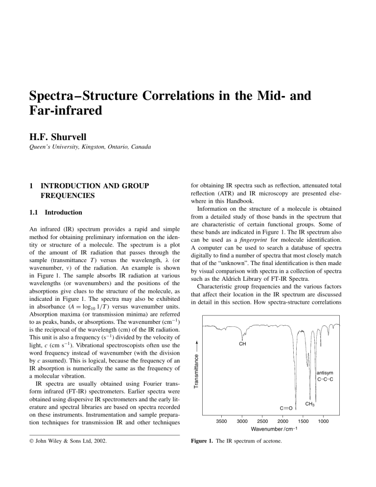
Spectra–Structure Correlations in the Mid- and Far-infrared H.F. Shurvell Queen’s University, Kingston, Ontario, Canada 1.1 Introduction An infrared (IR) spectrum provides a rapid and simple method for obtaining preliminary information on the identity or structure of a molecule. The spectrum is a plot of the amount of IR radiation that passes through the sample (transmittance T) versus the wavelength, l (or wavenumber, n) of the radiation. An example is shown in Figure 1. The sample absorbs IR radiation at various wavelengths (or wavenumbers) and the positions of the absorptions give clues to the structure of the molecule, as indicated in Figure 1. The spectra may also be exhibited in absorbance A D log10 1/T versus wavenumber units. Absorption maxima (or transmission minima) are referred to as peaks, bands, or absorptions. The wavenumber (cm1 ) is the reciprocal of the wavelength (cm) of the IR radiation. This unit is also a frequency (s1 ) divided by the velocity of light, c (cm s1 ). Vibrational spectroscopists often use the word frequency instead of wavenumber (with the division by c assumed). This is logical, because the frequency of an IR absorption is numerically the same as the frequency of a molecular vibration. IR spectra are usually obtained using Fourier transform infrared (FT-IR) spectrometers. Earlier spectra were obtained using dispersive IR spectrometers and the early literature and spectral libraries are based on spectra recorded on these instruments. Instrumentation and sample preparation techniques for transmission IR and other techniques for obtaining IR spectra such as reflection, attenuated total reflection (ATR) and IR microscopy are presented elsewhere in this Handbook. Information on the structure of a molecule is obtained from a detailed study of those bands in the spectrum that are characteristic of certain functional groups. Some of these bands are indicated in Figure 1. The IR spectrum also can be used as a fingerprint for molecule identification. A computer can be used to search a database of spectra digitally to find a number of spectra that most closely match that of the “unknown”. The final identification is then made by visual comparison with spectra in a collection of spectra such as the Aldrich Library of FT-IR Spectra. Characteristic group frequencies and the various factors that affect their location in the IR spectrum are discussed in detail in this section. How spectra-structure correlations CH Transmittance 1 INTRODUCTION AND GROUP FREQUENCIES antisym C−C−C C 3500 3000 2500 O 2000 Wavenumber /cm−1 John Wiley & Sons Ltd, 2002. Figure 1. The IR spectrum of acetone. CH3 1500 1000 2 Spectra–Structure Correlations may be used to identify a compound is the subject of Section 2. The material for these sections is based on Chapters 7, 8 and 9 of ‘Organic Structural Spectroscopy’.1 Cl C O C3H7 1.2 Vibrations of molecules IR spectroscopy gives information on molecular structure through the frequencies of the normal modes of vibration of the molecule. A normal mode is one in which each atom executes a simple harmonic oscillation about its equilibrium position. All atoms move in phase with the same frequency, while the center of gravity of the molecule does not move. There are 3N 6 normal modes of vibration (known as fundamentals) of a molecule (3N 5 for linear molecules), where N is the number of atoms. For a molecule with no symmetry, all 3N 6 fundamental modes are active in the IR and may give rise to absorptions. There is also the possibility of overtones and combinations of the fundamentals, some of which may absorb (usually weakly) and thereby increase the total number of peaks in the IR spectrum. Vibrations of certain functional groups such as OH, NH, CH3 , CDO, C6 H5 , and so on always give rise to bands in the IR spectrum within well-defined frequency ranges regardless of the molecule containing the functional group. The exact position of the group frequency within the range gives further information on the environment of the functional group. As an example, we might take the carbonyl stretching band (nCDO ) of simple aliphatic aldehydes or ketones as the standard (1730 cm1 ). Acid chlorides, carboxylic acid (monomers) and esters have their (nCDO ) bands at higher frequencies, while amides and aromatic ketones have lower CDO stretching frequencies. This is illustrated in Figure 2. The observation of a band in the spectrum within an appropriate frequency range can indicate the presence of one or more different functional groups in the molecule, because there is considerable overlap of the ranges of many functional groups. It is therefore necessary to examine other regions of the spectrum for confirmation of a particular group. Examples of this procedure are given in Section 2. Comparison of the spectrum of an unknown material with a reference spectrum, or with the spectrum of a known compound, can provide absolute proof of the identity of the unknown substance. 1.3 The far-infrared The spectral region called the far-infrared is not very well defined. For the present purposes the mid-infrared range will be assumed to be 4000–400 cm1 . With the use (a) 1800 H C O C3H7 1730 (b) NH2 C O (CH3)3C 1655 2000 (c) 1600 1200 Wavenumber /cm−1 Figure 2. The carbonyl stretching bands of (a) an acid chloride, (b) an aldehyde and (c) an amide. (Reproduced by permission from Sigma-Aldrich Co.) of special beam-splitters, detectors and window materials, some IR spectrometers can be configured to cover a wider range, which extends below 400 cm1 . The region below 400 cm1 down to 10 cm1 is defined as the far-infrared. The region below 200 cm1 is not readily accessible and there are not many useful spectra-structure correlations in this region. However, compounds containing halogen atoms, organometallic compounds and inorganic compounds absorb in the far-infrared and torsional vibrations and hydrogen bond stretching modes are found in this region. The far-infrared also is important for studies of external (lattice) modes of crystalline solids. Spectra–Structure Correlations in the Mid- and Far-infrared A useful discussion of some spectra-structure correlations in the far-infrared is given by Stewart.2 1.4 Introduction to group frequencies IR spectra of a large number of compounds containing a particular functional group such as carbonyl, amino, phenyl, nitro, and so on are found to have certain features that appear at more or less the same frequency for every compound containing the group. It is reasonable, then, to associate these spectral features with the functional group, provided a sufficiently large number of different compounds containing the group have been studied. For example, the IR spectrum of any compound that contains a CDO group has a strong band between 1800 and 1650 cm1 . Compounds containing –NH2 groups have two IR bands between 3400 and 3300 cm1 . The spectrum of a compound containing the C6 H5 – group has sharp peaks near 1600 and 1500 cm1 due to stretching modes of the benzene ring. These are just three examples of the many characteristic frequencies of chemical groups observed in IR spectra. A simple calculation can give an idea of where to expect a band due to stretching of a bond between a pair of atoms in a molecule. The stretching frequency (cm1 ) of such a diatomic group may be calculated using equation (1): p k 1 ncm D 130.3 1 m where k is the force constant (N m1 ) and m is the reduced mass, m1 m2 /m1 C m2 in atomic mass units (amu). The p numerical constant 130.3 D 1/2cp N ð 101 (N is Avogadro’s number, 6.022 ð 1023 , and c is the velocity of light, 2.998 ð 108 m s1 ). This simple approach gives surprisingly good results when one of the atoms of the pair is a light atom, not bonded to any other atom, for example, C–H and N–H in CH3 NH2 or C–H and CDO in (CH3 )2 CO. The stretching frequencies (cm1 ) of these and other diatomic groups, including CC and CDC can be calculated from equation (1). A band characteristic of the group will be observed in the IR spectrum in the predicted region, provided the vibrational frequency of the group is not close to that of another group in the molecule. Calculated frequencies for some diatomic groups are given in Table 1. These are all examples of characteristic group frequencies and can be used to establish the presence of the functional group in the molecule. The explanation of these characteristic diatomic group frequencies lies in the approximately constant values of the stretching force constant of a bond in different molecules. It can be seen from Table 1 that the values of the force constants of double and triple bonds are approximately 3 Table 1. Calculated frequencies of some diatomic groups. Group Reduced mass (amu) Force constant (N m1 ) Frequency (cm1 ) O–H N–H C–H C–C CDC CDO CC CN 0.94 0.93 0.92 6.00 6.00 6.86 6.00 6.46 700 600 500 425 960 1200 1600 2100 3600 3300 3000 1100 1650 1725 2100 2350 twice and three times those of single bonds, respectively. Carbon–carbon single bonds are included in Table 1, but C–C stretching does not usually give a well-defined group frequency. Most organic molecules contain several C–C single bonds and other groups that have similar vibrational frequencies to that of the C–C stretching mode. These vibrations interact with each other, and the simple model does not apply. Vibrational interactions can take several forms and are discussed later in this section. When two or more identical groups are present in a molecule, one might expect to observe two or more bands in the IR spectrum at similar wavenumbers. However, if the groups are attached to the same carbon atom, or to two adjacent atoms, the observed bands may be spread over a few hundred wavenumbers by interactions between the vibrations of the groups. The four CH groups in ethylene (C2 H4 ) provide an example of this behavior. The four observed CH stretching frequencies are 3270, 3105, 3020, and 2990 cm1 . Polyatomic groups also have characteristic frequencies, which involve both stretching and bending vibrations or combinations of these. No simple equation like equation (1) can be found to calculate the bending frequencies of polyatomic groups, and the best way to establish whether or not a particular group such as –CH2 , –CH3 , –NH2 , or –C6 H5 has characteristic bending in addition to stretching frequencies is to examine the vibrational spectra of a large number of compounds containing these groups. 1.5 Factors affecting group frequencies 1.5.1 Symmetry The vast majority of organic molecules have little or no symmetry. Nevertheless, some knowledge of symmetry can be of considerable help in understanding the factors that affect intensities of group frequencies. Occasionally, a group frequency is not observed in the IR spectrum. This is usually a consequence of symmetry. If a molecule possesses a center of symmetry, all vibrations 4 Spectra–Structure Correlations H H H H + − + + H H H H C C C C H stretch (sym) HCH bend CH2 twist C − CH2 wag H H C C H stretch (antisym) H H C CH2 rock Figure 3. CH2 group vibrations. The arrows show the direction of motion of atoms in the plane of the CH2 group. The C and signs denote motion above and below the plane, respectively. that are symmetric with respect to that center are inactive in the IR, because they do not produce a change in the dipole moment. An example of the effect of a center of symmetry is given by the CC stretching mode. In methylacetylene (CH3 CCH) the vibration is IR active, and a strong IR band is observed at 2150 cm1 . On the other hand, in dimethylacetylene (CH3 CCCH3 ), which has a center of symmetry, no band is observed in the IR near 2150 cm1 . In larger, more complicated molecules, a local symmetry may exist for a homonuclear diatomic group such as CDC or S–S, so that the IR absorption from the group vibration may be weak or absent. Molecules of high symmetry have simple IR spectra. As an example consider the benzene molecule. It has 12 atoms and therefore has 3N 6 D 30 normal modes of vibration. The first effect of the high symmetry is to make 10 pairs of these vibrations have identical frequencies (degenerate modes). This leaves 20 different normal frequencies. The second effect of the high symmetry is to reduce the number of modes for which there is a change in dipole moment (IR active). In fact, the IR spectrum of benzene contains only four bands due to fundamentals. When the symmetry of benzene is reduced, as in 1,3,5-trichlorobenzene, the number of IR active modes increases, but there are still some degenerate modes and the spectra are relatively simple. When the symmetry is completely removed, as in 1-chloro-2-bromobenzene, all 30 normal modes are active. However, because of the residual symmetry of the benzene ring, some of these vibrations, although allowed, appear only very weakly and are hard to distinguish from the weak bands that are due to overtones and combinations. Vibrations of the methyl group (–CH3 ) in an unsymmetrical molecule can be described in terms of the local symmetry of the free group, which has a threefold axis and three planes of symmetry. A free methyl group would have 3N 6 D 6 normal modes of vibration comprising symmetric and degenerate antisymmetric (with respect to the threefold axis) stretching and bending modes. When the methyl group is attached to a molecule, three new modes appear, a torsional mode and a degenerate pair of rocking vibrations. These motions would be rotations in the free methyl group. Thus, there are four regions of the spectrum where we expect to find group vibrations of the methyl group. This conclusion is amply supported experimentally. The presence of the methyl group also contributes three skeletal modes to the vibrations of the molecule. These correspond to translations of the free methyl group. When the methyl group is part of a molecule with lower symmetry, the degeneracies are removed, leading to the observation of doublets in some of the regions of the spectrum where the methyl group frequencies are expected. The torsional mode is actually inactive in the IR, because it produces no change in dipole moment. However, it may be allowed by the low symmetry of the whole molecule and, in fact, methyl torsions are sometimes observed as weak bands in the far-infrared. The vibrations of a methylene group (CH2 ) can also be described in terms of the local symmetry of the group, which has a twofold axis and two planes of symmetry. Figure 3 shows the vibrations associated with a CH2 group, when it is attached to a molecule. The free CH2 group would have three modes, symmetric and antisymmetric (with respect to the twofold axis) stretching and the bending (scissors) vibration. When the group is part of a larger molecule, three additional modes described as twisting, wagging and rocking are produced. Of these, the twisting mode produces no change in dipole moment and hence is not allowed in the IR. However, it can give rise to a very weak band in the spectrum of an unsymmetrical molecule. The terms twofold and threefold axis, plane of symmetry, and center of symmetry are examples of symmetry elements. The collection of all symmetry elements that a molecule possesses is known as a point group and provides a way of classifying the symmetry of the molecule. This, in turn, leads to an understanding of the symmetry of the normal vibrations of a molecule and to a prediction of the number of frequencies expected in the IR spectrum. 1.5.2 Mechanical coupling of vibrations Two completely free identical diatomic molecules will, of course, vibrate with identical frequencies. When the two diatomic groups are part of a molecule, however, they can no longer vibrate independently of each other because the vibration of one group causes displacements of the other atoms in the molecule. These displacements Spectra–Structure Correlations in the Mid- and Far-infrared are transmitted through the molecule and interact with the vibration of the second group. The resulting vibrations appear as in-phase and out-of-phase combinations of the two diatomic vibrations. When the groups are widely separated in the molecule, the coupling is very small and the two frequencies may not be resolved. Consider the two C–H stretching modes in acetylene (H–CC–H). These occur at 3375 cm1 (inphase) and 3280 cm1 (out-of-phase). In diacetylene (H–CC–CC–H), the two C–H stretching vibrations have closer frequencies, near 3330 and 3295 cm1 . The vibrations of two different diatomic groups are not coupled unless the uncoupled frequencies are similar as the result of a combination of mass and force constant effects. For example, in thioamides and xanthates, the CDS group has a force constant of about 650 N m1 and the reduced mass is 8.72 amu, so that the vibrational frequency calculated from equation (1) is approximately 1120 cm1 . The C–N and C–O groups have force constants of about 480 and 510 N m1 , respectively, and the reduced masses are 6.46 and 6.86 amu. The calculated frequencies are both approximately 1120 cm1 . Consequently, in any compound containing a CDS group adjacent to a C–O or C–N group, there may be an interaction between the stretching vibrations of the groups. In compounds such as thioamides and xanthates, where the carbon atom is common to both groups, the coupling is large and the two vibrations interact with each other to produce two new frequencies, neither of which is in the expected region of the spectrum. The way in which such mechanical coupling occurs can be illustrated for the case of two CDC groups coupled through a common atom, as in the allene molecule, CH2 DCDCH2 . In the absence of strong coupling one might expect to observe a band in the IR spectrum near 1600 cm1 due to the antisymmetric (out-of-phase) combination of the vibrations of the two CDC groups. For the 1,3-butadiene molecule (CH2 DCH–CHDCH2 ), this vibration gives rise to a band at 1640 cm1 . However, for allene the observed frequency is near 1960 cm1 . This result can be understood in terms of mechanical coupling of the two CDC group vibrations. When such coupling occurs, it is usually found that the antisymmetric (out-of-phase) vibration occurs at a higher frequency and is IR active, while the symmetric (in-phase) vibration occurs at lower frequency and is IR inactive. It is possible for coupling to occur between dissimilar modes such as stretching and bending vibrations, when the frequencies of the vibrations are similar and the two groups involved are adjacent in the molecule. An example is found in secondary amides, in which the C–N stretching vibration is of a similar frequency to that of the NH bending mode. Interaction of these two vibrations gives rise to two bands in the spectrum, one at a higher and one at a lower frequency 5 than the uncoupled frequencies. These bands are known as amide II and amide III bands. (The CDO stretching mode is known as the amide I band.) Singly bonded carbon atom chains are, of course, not linear, so that the simple model used for the allene molecule would have to be modified. In addition, we were able to ignore the bending of the CDCDC group in allene, which cannot couple with the stretching modes, because it takes place at right angles to the stretching vibrations. Mechanical coupling will always occur between C–C single bonds in an organic molecule, so that there is no simple C–C group stretching frequency. One can expect that there will always be several bands in the IR spectra in the 1200–800 cm1 range in compounds containing saturated carbon chains. 1.5.3 Fermi resonance A special case of mechanical coupling, known as Fermi resonance, often occurs. This phenomenon, which results from coupling of a fundamental vibration with an overtone or combination, can shift group frequencies and introduce extra bands. For a polyatomic molecule there are 3N 6 energy levels for which only one vibrational quantum number (v1 ) is 1 when all the rest are zero. These are called the fundamental levels and a transition from the ground state to one of these levels is known as a fundamental. In addition, there are the levels for which one v1 is 2, 3, and so on (overtones) or for which more than one v1 is nonzero (combinations). There are therefore a very large number of vibrational energy levels, and it quite often happens that the energy of an overtone or combination level is very close to that of a fundamental. This situation is termed accidental degeneracy, and an interaction known as Fermi resonance can occur between these levels provided that the symmetries of the levels are the same. Since most organic molecules have no symmetry, all levels have the same symmetry and Fermi resonance effects occur frequently in vibrational spectra. Normally, an overtone or combination band is very weak in comparison with a fundamental, because these transitions are not “allowed”. However, when Fermi resonance occurs there is a sharing of intensity and the overtone can be quite strong. The result is the same as that produced by two identical groups in the molecule. As an example, two peaks are observed in the carbonyl stretching band of benzoyl chloride, near 1760 and 1720 cm1 (Figure 4). If this were an unknown compound, one might be tempted to suggest that there were two nonadjacent carbonyl groups in the molecule. However, the lower frequency band is due to the overtone of the CH out-of-plane bending mode at 865 cm1 in Fermi resonance with the CDO stretching fundamental. Numerous other well-characterized examples of Fermi resonance are known. The N–H stretching mode of the 6 Spectra–Structure Correlations group frequencies through solvent–solute interactions, such as molecular association through hydrogen bonding. Transmission 1.5.5 Hydrogen bonding O COCl Fermi doublet 3500 3000 2500 2000 1500 1000 500 Wavenumber / cm−1 Figure 4. The IR spectrum of benzoyl chloride showing the Fermi doublet at 1760 cm1 and 1720 cm1 . –CO–NH– group in polyamides (nylons), peptides, proteins, appears as two bands near 3300 and 3205 cm1 . The N–H stretching fundamental and the overtone of the N–H deformation mode near 1550 cm1 combine through Fermi resonance to produce the two observed bands. The CH stretching region of the –CHO group in aldehydes provides another example of Fermi resonance. Two bands are often observed near 2900 and 2700 cm1 in the IR spectra of aldehydes. This doublet is attributed to Fermi resonance between the overtone of the C–H deformation mode, which occurs near 1400 cm1 , and the C–H stretching mode, which would also occur near 2800 cm1 in the absence of Fermi resonance. In many molecules, mechanical coupling of the group vibrations is so widespread that there are few, if any, frequencies that can be assigned to functional groups. Many such examples are found in aliphatic fluorine compounds, in which the CF and CC stretching modes are coupled with each other and with FCF and CCF bending vibrations. The presence of fluorine can be deduced from several very strong IR bands in the region between 1400 and 900 cm1 . Hydrogen bonding (written X–HÐ Ð ÐY) occurs between the hydrogen atom of a donor X–H group such as OH or NH and an acceptor atom Y which is usually O or N. The main effects are broadening of bands in the IR spectra and shifts of group frequencies. X–H stretching frequencies are lowered by hydrogen bonding, and X–H bending frequencies are raised. Hydrogen bonding also affects the frequencies of the acceptor group, but the shifts are less than those of the X–H group. Inert solvents can reduce the extent of hydrogen bonding and even eliminate the effect in very dilute solutions. A very broad band centered near 3100 cm1 in the spectrum of pure carboxylic acids is due mainly to OH stretching of hydrogen-bonded carboxylic acid polymers. In the solution spectra of these compounds the bands are sharper. However, hydrogen-bonding persists even in dilute solution and the carboxylic acid is present mainly as a cyclic dimer. Hydrogen bonding manifests itself in very broad OH and NH stretching bands at frequencies considerably lower than normal. Changes in the intensity of these bands can be brought about by changes in temperature and concentration. Stretching of the hydrogen bond itself (XHÐ Ð ÐY) has been observed in the far IR in many cases between 200 and 50 cm1 , and bending of the hydrogen bond occurs at very low frequencies, usually below 50 cm1 . Intramolecular hydrogen bonding can occur between OH groups in alcohols or phenols and halogen atoms. In 2-chloroethanol, for example, an intramolecular hydrogen bond stabilizes the gauche rotational isomer. The free nOH in the trans conformation absorbs at 3623 cm1 , whereas for the hydrogen-bonded isomer the frequency is 3597 cm1 . Halophenols also show two nOH bands separated by 50–100 cm1 due to bonded and nonbonded conformations. IR spectra also indicate that intermolecular OHÐ Ð Ðhalogen bonding occurs between alkyl halides and phenols or alcohols. 1.5.4 Chemical and environmental effects 1.5.6 Steric effects Hydrogen bonding, electronic and steric effects, physical state, and solvent and temperature effects all contribute to the position, intensity, and appearance of the bands in the IR spectrum of a compound. Lowering the temperature usually makes the bands sharper and shifts them to higher frequencies. There is also a possibility of splittings arising due to crystal effects, which must be considered when examining the spectra of solids under moderately high resolution. Polar solvents can cause significant shifts of Ring strain increases the frequency of the carbonyl stretching frequency in alicylic ketones. For cyclohexanone, cyclopentanone, and cyclobutanone, the observed carbonyl stretching frequencies are 1714, 1746, and 1783 cm1 , respectively. This increase in frequency with increasing angle strain is generally observed for double bonds directly attached (exocyclic) to rings. Similar frequency changes are observed in the series of compounds, methylenecyclohexane, methylenecyclopentane, and methylenecyclobutane, in Spectra–Structure Correlations in the Mid- and Far-infrared 1.5.7 Electronic effects Effects arising from the change in the distribution of electrons in a molecule produced by a substituent atom or group can often be detected in the vibrational spectrum. There are several mechanisms such as inductive and resonance effects, which can be used to explain observed shifts and intensity changes in a qualitative way. These effects involve changes in electron distribution in a molecule and cause changes in the force constants that are, in turn, responsible for changes in group frequencies. Inductive and resonance effects have been used successfully to explain the shifts observed in CDO stretching frequencies produced by various substituent groups in compounds such as acid chlorides and amides. High CDO stretching frequencies are usually attributed to inductive effects and low frequencies arise when delocalized structures are possible. For example, in acid chlorides the CDO frequency is near 1800 cm1 , which is high compared with a normal CDO frequency such as that observed for aldehydes or ketones (near 1730 cm1 ). On the other hand, in amides the carbonyl frequency is lower (near 1650 cm1 ). In acid chlorides the electronegative chlorine atom adjacent to the carbonyl group causes the increase in frequency, whereas in amides the delocalized electronic structure lowers the CDO stretching frequency. Conjugation of double bonds tends to lower the double bond character and increase the bond order of the intervening single bond. For compounds in which a carbonyl group can be conjugated with an ethylenic double bond, the CDO stretching frequency is lowered by 20–30 cm1 . 1.5.8 Structural isomerism Structural (constitutional) isomers often differ in the functional groups present, and their vibrational spectra then differ considerably. Some examples are the a-amino acid alanine, the ester ethyl carbamate (urethane), and the nitro compound 1-nitropropane. All three compounds have the empirical formula C3 H7 O2 N. IR spectra of these three compounds are shown in Figure 5. A good example of the importance of vibrational spectra in differentiating between structural isomers is found in ortho-, meta-, and para-disubstituted benzenes. The CH D,L-Alanine Ethyl carbamate Transmission which a CDCH2 group replaces the CDO group in the ring. The observed CDC stretching frequencies are 1649, 1656, and 1677 cm1 , respectively. When the double bond is a part of the ring, a decrease in the ring angle causes a lowering of the CDC stretching frequency. The observed frequencies for cyclohexene, cyclopentene, and cyclobutene are 1650, 1615, and 1565 cm1 , respectively. 7 1-Nitropropane 3500 3000 2500 2000 1500 1000 500 Wavenumber /cm−1 Figure 5. IR spectra of three compounds with the empirical formula C3 H7 O2 N. out-of-plane deformation patterns in the 850–700 cm1 region are different for each isomer. Substituted pyridines, pyrimidines, and other heterocyclic compounds provide further examples of structural isomers that can be distinguished by their vibrational spectra. 1.5.9 Geometrical (cis–trans) isomerism IR spectroscopy is useful in distinguishing between cis and trans isomers. Absorption of IR radiation by a molecule can only occur if there is a change of dipole moment accompanying a vibration. For cis isomers a dipole moment change occurs for most of the normal vibrations. However, trans isomers usually have higher symmetry, which leads to a zero or very small dipole moment change for some vibrations, so that they are not observed in the IR spectrum. Thus, we can conclude that trans compounds often have simpler IR spectra than their cis isomers. 1.5.10 Rotational isomerism In open chain compounds, the barriers to internal rotation about one or more carbon–carbon single bonds may be too high for rapid interconversion between different configurations. In such cases, two or more different isomers can exist and their presence may be detected in the IR spectrum. The restriction of rotation about double bonds can be thought of as an extension of the above concept. In this case, very high barriers are involved and cis and trans compounds result. The axial-equatorial conformations of cyclohexane and cyclopentane derivatives are examples of another kind of conformational (rotational) isomerism. In noncyclic structures, rotation about a single bond can produce an infinite number of configurations. Some of Spectra–Structure Correlations these are energetically favored (energy minima). The simplest examples are the substituted ethanes, CH2 XCH2 Y, for which there are several preferred staggered conformations. When there is a stabilizing interaction, the eclipsed form may be one of the stable conformations. Many such examples can be found in a-halo ketones, esters, acid halides, and amides. In these compounds, the halogen atom is believed to be either cis (eclipsed) or gauche (staggered) with respect to the carbonyl group. Two CDO stretching bands are observed in such cases. One is at higher frequencies, owing to the eclipsed interaction between the halogen atom and the CDO group. The other is found at the normal frequency. For a-halocarboxylic acids, multiple carbonyl bands are not usually observed because of complications from hydrogen bonding. In cyclic compounds, the possibility of axial and equatorial conformations exists. For example, in a-chloro substituted cyclopentanones or cyclohexanones, two distinct carbonyl stretching frequencies can be observed. One band is found near 1745 cm1 , due to the equatorial conformation, in which interaction between the Cl and CDO groups can occur. A second band near 1725 cm1 is attributed to the axial isomer, in which interaction is minimized. The relative proportions of axial and equatorial forms change with phase, temperature, and solvent, and such changes can be readily followed in the vibrational spectra. Ortho-halogenated benzoic acids also show two carbonyl stretching frequencies, due to the two rotational isomers, which could be described as cis and trans with respect to the halogen and CDO groups. Vinyl ethers show a doublet for the CDC stretching mode at 1640–1620 and 1620–1610 cm1 . These bands correspond to rotational isomers about the CDC bond. The two bands show variations in intensity with temperature. The CH2 deformation band also is found to be a doublet due to the two different rotational isomers. 1.5.11 Tautomerism IR spectroscopy offers a useful means of distinguishing between possible tautomeric structures. A simple example is found in b-diketones. The keto form has two CDO groups, which have separate stretching frequencies, and a doublet is often observed in the usual ketone carbonyl stretching region, near 1730 cm1 . The enol form, on the other hand, has only one carbonyl group, the frequency of which is lowered by hydrogen bonding and conjugation by 80–100 cm1 . This structure also has an alkenic double bond that should give a band between 1650 and 1600 cm1 . The CDO and CDC peaks may then appear as a doublet. An example of tautomerism is shown in Figure 6. The IR spectrum of the compound ethyl propionylacetate clearly shows both keto and enol forms. Transmission 8 Enol Keto 3500 3000 2500 2000 1500 1000 500 Wavenumber / cm−1 Figure 6. The IR spectrum of ethyl propionylacetate. 1.6 IR group frequencies A good group frequency is one that falls within a fairly restricted range regardless of the compound in which the group is found. Mechanical coupling, symmetry, or other effects discussed in the previous sections may occasionally cause group frequencies to fall outside the normal range, so one should be aware of this possibility. A list of frequency ranges from 4000 down to 50 cm1 is presented in Table 2, with possible groups that could absorb within a given range. Table 2 is by no means comprehensive and in order to make full use of the group frequency method for structure determination, the literature should be consulted.2 – 9 2 SPECTRA–STRUCTURE CORRELATIONS 2.1 Introduction A preliminary examination of the IR spectrum of a compound will indicate the various X–H bonds present in the molecule and the presence or absence of triple or double bonds, carbonyl groups, aromatic rings, and other functional groups. This information will suggest various possible structures. The IR spectrum can then be searched in detail for bands due to the various groups in the postulated structures. Comparison with published IR spectra of compounds having the suggested or a similar structure may then be made. Published spectra are found in the literature.10 – 14 Also, the computer attached to the FT-IR spectrometer usually has search software and a library of IR spectra which may be searched for spectra that most closely match the spectrum of the unknown compound. Spectra–Structure Correlations in the Mid- and Far-infrared 9 Table 2. Wavenumber ranges in which some functional groups and classes of compounds absorb in the mid- and far-infrared. Range (cm1 ) and intensitya 3700–3600 3520–3320 3420–3250 3400–3300 3360–3340 3320–3250 3300–3250 3300–3280 3200–3180 3200–3000 3100–2400 3100–3000 2990–2850 2850–2700 2750–2650 2750–2350 2720–2560 2600–2540 2410–2280 2300–2230 2285–2250 2260–2200 2260–2190 2190–2130 2175–2115 2160–2080 2140–2100 2000–1650 1980–1950 1870–1650 1870–1830 1870–1790 1820–1800 1780–1760 1765–1725 1760–1740 1750–1730 1750–1740 1740–1720 1720–1700 1710–1690 1690–1640 1680–1620 1680–1635 1680–1630 1680–1630 1670–1640 1670–1650 1670–1630 1655–1635 1650–1620 1650–1580 1640–1580 1640–1580 1620–1610 (s) (m-s) (s) (vs) (m) (m) (m-s) (s) (s) (vs) (vs) (m) (m-s) (m) (w-m) (br) (m) (w) (m) (m) (s) (m-s) (w-m) (m) (s) (m) (w-m) (w) (s) (vs) (s) (vs) (s) (s) (vs) (vs) (s) (vs) (s) (s) (s) (s) (s) (s) (m-s) (vs) (s-vs) (vs) (vs) (vs) (w-m) (m-s) (s) (vs) (s) Group and class –OH in alcohols and phenols –NH2 in aromatic amines, primary amines and amides –OH in alcohols and phenols –OO–H in hydroperoxides –NH2 in primary amides –OH in oximes CH in acetylenes –NH in secondary amides –NH2 in primary amides –NH3 C in amino acids –OH in carboxylic acids DCH in aromatic and unsaturated hydrocarbons –CH3 and – CH2 – in aliphatic compounds –CH3 attached to O or N –CHO in aldehydes –NH3 C in amine hydrohalides –OH in phosphorus oxyacids –SH in alkyl mercaptans –PH in phosphines –NN in diazonium salts –NDCDO in isocyanates –CN in nitriles –CC in disubstituted alkynes –CN in thiocyanates –NC in isonitriles NDNDN in azides –CC in monosubstituted alkynes Substituted benzene rings CDCDC in allenes CDO in carbonyl compounds CDO in b-lactones CDO in anhydrides CDO in acid halides CDO in g-lactones CDO in anhydrides CDO in a-keto esters CDO in υ-lactones CDO in esters CDO in aldehydes CDO in ketones CDO in carboxylic acids CDN in oximes CDO and NH2 in primary amides CDO in ureas CDC in alkenes, etc. CDO in secondary amides CDO in benzophenones CDO in primary amides CDO in tertiary amides CDO in b-keto esters N–H in primary amides NH2 in primary amines NH3 C in amino acids CDO in b-diketones CDC in vinyl ethers Assignment and remarks OH stretch; (dilute solution) NH stretch; (dilute solution) OH stretch; (solids and liquids) Hydrogen bonded OO–H stretch NH2 antisymmetric stretch; (solids) O–H stretch C–H stretch NH stretch; (solids); also in polypeptides and proteins NH2 symmetric stretch; (solids) NH3 C antisymmetric stretching; very broad band H-bonded OH stretch ; very broad band DC–H stretch CH antisymmetric and symmetric stretching CH stretching modes Overtone of CH bending; (Fermi resonance) NH stretching modes Associated OH stretching S–H stretch P–H stretch; sharp peak NN stretch; (in aqueous solution) NDCDO antisymmetric stretch CN stretch CC stretch CN stretch NC stretch NDNDN antisymmetric stretch CC stretch Several bands from overtones and combinations CDCDC antisymmetric stretch CDO stretch CDO stretch CDO antisymmetric stretch; part of doublet CDO stretch; lower for aromatic acid halides CDO stretch CDO symmetric stretch; part of doublet CDO stretch; enol form CDO stretch CDO stretch; 20 cm1 lower if unsaturated CDO stretch; 30 cm1 lower if unsaturated CDO stretch; 20 cm1 lower if unsaturated CDO stretch; fairly broad CDN stretch; also imines Two bands; (CDO stretch and NH2 deformation) CDO stretch; (broad band) CDC stretch CDO stretch; (amide I band) CDO stretch CDO stretch; (amide I band) CDO stretch CDO stretch; enol form NH deformation; (amide II band) NH2 deformation NH3 C deformation CDO stretch; enol form CDC stretch; doublet due to rotational isomerism (continued overleaf ) 10 Spectra–Structure Correlations Table 2. (continued ) Range (cm1 ) and intensitya 1615–1590 (m) 1615–1565 (s) 1610–1580 (s) 1610–1560 (vs) 1590–1580 (m) 1575–1545 (vs) 1565–1475 (vs) 1560–1510 (s) 1550–1490 (s) 1530–1490 (s) 1530–1450 (m-s) 1515–1485 (m) 1475–1450 (vs) 1465–1440 (vs) 1440–1400 (m) 1420–1400 (m) 1400–1370 (m) 1400–1310 (s) 1390–1360 (vs) 1380–1370 (s) 1380–1360 (m) 1375–1350 (s) 1360–1335 (vs) 1360–1320 (vs) 1350–1280 (m-s) 1335–1295 (vs) 1330–1310 (m-s) 1300–1200 (vs) 1300–1175 (vs) 1300–1000 (vs) 1285–1240 (vs) 1280–1250 (vs) 1280–1240 (m-s) 1280–1180 (s) 1280–1150 (vs) 1255–1240 (m) 1245–1155 (vs) 1240–1070 (s-vs) 1230–1100 (s) 1225–1200 (s) 1200–1165 (s) 1200–1015 (vs) 1170–1145 (s) 1170–1140 (s) 1160–1100 (m) 1150–1070 (vs) 1120–1080 (s) 1120–1030 (s) 1100–1000 (vs) 1080–1040 (s) 1065–1015 (s) 1060–1025 (vs) 1060–1045 (vs) 1055–915 (vs) 1030–950 (w) Group and class Benzene ring in aromatic compounds Pyridine derivatives NH2 in amino acids COO in carboxylic acid salts NH2 primary alkyl amide NO2 in aliphatic nitro compounds NH in secondary amides Triazine compounds NO2 in aromatic nitro compounds NH3 C in amino acids or hydrochlorides NDN–O in azoxy compounds Benzene ring in aromatic compounds CH2 in aliphatic compounds CH3 in aliphatic compounds OH in carboxylic acids C–N in primary amides tert-Butyl group COO group in carboxylic acid salts SO2 in sulfonyl chlorides CH3 in aliphatic compounds Isopropyl group NO2 in aliphatic nitro compounds SO2 in sulfonamides NO2 in aromatic nitro compounds NDN–O in azoxy compounds SO2 in sulfones CF3 attached to a benzene ring N–O in pyridine N-oxides PDO in phosphorus oxyacids and phosphates C–F in aliphatic fluoro compounds Ar–O in alkyl aryl ethers Si–CH3 in silanes C–O–C in epoxides C–N in aromatic amines C–O–C in esters, lactones tert-Butyl in hydrocarbons SO3 H in sulfonic acids C–O–C in ethers C–C–N in amines C–O–C in vinyl ethers SO2 Cl in sulfonyl chlorides C–OH in alcohols SO2 NH2 in sulfonamides SO2 – in sulfones CDS in thiocarbonyl compounds C–O–C in aliphatic ethers C–OH in secondary or tertiary alcohols C–NH2 in primary aliphatic amines Si–O–Si in siloxanes SO3 H in sulfonic acids CH–OH in cyclic alcohols CH2 –OH in primary alcohols SDO in alkyl sulfoxides P–O–C in organophosphorus compounds Carbon ring in cyclic compounds Assignment and remarks Ring stretch; sharp peak Ring stretch; doublet NH2 deformation; broad band –COO antisymmetric stretch NH2 deformation; (amide II band) NO2 antisymmetric stretch NH deformation; (amide II band) Ring stretch; sharp band NO2 antisymmetric stretch NH3 C deformation NDN–O antisymmetric stretch Ring stretch, sharp band CH2 bending (scissors) vibration Antisymmetric CH3 deformation In-plane OH bending C–N stretch; (amide III band) Symmetric CH3 deformations; (two bands) COO symmetric stretch; (broad band) SO2 antisymmetric stretch CH3 symmetric deformation CH3 symmetric deformations; (two bands) NO2 symmetric stretch SO2 antisymmetric stretch NO2 symmetric stretch NDN–O symmetric stretch SO2 antisymmetric stretch CF3 antisymmetric stretch N–O stretch PDO stretch C–F stretch C–O stretch CH3 symmetric deformation C–O stretch C–N stretch C–O–C antisymmetric stretch Skeletal vibration; second band near 1200 cm1 SDO stretch C–O–C stretch; also in esters C–C–N bending C–O–C antisymmetric stretch SO2 symmetric stretch C–O stretch SO2 symmetric stretch SO2 symmetric stretch CDS stretch C–O–C antisymmetric stretch C–O stretch C–N stretch Si–O–Si antisymmetric stretch SO3 symmetric stretch C–O stretch C–O stretch SDO stretch P–O–C antisymmetric stretch Ring breathing mode Spectra–Structure Correlations in the Mid- and Far-infrared 11 Table 2. (continued ) Range (cm1 ) and intensitya Group and class 1000–950 (s) 980–960 (vs) 950–900 (vs) 900–865 (vs) 890–830 (w) 890–805 (vs) 860–840 (w) 860–760 (vs) 860–720 (vs) 850–830 (vs) 850–810 (vs) 850–790 (m) 850–550 (m) 830–810 (vs) 825–805 (vs) 820–800 (s) 815–810 (s) 810–790 (vs) 800–690 (vs) 785–680 (vs) 775–650 (m) 770–690 (vs) 760–740 (s) 760–510 (s) 740–720 (w-m) CHDCH2 in vinyl compounds CHDCH– in trans disubstituted alkenes CHDCH2 in vinyl compounds CH2 DCRR0 in vinylidenes Peroxides 1,2,4-Trisubstituted benzenes Hydroperoxides R–NH2 primary amines Si–C in organosilicon compounds 1,3,5-Trisubstituted benzenes Si–CH3 in silanes CHDC in trisubstituted alkenes C–Cl in chloro compounds p-Disubstituted benzenes 1,2,4-Trisubstituted benzenes Triazines CHDCH2 in vinyl ethers 1,2,3,4-tetrasubstituted benzenes m-Disubstituted benzenes 1,2,3-Trisubstituted benzenes C–S in sulfonyl chlorides Monosubstituted benzenes o-Disubstituted benzenes C–Cl alkyl chlorides –(CH2 )n – in hydrocarbons 730–665 720–600 710–570 700–590 695–635 680–620 680–580 650–600 650–600 650–500 650–500 645–615 645–575 640–630 635–605 630–570 630–565 615–535 610–565 610–545 600–465 580–530 580–520 580–430 580–420 570–530 565–520 565–440 560–510 CHDCH in cis disubstituted alkenes Ar–OH in phenols C–S in sulfides O–CDO in carboxylic acids C–C–CHO in aldehydes C–OH in alcohols CDC–H in alkynes S–CDN in thiocyanates NO2 in aliphatic nitro compounds Ar–CF3 in aromatic trifluoro-methyl compounds C–Br in bromo compounds Naphthalenes O–C–O in esters DCH2 in vinyl compounds Pyridines N–CDO in amides C–CO–C in ketones CDO in amides O2 in sulfonyl chlorides SO2 in sulfones C–I in iodo compounds C–C–CN in nitriles NO2 in aromatic nitro compounds Ring in cycloalkanes Ring in benzene derivatives SO2 in sulfonyl chlorides C–CDO in aldehydes Cn H2nC1 in alkyl groups C–CDO in ketones (s) (s) (m) (s) (s) (s) (s) (w) (s) (s) (s) (m-s) (s) (s) (m-s) (s) (s) (s) (vs) (m-s) (s) (m-s) (m) (s) (m-s) (vs) (s) (w-m) (s) Assignment and remarks DCH out-of-plane deformation DCH out-of-plane deformation CH2 out-of-plane wag CH2 out-of-plane wag O–O stretch CH out-of-plane deformation (two bands) O–OH stretch NH2 wag; broad band Si–C stretch CH out-of-plane deformation Si–CH3 rocking CH out-of-plane deformation C–Cl stretch CH out-of-plane deformation CH out-of-plane deformation CH out-of-plane deformation CH2 out-of-plane wag CH out-of-plane deformation CH out-of-plane deformation (two bands) CH out-of-plane deformation (two bands) C–S stretch; strong in Raman CH out-of-plane deformation (two bands) CH out-of-plane deformation C–Cl stretch CH2 rocking in methylene chains; intensity depends on chain length CH out-of-plane deformation OH out-of-plane deformation; broad band C–S stretch O–CDO bending C–C–CHO bending C–O–H bending CDC–H bending S–C stretch NO2 deformation CF3 deformation (two or three bands) C–Br stretch In-plane ring deformation O–C–O bend DCH2 twisting In-plane ring deformation N–CDO bend C–CO–C bend CDO out-of-plane bend SO2 deformation SO2 scissoring C–I stretch C–C–CN bend NO2 deformation Ring deformation In-plane and out-of-plane ring deformations (two bands) SO2 rocking C–CDO bend Chain deformation modes (two bands) C–CDO bend (continued overleaf ) 12 Spectra–Structure Correlations Table 2. (continued ) Range (cm1 ) and intensitya Group and class 560–500 (s) 555–545 (s) 550–465 (s) 545–520 (s) 530–470 (m-s) 520–430 (m-s) 510–400 (s) 490–465 (variable) 440–420 (s) 405–400 (s) 395–360 (variable) 385–355 (m-s) 380–330 (m-s) 360–305 (m-s) 355–330 (variable) 330–230 (variable) 330–175 (variable) 270–220 (w) 265–180 (variable) 200–50 (w) 185–160 (variable) a s, Assignment and remarks CO2 in amino acids DCH2 in vinyl compounds C–CDO in carboxylic acids Naphthalenes NO2 in nitro compounds C–O–C in ethers C–N–C in amines Naphthalenes Cl–CDO in acid chlorides S–CDN in thiocyanates C–S–O in sulfoxides Nitriles Secondary amides Primary amides Alkynes Halogenated aromatic compounds Halogenated aromatic compounds C–CH3 , O–CH3 , N–CH3 Dichloro alkenes Hydrogen-bonded OH group Aliphatic halogeno acetylenes CO2 rocking DCH2 twisting C–CDO bend In-plane ring deformation NO2 rocking C–O–C bend C–N–C bend Out-of-plane ring bending Cl–CDO in-plane deformation S–CDN bend Symmetric C–S–O bend C–CN bend C–CDO bend C–CDO bend C–CC bend In plane phenyl-halogen bend (Cl and Br) Out-of-plane phenyl-halogen bend (Cl and Br) Methyl torsion Two bands due to DCCl2 deformation and rocking OH . . . Y stretch (Y is the hydrogen acceptor atom) CC–X bend (X D Cl, Br, I) strong; m, medium; w, weak; v, very; br, broad. Table 3. Regions of the IR spectrum for preliminary analysis. Region (cm1 ) 3700–3100 Group 1870–1650 –OH –NH C–H DCH DCH2 or –CHDCH– –CH, –CH2 , or –CH3 –CHO –POH –SH –PH –CN –NDNDN –CC– CDO 1650–1550 CDC, CDN, NH 1550–1300 NO2 CH3 and CH2 C–O–C and C–OH SDO, PDO, C–F Si–O and P–O DC–H –NH C–halogen Aromatic rings 3100–3000 3000–2800 2800–2600 2700–2400 2400–2000 1300–1000 1100–800 1000–650 800–400 a Band may be absent owing to symmetry. Possible compounds present (or absent) Alcohols, aldehydes, carboxylic acids Amides, amines Alkynes Aromatic compounds Alkenes or unsaturated rings Aliphatic groups Aldehydes (Fermi doublet) Phosphorus compounds Mercaptans and thiols Phosphines Nitriles Azides Alkynesa Acid halides, aldehydes, amides, amino acids, anhydrides, carboxylic acids, esters, ketones, lactams, quinones Unsaturated aliphatics,a aromatics, unsaturated heterocycles, amides, amines, amino acids Nitro compounds Alkanes, alkenes, etc. Ethers, alcohols, sugars Sulfur, phosphorus, and fluorine compounds Organosilicon and phosphorus compounds Alkenes and aromatic compounds Aliphatic amines Halogen compounds Aromatic compounds Spectra–Structure Correlations in the Mid- and Far-infrared 13 2.2 Preliminary analysis (a) (d) CH3 (C2H5)2CHCH2NH2 C2H5CHCH2OH (b) (e) Transmission The IR spectrum can be arbitrarily divided into the regions shown in Table 3. The presence of bands in these regions gives immediate information on structural groups in the compound. The absence of bands in these regions is also important information, since many groups can be excluded from further consideration, provided the factors affecting group frequencies (Section 1.5) have been considered. Certain types of compounds give strong broad absorptions, which are very prominent in the IR spectrum. The hydrogen-bonded OH stretching bands of alcohols, phenols, and carboxylic acids are easily recognized at the high frequency end of the spectrum (3600–3200 cm1 ). The stretching of the NH3 C group in amino acids gives a very broad asymmetric band, which extends over several hundred wavenumbers (3300–2300 cm1 ). Broad bands associated with bending of NH2 or NH groups of primary or secondary amines are found at the low frequency end of the spectrum (850–650 cm1 ). Amides also give a broad band in this region. Some examples of these characteristic broad absorptions are illustrated in Figure 7. These and other broad bands are listed in Table 4. C4H9NHCH3 CHCl2COOH (c) (f) O C3H7CNH2 CH2ClC2H4NH2 • HCl 3800 3000 2200 1000 600 Wavenumber /cm−1 Figure 7. Some characteristic broad IR bands. Table 4. Characteristic broad absorption bands. Range or band center (cm1 ) and intensitya Possible compounds Assignment and remarks 1700–1250 (vs) Alcohols, phenols, oximes Primary amides Carboxylic acids and other compounds with –OH groups Amino acids (zwitterion), amine hydrohalides Hydrocarbons, all compounds containing CH3 and CH2 groups Amino acids 1650–1500 (vs) ca 1250 (vs) Salts of carboxylic acids Perfluoro compounds ca 1200 (vs) Esters ca 1200 (vs) ca 1150 (vs) 1150–950 (vs) ca 1100 (vs) 1100–1000 (s) ca 1050 (vs) 1050–950 (vs) ca 920 (ms) ca 830 (vs) ca 730 (s) ca 650 (s) 800–500 (w) Phenols Sulfonic acids Sugars Ethers Alcohols Anhydrides Phosphites and phosphates Carboxylic acids Primary aliphatic amines Secondary aliphatic amines Amides Alcohols OH stretch (hydrogen-bonded) NH2 stretch; usually a doublet H-bonded OH stretch of dimers and polymers NH3 C stretching; a very broad asymmetric band CH stretch; bands due to Nujol obscure this region when spectra are obtained from Nujol mulls CDO stretch; a broad region of absorption with much structure –COO antisymmetric stretch CF stretches; may cover the whole region from 1400–1100 cm1 with several bands C–O–C stretch; ester linkage (not always broad) with much structure C–OH stretch SDO stretch; with structure Very broad band with structure C–O–C stretch C–OH stretch Not always reliable PDO stretch; often two bands H-bonded C–OH deformation May cover the region 1000–700 cm1 May cover the region 850–650 cm1 May cover the region 750–550 cm1 A weak, broad band 3600–3200 3400–3000 3400–2400 3200–2400 3000–2800 a s, (vs) (vs) (vs) (vs) (vs) strong; m, medium; w, weak; v, very. 14 Spectra–Structure Correlations Table 5. Some characteristic overtone or combination bands. Range (cm1 ) ca 3450 3100–3060 ca 2700 2200–2000 2000–1650 1990–1960 and 1830–1800 1800–1780 Classes of compounds Assignment and remarks Esters Secondary amides Aldehydes Amino acids and amine hydrohalides Aromatic compounds Vinyl compounds Overtone of CDO stretch Overtone of NH deformation Part of a Fermi doublet between 2800–2700 cm1 Combination of NH3 C torsion and NH3 C antisymmetric deformation Overtones and combinations of CH out-of-plane deformations Overtones of CH and CH2 out-of-plane deformations (high frequency band is stronger) Overtone of CH2 wag Vinylidine compounds O Br (a) CH3CH2CH2COCH2CH3 (d) Br O Transmission (b) H3CCNHCH2CH2OH (e) CH2 CH(CH2)3CH3 O (c) CH3(CH2)4CH CH3 (f) CH2 4000 3000 Wavenumber /cm−1 2000 2000 CCH2CH2CH3 1500 Wavenumber /cm−1 Figure 8. Some characteristic weak IR bands due to overtones or combinations: (a) an ester, (b) a secondary amide, (c) an aldehyde, (d) a substituted benzene, (e) a vinyl compound, (f) a vinylidene compound. Spectra–Structure Correlations in the Mid- and Far-infrared In the IR spectra of certain compounds there are weak but characteristic bands that are known to be due to overtones or combinations. Some of these bands are listed in Table 5 with assignments and are shown in Figure 8. 2.3.1 CH stretching bands Transmission The nature of a hydrocarbon or the hydrocarbon part of a molecule can be identified by first looking in the region between 3100 and 2800 cm1 . If there is no absorption between 3000 and 3100 cm1 , the compound contains no aromatic or unsaturated aliphatic CH groups. Cyclopropanes, which absorb above 3000 cm1 , are an exception. If the absorption is entirely above 3000 cm1 , the compound is probably aromatic or contains DCH or DCH2 groups only. Absorption both above and below 3000 cm1 indicates both saturated and unsaturated or cyclic hydrocarbon moieties. This is illustrated for sec-butylbenzene in Figure 9. CH stretching bands are occasionally observed outside the 3100–2800 cm1 range. A band near 3300 cm1 indicates the presence of an acetylenic hydrogen atom, while a band near 2710 cm1 usually means that there is an aldehyde group in the molecule. The CH stretching mode of the aldehyde group appears as a Fermi doublet (see Section 1.5.3) near 2820 and 2710 cm1 . The 2710 cm1 absorption is very useful and characteristic of aldehydes, but the higher frequency component often appears as a shoulder on the 2850 cm1 CH2 stretching band. In the spectrum of a purely aromatic aldehyde there is no overlap of other CH stretching bands 4000 CH2CH(CH3)2 Aromatic CH Aliphatic CH 3500 3000 2500 2000 1500 Wavenumber /cm−1 Figure 9. The IR spectrum of sec-butylbenzene. 1000 Fermi doublet Transmission 2.3 Hydrocarbons or hydrocarbon groups 500 15 O C H Cl 3500 3000 2500 2000 1500 1000 500 Wavenumber /cm−1 Figure 10. The IR spectrum of an aromatic aldehyde showing the Fermi doublet. and the doublet is clearly resolved. An example is given in Figure 10. It should be noted that, when IR spectra of solids are recorded from paraffin oil mulls, the CH stretching region is obscured by strong bands from the mulling material. Other mulling materials such as hexachlorobutadiene can be used for this region, or spectra can be recorded from KBr disks. Saturated compounds can have methyl, methylene, or methine groups, each of which has characteristic CH stretching frequencies. CH3 groups absorb near 2960 and 2870 cm1 and the CH2 bands are at 2930 and 2850 cm1 . In many cases, only one band in the 2870–2850 cm1 region can be resolved when both CH3 and CH2 groups are present in the molecule. Figure 11 shows the CH stretching region of three normal saturated molecules. As the carbon chain becomes longer, the CH2 group bands increase in intensity relative to the CH3 group absorptions. The doublet at 2960 cm1 is due to the antisymmetric CH3 stretching mode, which would be degenerate in a free CH3 group or in a molecule in which the three-fold symmetry of the group was maintained, for example, CH3 Cl. The degeneracy is removed in the saturated molecules, and consequently two individual antisymmetric CH3 stretching bands are observed. The methine CH stretch can only be observed (as a weak peak near 2885 cm1 ) when CH3 and CH2 groups are absent. The methoxy group CH3 O– has a characteristic sharp band of medium intensity near 2830 cm1 separate from other CH stretching bands. Saturated cyclic hydrocarbon groups also have characteristic CH stretching bands in their IR spectra. The frequencies range from 3100 cm1 in cyclopropanes down to 2900 cm1 in cyclohexanes and larger rings. Spectra–Structure Correlations 16 CH3 C4H10 Table 6. Frequencies of the symmetric CH3 deformation vibration in various compounds. Compounds Esters, ethers, etc. Amines, amides Hydrocarbons Sulfoxides, thiophenes, etc. Phosphines Silanes Organomercury compounds Group O–CH3 N–CH3 C–CH3 S–CH3 P–CH3 Si–CH3 Hg–CH3 Range (cm1 ) 1460–1430 1440–1410 1380–1360 1330–1290 1310–1280 1280–1250 1210–1190 Transmission n-C8H18 n-C20H42 Section 2.3.3). When the methyl group is attached to an atom other than carbon, there is a significant shift in the symmetric CH3 deformation frequency. Table 6 lists some typical wavenumber ranges. The CH3 rocking vibrations are usually coupled with skeletal modes and may be found anywhere between 1240 and 800 cm1 . Medium to strong bands in the IR spectrum may be observed, but these are of little use for structure determination. The methyl torsion vibration has a frequency in the far-infrared between 200 and 50 cm1 , but is often not observed. In cases where torsional frequencies can be observed or estimated, information on rotational isomerism, conformation, and barriers to internal rotation can be obtained. 2.3.3 Isopropyl and tert-butyl groups Figure 11. The C–H stretching region of n-C4 H10 , n-C8 H18 , and n-C20 H42 . Isopropyl and tertiary butyl groups give characteristic doublets in the symmetric CH3 deformation region of the IR spectrum. The isopropyl group gives a strong doublet at 1385/1370 cm1 , while the tert-butyl group gives a strong band at 1370 cm1 with a weaker peak at 1395 cm1 . Examples of these doublets are seen in Figure 12. 2.3.2 Methyl groups 2.3.4 Methylene groups The vibrations of a methyl group: stretching, bending (deformation), rocking, and torsion give rise to IR absorption in four different regions of the spectrum. The frequencies of CH stretching vibrations of methyl groups have been discussed above. Antisymmetric deformation of the HCH angles of a CH3 group gives rise to very strong IR absorption in the 1470–1440 cm1 region. Bending of methylene (–CH2 –) groups also gives rise to a band in the same region. The symmetric CH3 deformation gives a strong, sharp IR band between 1380 and 1360 cm1 . This band appears as a doublet when more than one CH3 group is attached to the same carbon atom and gives a good indication of the presence of isopropyl or tert-butyl groups (see There are two kinds of methylene groups, the –CH2 – group in a saturated chain and the terminal DCH2 group in vinyl, allyl, or vinylidene compounds. Diagrams of stretching, bending, wagging, twisting, and rocking motions of a CH2 group were given in Figure 3. The CH stretching vibrations were covered in Section 2.3.1. However, bending, wagging and rocking modes of CH2 groups also give rise to important group frequencies. CH2 3000 2900 2800 Wavenumber /cm−1 CH2 bending (scissoring). The bending (sometimes called scissoring) motion of saturated –CH2 – groups gives a band of medium to strong intensity between 1480 and 1440 cm1 . When the –CH2 – group is adjacent to a carbonyl or nitro Spectra–Structure Correlations in the Mid- and Far-infrared H CH3 CH3 C CH2NH2 C CH3CH2CH2 (a) 17 H C CH2CH3 CH3 (a) CH2CH3 H C CH3CH2CH2 C H CH3 CH3CHCH2CH2NH2 (b) 1000 800 600 Wavenumber /cm−1 (b) 2000 1600 1200 Wavenumber /cm−1 Figure 12. Examples of the doublets observed in the symmetric CH3 deformation region for (a) an isopropyl group and (b) a tertbutyl group. (Reproduced by permission from Sigma-Aldrich Co.) group, the frequency is lowered to 1430–1420 cm1 . A vinyl DCH2 group gives a band of medium intensity between 1420 and 1410 cm1 . This band is sometimes assigned as an in-plane deformation, since the two hydrogen atoms are in the same plane as the CDC group. CH2 wagging and twisting. The –CH2 – wagging and twisting frequencies in saturated groups are observed between 1350 and 1150 cm1 . The IR bands are weak unless an electronegative atom such as a halogen or sulfur is attached to the same carbon atom. The CH2 twisting modes occur at the lower end of the frequency range and give very weak IR absorption. CH2 rocking. A small band is observed near 725 cm1 in the IR spectrum when there are four or more –CH2 – groups in a chain. The intensity increases with the increasing chain length. This band is assigned to the rocking of the CH2 groups in the chain. However, many compounds have bands in this region, so the CH2 rocking band is only useful for Figure 13. Portions of IR spectra of (a) cis- and (b) transdisubstituted alkenes showing the out-of-plane CH bending bands. (Reproduced by permission from Sigma-Aldrich Co.) aliphatic molecules. The band will always be present in spectra recorded from Nujol mulls. DCH and DCH2 bending modes. The CH and CH2 wagging or out-of-plane bending modes are very important for structure identification in unsaturated compounds. They occur in the region between 1000 and 650 cm1 . The trans-CH bending of a vinyl group gives rise to a strong IR band between 1000 and 980 cm1 , while a trans-disubstituted alkene is characterized by a very strong band in the 980–950 cm1 frequency range. Electronegative groups tend to lower this frequency. The cis disubstituted alkenes give a medium to strong, but less reliable band between 750 and 650 cm1 . IR spectra of trans- and cis-disubstituted alkenes are shown in Figure 13. CH2 wagging in vinyl and vinylidene compounds. The CH2 wagging modes in vinyl and vinylidene compounds are found at much lower frequencies than in the saturated groups. In vinyl compounds, a strong band is observed in the IR spectrum between 910 and 900 cm1 . For vinylidene compounds the frequency range is 10 cm1 lower. The overtone of the CH2 wag often can be clearly seen as a band of medium intensity near 1820 cm1 for vinyl Spectra–Structure Correlations 18 Table 7. CH2 wagging and out-of-plane bending frequencies of alkenes. Range (cm1 ) and intensitya Group or class Assignment and remarks 1000–980 (s) 980–950 (vs) 920–900 (s) Vinyl group (–CHDCH2 ) trans-Disubstituted alkenes (vinylenes) Vinyl group (–CHDCH2 ) 900–880 (s) 750–650 (m-s) Vinylidene group (CDCH2 ) cis-Disubstituted and cyclic alkenes trans CHDCH bending (lower in vinyl ethers) CHDCH bending (frequency lowered by halogen substituents) DCH2 out-of-plane wagging (frequency higher in vinyl ketones but lower in vinyl ethers) Terminal DCH2 out-of-plane wagging cis CHDCH bending a s, strong; m, medium; v, very. and 1780 cm1 for vinylidene compounds. These frequencies are raised above the normal range by halogens or other functional groups on the carbon atom. The CH2 out-of-plane wagging vibration of vinyl and vinylidene compounds gives a strong band between 910 and 890 cm1 . This band coupled with the trans CH bending mode gives a very characteristic doublet (1000 and 900 cm1 ) and distinguishes the vinyl group from the vinylidene group (900 cm1 only). Examples of IR spectra of vinyl and vinylidene compounds are seen in Figure 14. The overtones of the out-of-plane CH bend and the CH2 wagging modes give weak but characteristic bands near 1950 and 1800 cm1 , respectively. These are clearly seen 100 in Figure 14 and provide a useful confirmation of the structural grouping. The unsaturated DCH2 stretch can be observed near 3100 cm1 . CH2 vibrations in cyclic alkenes. Cyclic alkenes usually have a strong band between 750 and 650 cm1 due to out-of-plane bending of the two CH groups in a cis arrangement. However, when another group such as methyl substitutes one of these hydrogens, the band between 750 and 650 cm1 is absent. There are always several bands of medium intensity between 1200 and 800 cm1 in the IR spectra of cycloalkyl and cycloalkene compounds due to –CH2 – rocking. A summary of the out-of-plane CH2 bending or wagging modes in alkenic compounds is given in Table 7. Vinyl 80 2.4 Aromatic compounds 60 40 Transmission (%) 20 0 (a) Vinylidene 80 60 40 20 0 4000 (b) 3000 2000 1600 1200 800 400 Wavenumber /cm−1 Figure 14. IR spectra of (a) 3,4-dimethyl-1-hexene, a compound containing a vinyl group, and (b) 2,3-dimethyl-1-pentene, a compound containing a vinylidene group. Bands characteristic of aromatic compounds can be found in five regions of the IR spectrum. These are 3100–3000 cm1 (CH stretching), 2000–1700 cm1 (overtones and combinations), 1650–1430 cm1 (CDC stretching), 1275–1000 cm1 (in-plane CH deformation) and 900–690 cm1 (out-of-plane CH deformation). Examples of IR spectra of three aromatic compounds can be seen in Figures 9, 10 and 15. In these spectra, there are bands in all five regions. However, the intensities of the bands vary widely. The intensities of the CH stretching bands range from medium, to weak. Occasionally, these bands are seen only as shoulders on a strong aliphatic CH stretching band. There are usually sharp bands near 1600, 1500, and 1430 cm1 in benzene derivatives. The 1600 cm1 absorption can be hidden by a strong CDO stretching band in some carbonyl compounds, or an NH deformation band in amines. There are also several sharp bands between 1275 and 1000 cm1 in aromatic compounds. In the 900–690 cm1 region, one or two strong bands are observed. These bands, together with the pattern of weak bands between 2000 and Spectra–Structure Correlations in the Mid- and Far-infrared Transmission C4 H9 In addition, monosubstitution is indicated by four weak but clear absorptions between 2000 and 1700 cm1 . Again, Figure 9 illustrates this pattern. The polystyrene test film supplied by the instrument manufacturers also provides a “text book” example of monosubstitution. The patterns of bands in these two regions can also give an indication of the positions of substitution in di- and trisubstituted benzene rings. These regions are less useful for highly substituted derivatives. The characteristic bands of mono-, di-, and trisubstituted aromatic compounds are summarized in Table 8. Examples of the three disubstitution patterns are shown in Figure 16. NH 2 NH2 str. NH2 def. 3500 3000 2500 2000 19 1500 1000 2.5 Compounds containing C=C and C≡C bonds 500 Wavenumber /cm−1 Figure 15. The IR spectrum of an aniline derivative, p-butylaniline. 1700 cm1 , give an indication of the type of substitution on the benzene ring. 2.4.1 Substituted aromatic compounds The monosubstitution pattern is easily recognized by two very strong bands near 750 and 700 cm1 in the IR spectrum due to out-of-plane CH bending. These are very prominent in the spectrum of Figure 9, which will now be recognized as a monosubstituted benzene derivative. 2.5.1 The CDC stretching mode Stretching of a CDC bond gives rise to a sharp but weak peak near 1650 cm1 . The corresponding IR band is often weak and sometimes is not observed at all in symmetrical molecules. Weak absorption due to overtones or combinations may occur in this region and could be mistaken for a CDC stretching mode. The exact frequency of the CDC stretching mode gives some additional information on the environment of the double bond. Tri- or tetralkyl-substituted groups and trans-disubstituted alkenes have frequencies in the 1690–1660 cm1 Table 8. Absorption bands characteristic of substitution in benzene rings. Range (cm1 ) and intensitya Remarks Monosubstitution 770–730 (vs) and 710–690 (s) 2000–1700 (w) Ortho-disubstitution 770–730 (vs) 1950–1650 (vw) 810–750 (vs) and 725–680 (s) Two bands; very characteristic of monosubstitution Four weak but prominent bands; also very characteristic of monosubstitution A single strong band Several bands, the two most prominent near 1900 and1800 cm1 Two bands similar to monosubstitution, but the higher frequency band is 50 cm1 higher, and the lower is more variable in position and intensity than in monosubstituted benzenes Three weak but prominent bands; the lowest frequency band may be broader with some structure A single strong band, similar to ortho-disubstitution, but 50 cm1 higher in frequency Two bands; the higher frequency one is usually stronger Two bands with wider separation than mono- or m-disubstitution One fairly broad band with a much weaker one near 1900 cm1 Two bands, similar to m-disubstitution, but closer in frequency Similar to monosubstitution, but only three bands Two bands at higher frequencies than mono-, m-di-, and the other trisubstituted compounds Two prominent bands near 1880 and 1740 cm1 with a much weaker one between Type of substitution Meta-disubstitution 1930–1740 (vw) Para-disubstitution 1,3,5-Trisubstitution 1,2,3-Trisubstitution 1,2,4-Trisubstitution 860–800 (vs) 1900–1750 (w) 865–810 (s) and 765–730 (s) 1800–1700 (w) 780–760 (s) and 745–705 (s) 2000–1700 (w) 885–870 (s) and 825–805 (s) 1900–1700 (w) a s, strong; m, medium; w, weak; v, very. 20 Spectra–Structure Correlations NH2 C2H5 Transmission (a) CH3 (b) CN OCH3 Br (c) 4000 3000 2000 1600 1200 800 400 Wavenumber/cm−1 Figure 16. IR spectra showing characteristic substitution patterns in (a) ortho-, (b) meta-, and (c) para-disubstituted benzenes. range. Vinyl and vinylidene compounds as well as cisalkenes absorb between 1660 and 1630 cm1 . Substitution by halogens may shift the CDC stretching band out of the usual range. Fluorinated alkenes have very high CDC stretching frequencies (1800–1730 cm1 ). Chlorine and other heavy substituents, on the other hand, usually lower the frequency. The CDC stretching frequencies of cyclic unsaturated compounds depend on ring size and substitution. Cyclobutene, for example, has its CDC stretching mode at 1565 cm1 , whereas cyclopentene, cyclohexene, and cycloheptene have CDC stretching frequencies of 1610, 1645, and 1655 cm1 , respectively. These frequencies are increased by substitution. Spectra–Structure Correlations in the Mid- and Far-infrared 2.5.2 The CC stretching mode A small sharp peak due to CC stretching is observed in the IR spectra of terminal alkynes near 2100 cm1 . In substituted alkynes the band is shifted by 100–150 cm1 to higher frequencies. If the substitution is symmetric, no band is observed in the IR because there is no change in dipole moment during the CC stretching vibration. Even when substitution is unsymmetrical, the band may be very weak in the IR and could be missed. 2.6 Compounds containing oxygen There are three regions of the IR spectrum that may contain bands due to oxygen-containing functional groups. A strong broad band between 3500 and 3200 cm1 is likely due to the hydrogen-bonded OH stretching mode of an alcohol or a phenol (water also absorbs in this region). (a) C5H11COOCH3 sym C O C antisym C O C Transmission (b) C4H9 O C4H9 antisym C O C (c) C4H9CHCH2OH C2H5 C OH 4000 3000 2000 1600 1200 800 Wavenumber /cm−1 Figure 17. The IR spectra of (a) an ester, (b) an ether, and (c) an alcohol, showing the C–O stretching bands between 1300 and 1000 cm1 . 21 If hydrogen bonding is absent, the OH stretching band will be sharp and at higher frequencies (3650–3600 cm1 ). Carboxylic acids give very broad OH stretching bands between 3200 and 2700 cm1 . Several sharp peaks may be seen on this broad band near 3000 cm1 due to CH stretching vibrations. A very strong band between 1850 and 1650 cm1 is almost certainly due to the CDO stretching of a carbonyl group. (Poly(vinyl alcohol) has a band in this region, but this is probably due to the presence of the precursor polymer, poly(vinyl acetate), which has not been fully hydrolyzed). The CO3 2 antisymmetric stretching mode in carbonates gives rise to a very strong broad band near 1450 cm1 . Some examples of CDO stretching bands were shown earlier in Figure 2. The third region is between 1300 and 1000 cm1 , where bands due to C–OH stretching of alcohols and carboxylic acids and C–O–C stretching modes of ethers and esters are observed. Some examples of C–O stretching bands are shown in Figure 17. The presence or absence of the bands due to OH, CDO, and C–O stretching gives a good indication of the type of oxygen-containing compound. IR spectra of aldehydes and ketones show only the CDO band, while spectra of ethers have only the C–O–C band. Esters have bands due to both CDO and C–O–C groups, but none due to the OH group. Alcohols have bands due to both OH and C–O groups, but no CDO stretching band. Spectra of carboxylic acids contain bands in all three regions mentioned above. 2.6.1 The carbonyl stretching region (1850–1650 cm1 ) A very important region of the spectrum for structural analysis is the carbonyl stretching region. If we were to include metal carbonyls and salts of carboxylic acids, the region of the CDO stretching mode would extend from 2200 cm1 down to 1350 cm1 . However, most organic compounds containing the CDO group show very strong IR absorption in the range 1850–1650 cm1 . The actual position of the peak (or peaks) within this range is characteristic of the type of compound. At the upper end of the range are found bands due to anhydrides (two bands) and four-membered rings (b-lactones), while acyclic amides and substituted ureas absorb at the lower end of the range. In compounds that have two interacting carbonyl groups outof-phase and in-phase (antisymmetric and symmetric) CDO stretching vibrations can occur (see Section 1.5.2). Usually, the out-of-phase mode is found at higher frequencies than the in-phase mode. The type of functional group usually cannot be identified from the CDO stretching band alone, because there may be several carbonyl-containing functional groups that absorb within a given frequency range. However, an initial 22 Spectra–Structure Correlations Table 9. Carbonyl stretching frequencies for compounds having one carbonyl group. Range (cm1 ) and intensitya Classes of compounds 1840–1820 1810–1790 1800–1750 1800–1740 1790–1740 1790–1760 (vs) (vs) (vs) (s) (vs) (vs) b-Lactones Acid chlorides Aromatic and unsaturated esters Carboxylic acid monomer g-Lactones Aromatic or unsaturated acid chlorides 1780–1700 1770–1745 1750–1740 1750–1730 1750–1700 1745–1730 1740–1720 1740–1720 1730–1705 1730–1700 1720–1680 1720–1680 1710–1640 1700–1680 1700–1680 1700–1670 1700–1650 1690–1660 1680–1630 1670–1660 1670–1640 1670–1630 (s) (vs) (vs) (vs) (s) (vs) (vs) (vs) (vs) (vs) (vs) (vs) (vs) (vs) (vs) (s) (vs) (vs) (vs) (s) (s) (vs) Lactams a-Halo esters Cyclopentanones Esters and υ-lactones Urethanes a-Halo ketones Aldehydes a-Halo carboxylic acids Aryl and a, b-unsaturated aliphatic esters Ketones Aromatic aldehydes Carboxylic acid dimer Thiol esters Aromatic ketones Aromatic carboxylic acids Primary and secondary amides Conjugated ketones Quinones Amides (solid state) Diaryl ketones Ureas Aromatic ketones with ortho –OH or –NH2 group a s, Remarks Four-membered ring Saturated aliphatic compounds The CDC stretch is longer than normal Only observed in dilute solution Five-membered ring Second weaker band near 1740 cm1 due to Fermi resonance (see text) Position of band depends on ring size Higher frequency due to electronegative halogen Unconjugated structure Aliphatic compounds R–O–(CDO)–NHR compounds Noncyclic compounds Aliphatic compounds 20 cm1 higher frequency if halogen is fluorine Conjugated carbonyl group Aliphatic and large ring alicyclic ketones Also a, b-unsaturated aliphatic aldehydes Broader band Lower than in normal esters Position affected by substituents on ring Dimer band Observed in dilute solution Check CDC stretch region Position affected by substituents on ring Note second peak due to NH deformation near 1625 cm1 Position affected by substituents on ring Second peak due to NH deformation near 1590 cm1 CDO frequency has been lowered by chelation with the ortho –OH or –NH2 group strong; m, medium; v, very. separation into possible compounds can be achieved using Tables 9 and 10. For example, a single carbonyl peak in the region 1750–1700 cm1 could indicate an ester, an aldehyde, a ketone (including cyclic ketones), a large ring lactone, a urethane derivative, an a-halo ketone, or an a-halo carboxylic acid. The presence of the halogen could be checked from the elemental analysis, an aldehyde would have a peak near 2700 cm1 and esters and lactones give a strong band near 1200 cm1 , which is often quite broad in esters. Urethanes would have an NH stretching band near 3300 cm1 if hydrogen bonding is present or near 3400 cm1 if it is absent. 2.6.2 The C–O–C and C–OH stretching region (1400–900 cm1 ) Carboxylic acids and anhydrides, alcohols, phenols, and carbohydrates all give strong, often broad IR absorption bands somewhere between 1400 and 900 cm1 . These bands are associated with stretching of the C–O–C or C–OH bonds or bending of the C–O–H group. The position and multiplicity of the absorption together with evidence from other regions of the spectrum can help to distinguish the particular functional group. The usual frequency ranges for these groups in various compounds are summarized in Table 11. 2.6.3 Ethers The simplest structure with the C–O–C link is the ether group. Aliphatic ethers absorb near 1100 cm1 , while alkyl aryl ethers have a very strong band between 1280 and 1220 cm1 and another strong band between 1050 and 1000 cm1 . In vinyl ethers the C–O–C stretching mode is found near 1200 cm1 . Vinyl ethers can be further distinguished by a very strong CDC stretching band and the out-of-plane CH bending and CH2 wagging bands, which are observed near 960 and 820 cm1 , respectively. These frequencies are below the usual ranges discussed in Section 2.3.4. Examples of these three types of ether Spectra–Structure Correlations in the Mid- and Far-infrared 23 Table 10. Carbonyl stretching frequencies for compounds having two interacting carbonyl groups. Range (cm1 ) and intensitya Classes of compounds Remarks 1870–1840 (m-s) and 1800–1770 (vs) 1825–1815 (vs) and 1755–1745 (s) 1780–1760 (m) and 1720–1700 (vs) Cyclic anhydrides Non-cyclic anhydrides Imides 1760–1740 1740–1730 1660–1640 1710–1690 a-Keto esters b-Keto esters (keto form) b-Keto esters (enol form) Diketones (vs) (vs) (vs) (vs) and 1640–1540 (vs) 1690–1660 (vs) 1650–1550 (vs) and 1440–1350 (s) a s, Quinones Carboxylic acid salts Two bands, low-frequency band is stronger Two bands, high-frequency band is stronger Two bands, low frequency band is broad; high frequency band may be obscured Usually only one band May be a doublet due to two CDO groups May be a doublet due to a CDO and a CDC group Two bands, high-frequency band due to keto form; low-frequency band due to enol form Frequency depends on substituents Two broad bands due to antisymmetric and symmetric –COO stretches strong; m, medium; v, very. Table 11. C–O–C and C–O–H group vibrations. Range (cm1 ) and intensitya 1440–1400 (m) 1430–1280 (m) 1390–1310 (m-s) 1340–1160 (vs) 1310–1250 (vs) 1300–1200 (s) 1300–1100 (vs) 1160–1000 (s) 1280–1220 (vs) 1270–1200 (s) 1265–1245 (vs) 1250–900 (s) 1230–1000 (s) 1200–1180 (vs) 1180–1150 (m) 1150–1050 (vs) 1150–1130 (s) 1110–1090 (s) 1060–1040 (s-vs) 1060–1020 (s) 1050–1000 (s) 960–900 (m-s) a s, Group or class Assignment and remarks Aliphatic carboxylic acids Alcohols Phenols Phenols Aromatic esters Aromatic carboxylic acids Aliphatic esters Aliphatic esters Alkyl aryl ethers Vinyl ethers Acetate esters Cyclic ethers Alcohols Formate and propionate esters Alkyl-substituted phenols Aliphatic ethers Tertiary alcohols Secondary alcohols Primary alcohols Saturated cyclic alcohols Alkyl aryl ethers Carboxylic acids C–O–H deformation; may be obscured by CH3 and CH2 deformation bands C–O–H deformation; broad band C–O–H deformation C–O stretch; broad band with structure C–O–C antisymmetric stretch C–O stretch C–O–C antisymmetric stretch C–O–C symmetric stretch Aryl C–O stretch; a second band near 1030 cm1 C–O–C stretch; a second band near 1050 cm1 C–O–C antisymmetric stretch C–O–C stretch, position varies with compound C–O stretch; see below for more specific frequency ranges C–O–C stretch C–O stretch C–O–C stretch; usually centered near 1100 cm1 C–O stretch; lowered by chain branching or adjacent unsaturated groups C–O stretch; lowered 10–20 cm1 by chain branching C–O stretch; often fairly broad C–O stretch; not cyclopropanol or cyclobutanol Alkyl C–O stretch C–O–H deformation of dimer strong; m, medium; v, very. are compared in Figure 18. Cyclic saturated ethers such as tetrahydrofuran have a strong antisymmetric C–O–C stretching band in the range 1250–1150 cm1 , while unsaturated cyclic ethers have their C–O–C stretching modes at lower frequencies. 2.6.4 Alcohols and phenols Alcohols and phenols in the pure liquid or solid state have broad bands due to hydrogen-bonded OH stretching. For alcohols, this band is centered near 3300 cm1 , while in phenols the absorption maximum is 50–100 cm1 lower. A sterically hindered OH group will give rise to a sharp band in the free OH stretching region near 3600 cm1 . Phenols absorb near 1350 cm1 due to the OH deformation and give a second broader, stronger band due to C–OH stretching near 1200 cm1 . This second band often has fine structure due to underlying aromatic CH in-plane deformation vibrations. IR spectra of an alcohol and a phenol are compared in Figure 19. 24 Spectra–Structure Correlations C5H11 O found near 1700 cm1 , while in the monomer spectrum the band is located at higher frequencies (1800–1740 cm1 ). In addition to the very broad OH stretching band mentioned in Section 2.2, there are three vibrations associated with the C–OH group in carboxylic acids: a band of medium intensity near 1430 cm1 , a stronger band near 1240 cm1 , and another band of medium intensity near 930 cm1 . The presence of an anhydride is detected by the characteristic absorption in the CDO stretching region. This consists of a strong sharp doublet with one band at unusually high frequency (1840–1800 cm1 ) and a second band at about 60 cm1 lower (1780–1740 cm1 ). The C–O–C stretch gives rise to a broad band near 1150 cm1 in open chain anhydrides and at higher frequencies in cyclic structures. C5H11 (a) CH2 rock C O Transmission C6H5 O C C4H8Br (b) C2H4Cl O CH CH2 C O C (c) C Cl CH C O 2.6.6 Esters CH2 C 2000 1800 1600 1400 1200 1000 800 600 Wavenumber /cm−1 Figure 18. The IR spectra of three types of ethers: (a) a simple aliphatic ether; (b) an alkyl aryl ether; and (c) a vinyl ether. (a) The antisymmetric C–O–C stretching mode in esters gives rise to a very strong and often quite broad band near 1200 cm1 . The actual frequency of the maximum of this band can vary from 1290 cm1 in benzoates down to 1100 cm1 in aliphatic esters. There may be structure on this band due to CH deformation vibrations that absorb in the same region. The band may be even stronger than the CDO stretching band near 1750 cm1 . The symmetric C–O–C stretch also gives a strong band at lower frequencies between 1160 and 1000 cm1 in aliphatic esters. Transmission C2H5CH(CH3)CH2OH C OH OH (b) C2H5 OH OH 3500 C 3000 2500 2000 1500 OH 1000 Wavenumber /cm−1 Figure 19. IR spectra of (a) an alcohol, 2-methyl-1-pentanol, and (b) a phenol, 4-ethylphenol. In simple alcohols the frequency of the C–OH stretch is raised by substitution on the C–OH carbon atom. Sugars and carbohydrates give very broad absorption bands centered near 3300 cm1 (OH stretching), 1400 cm1 (OH deformation) and 1000 cm1 (C–OH stretching). 2.6.5 Carboxylic acids and anhydrides Carboxylic acids usually exist as dimers except in dilute solution. The carbonyl stretching band of the dimer is 2.6.7 Peroxides and hydroperoxides Ethers slowly form hydroperoxides (–OOH) on standing in contact with air. These compounds are often dangerously unstable and suppliers usually add small amounts of inhibitors to prevent the formation of hydroperoxides. The dominant feature of the IR spectra of hydroperoxides is a very strong broad band centered near 3350 cm1 , which is assigned to the hydrogen bonded OO–H stretching mode. The O–O stretching vibration gives rise to a weak absorption near 850 cm1 . The O–O stretching vibration in hydrogen peroxide occurs at 877 cm1 . A band in the range 890–830 cm1 often is observed in the spectra of organic peroxides. However the band is weak and difficult to differentiate from skeletal frequencies in large molecules. 2.7 Compounds containing nitrogen In an examination of the IR spectrum of a compound known to contain nitrogen, one or two bands in the region 3500–3300 cm1 indicate primary or secondary amines or amides. These bands may be confused with OH stretching Spectra–Structure Correlations in the Mid- and Far-infrared bands. Amides may be identified by a strong doublet in the IR spectrum centered near 1640 cm1 . These bands are associated with the stretching of the CDO group and bending of the NH2 group of the amide. A sharp band near 2200 cm1 is characteristic of a nitrile. When both nitrogen and oxygen are present, but no NH groups are indicated, two very strong bands near 1560 and 1370 cm1 provide evidence of the presence of nitro groups. An extremely broad band with some structure centered near 3000 cm1 and extending as low as 2200 cm1 is indicative of an amino acid or an amine hydrohalide, while a broad band between 850 and 650 cm1 suggests an amine or an amide. Figure 7 shows examples of these broad bands. The presence of primary or secondary amines and amides can be detected by absorption due to stretching of NH2 or NH groups between 3350 and 3200 cm1 . Tertiary amines and amides, on the other hand, are more difficult to identify, because they have no N–H groups. Nitriles and nitro compounds also give characteristic IR absorption bands near 2250 and 1530 cm1 , respectively. Isocyanates and carbodiimides have very strong IR bands near 2260 and 2140 cm1 , respectively, where very few absorptions due to other groupings occur. Oximes, imines, and azo compounds give weak IR bands in the 1700–1600 cm1 region due to stretching vibrations of the –CDN–, or –NDN– group. Some characteristic group frequencies of nitrogencontaining compounds are listed in Table 12. 2.7.1 Amino acids, amines, and amine hydrohalides Three classes of nitrogen-containing compounds (amino acids, amines, and amine hydrohalides) give rise to very characteristic broad absorption bands. Perhaps the most striking of these are found in the IR spectra of amino acids, which contain an extremely broad band centered near 3000 cm1 and often extending as low as 2200 cm1 , with some structure. An example is shown in Figure 20. Amine hydrohalides give a similar, very broad band, which has structure on the low frequency side. The center of the band tends to be lower than in amino acids, especially in the case of tertiary amine hydrohalides, the band center for which may be as low as 2500 cm1 . In fact, this band gives a very useful indication of the presence of a tertiary amine hydrohalide, and an example is shown in Figure 21. Both amino acids and primary amine hydrohalides have a weak but characteristic band between 2200 and 2000 cm1 , which is believed to be a combination of the –NH3 C deformation near 1600 cm1 and the –NH3 C torsion near 500 cm1 . Primary amines have a fairly broad band in their IR spectra centered near 830 cm1 , whereas the frequency for secondary amines is about 100 cm1 lower (see Figure 7d 25 and e). This band is not present in the spectra of tertiary amines or amine hydrohalides. 2.7.2 Anilines In anilines, the characteristic broad band shown by aliphatic amines in the 830–730 cm1 region is not present, so that the out-of-plane CH deformations of the benzene ring can be observed. These bands permit the ring substitution pattern to be determined. Of course, when an aliphatic amine is joined to a benzene ring through a carbon chain, both the characteristic amine band and the CH deformation pattern will be present. In Figure 15 the IR spectrum of an aniline derivative is shown. Figure 22 shows the spectrum of an aliphatic amine joined to a benzene ring. The presence of the benzene ring is identified in both compounds by CH stretching bands between 3100 and 3000 cm1 , out-of-plane CH bending bands between 850 and 700 cm1 , and bands diagnostic of the substitution patterns between 2000 and 1700 cm1 . In the out-of-plane bending region, the single band at 825 cm1 in Figure 15 indicates the presence of a para-disubstituted benzene ring, while the doublet at 740 and 700 cm1 in Figure 22 indicates monosubstitution (see Section 2.4.1). 2.7.3 Nitriles Saturated nitriles absorb weakly in the IR near 2250 cm1 . Unsaturated or aromatic nitriles for which the double bond or ring is adjacent to the CN group absorb more strongly in the IR than saturated compounds, and the band occurs at somewhat lower frequencies near 2230 cm1 . 2.7.4 Nitro compounds Nitro compounds have two very strong absorption bands in the IR due to the antisymmetric and symmetric NO2 stretching vibrations. In aliphatic compounds, the frequencies are near 1550 and 1380 cm1 , whereas in aromatic compounds the bands are observed near 1520 and 1350 cm1 . These frequencies are somewhat sensitive to nearby substituents. In particular, the 1350 cm1 band in aromatic nitro compounds is intensified by electron-donating substituents in the ring. The out-of-plane CH bending patterns of ortho-, meta-, and para-disubstituted benzene rings are often perturbed in nitro compounds. Other compounds containing N–O bonds have strong characteristic IR absorption bands. 2.7.5 Amides Secondary amides (N-monosubstituted amides) usually have their NH and CDO groups trans to each other. The 26 Spectra–Structure Correlations Table 12. Details of IR frequencies of some nitrogen-containing groups. Group and class The –NH2 group Primary amides (solid state) Primary amines The –NH– Group Secondary amides Secondary amines The CN group Saturated aliphatic nitriles Aromatic nitriles Isonitriles (alkyl) Isonitriles (aryl) The CDN group Oximes Pyridines The C–N group (amines and amides) Primary aliphatic Secondary aliphatic Primary aromatic Secondary aromatic The NO2 group Aliphatic nitro compounds Aromatic nitro compounds Nitrates R–O–NO2 The NO groups (NDO, N–O) Nitrites R–O–NO Oximes Nitrates R–O–NO2 Aromatic N-oxides Aliphatic N-oxides The NN and NDN groups Azides Azo compounds a s, Range (cm1 ) and intensitya Assignment and remarks 3360–3340 (vs) 3190–3170 (vs) 1680–1660 (vs) 1650–1620 (m) 3450–3250 (m) 1650–1590 (s) 850–750 (s) NH2 antisymmetric stretch NH2 symmetric stretch CDO stretch; (amide I band) NH2 deformation; (amide II band) NH2 stretching; broad, with structure NH2 deformation NH2 wagging; broad band 3300–3250 (m) 3100–3060 (w) 1680–1640 (vs) 1560–1530 (vs) 750–650 (s) 3500–3300 (m) 750–650 (s, br) NH stretch; solid state Overtone band CDO stretch; (amide I band) Coupled NH deformation and C–N stretch; (amide II band) NH wag, broad band NH stretch NH wag 2260–2240 2240–2220 2180–2110 2130–2110 CN stretch CN stretch; stronger than in saturated aliphatic nitriles –NC stretch –NC stretch (w) (variable) (w-m) (w-m) 1690–1620 (w-m) 1615–1565 (s) CDNOH stretch Two bands, due to CDC and CDN stretching in ring 1140–1070 1190–1130 1330–1260 1340–1250 C–C–N antisymmetric stretch C–N–C antisymmetric stretch Phenyl-N stretch Phenyl-N stretch (m) (m-s) (s) (s) antisymmetric stretch symmetric stretch antisymmetric stretch symmetric stretch antisymmetric stretch symmetric stretch deformation 1560–1530 (vs) 1390–1370 (m-s) 1540–1500 (vs) 1370–1330 (s-vs) 1660–1620 (vs) 1300–1270 (s) 710–690 (s) NO2 NO2 NO2 NO2 NO2 NO2 NO2 1680–1650 (vs) 965–930 (s) 870–840 (s) 1300–1200 (vs) 970–950 (vs) NDO stretch N–O stretch N–O stretch N–O stretch N–O stretch 2120–2160 (variable) 1450–1400 (vw) NN stretch NDN stretch strong; m, medium; w, weak; v, very. carbonyl stretching mode gives rise to a very strong IR band between 1680 and 1640 cm1 . This band is known as the amide I band. A second, very strong absorption that occurs between 1560 and 1530 cm1 is known as the amide II band. It is believed to be due to coupling of the NH bending and C–N stretching vibrations. The trans amide linkage also gives rise to absorption between 1300 and 1250 cm1 and to a broad band centered near 700 cm1 (Figure 7f). Occasionally, the amide linkage is cis in cyclic compounds such as lactams. In such cases, a strong NH stretching band Spectra–Structure Correlations in the Mid- and Far-infrared is seen near 3200 cm1 and a weaker combination band near 3100 cm1 involving simultaneous excitation of CDO stretching and NH bending. The amide II band is absent but a cis NH bending mode absorbs between 1500 and 1450 cm1 . This band may be confused with the CH2 or antisymmetric CH3 deformation bands. CH(NH2)COOH Transmission CH3 27 2.8 Compounds containing phosphorus and sulfur OH and NH2 C 3500 3000 2500 O 2000 1500 1000 500 Wavenumber /cm−1 Transmission Figure 20. The IR spectrum of an amino acid, L-alanine. CH3 N C2H4Cl • HCl CH3 Amine hydrochloride 3500 3000 2500 2000 1500 1000 500 The presence of phosphorus in organic compounds can be detected by the IR absorption bands arising from the P–H, P–OH, P–O–C, PDO, and PDS groups. A phosphorus atom directly attached to an aromatic ring is also well characterized. The usual frequencies of these groups in various compounds are listed in Table 13. Thomas9 is a good source of information on IR spectra of organophosphorus compounds. Most of these groups absorb strongly or very strongly in the IR, with the exception of PDS. There is no characteristic P–C group frequency in aliphatic compounds. The SO2 and SO groups give rise to very strong IR bands in various compounds between 1400 and 1000 cm1 . Other bonds involving sulfur, such as C–S, S–S, and S–H, give very weak IR absorptions. Characteristic frequencies of some sulfur-containing groups are also listed in Table 13. The CDS group has been omitted from the table because the CDS stretching vibration is invariably coupled with vibrations of other groups in the molecule. Frequencies in the 1400–850 cm1 range have been assigned to this group with thioamides at the low frequency end of the range. The IR bands involving CDS groups are usually weak. Wavenumber /cm−1 Figure 21. The IR spectrum of a tertiary amine hydrohalide, (2-chloroethyl)dimethylamine hydrochloride. Transmission NH2 Primary amine 3000 2500 2000 1500 Phosphorus acids have P–OH groups that give one or two broad bands of medium intensity between 2700 and 2100 cm1 . Esters and acid salts that have P–OH groups also absorb in this region. A small, sharp band near 2400 cm1 indicates the presence of a PH group. In ethoxy and methoxy phosphorus compounds, as well as other aliphatic compounds with a P–O–C linkage, a very strong and quite broad IR band is observed between 1050 and 950 cm1 . The presence of a PDO bond is indicated by a strong band close to 1250 cm1 . 2.8.2 Aromatic phosphorus compounds C2H4NH2 3500 2.8.1 Phosphorus acids and esters 1000 500 Wavenumber /cm−1 Figure 22. The IR spectrum of an aliphatic amine joined to a benzene ring, phenethylamine. Aromatic phosphorus compounds have several characteristic group frequencies. A fairly strong, sharp IR peak is observed near 1440 cm1 in compounds in which a phosphorus atom is attached directly to a benzene ring. A quaternary phosphorus atom attached to a benzene ring has a characteristic strong, sharp band near 1100 cm1 . The 28 Spectra–Structure Correlations Table 13. Characteristic IR frequencies of groups containing phosphorus or sulfur. Group and class The P–H group Phosphorus acids and esters Phosphines Phosphines Phosphines The P–OH group Phosphoric or phosphorus acids Esters and salts The P–O–C group Aliphatic compounds Aromatic compounds The P–C group Aromatic compounds Quaternary aromatic The PDO group Aliphatic compounds R–O–P(DO)– Aromatic compounds Ar–O–P(DO)– Phosphine oxides The S–H group Thiols (mercaptans) The C–S group General The S–S group Disulfides The SDO group Sulfoxides Dialkyl sulfites The SO2 group Sulfones, sulfonamides, sulfonic acids, sulfonates, sulfonyl chlorides Dialkyl sulfates and sulfonyl fluorides The S–O–C group Dialkyl sulfites Sulfates a s, Range (cm1 ) and intensitya Assignment and remarks 2425–2325 (m) 2320–2270 (m) 1090–1080 (m) 990–910 (m-s) P–H stretch P–H stretch; sharp band PH2 deformation P–H wag 2700–2100 (w) OH stretch; one or two broad and often weak bands P–OH stretch 1040–929 (s) 1050–950 (vs) 830–750 (s) 1250–1160 (vs) 1050–870 (vs) Antisymmetric P–O–C stretch Symmetric P–O–C stretch (methoxy and ethoxy phosphorus compounds only) Aromatic C–O stretch P–O stretch 1450–1430 (s) 1110–1090 (s) P joined directly to a ring; sharp band PC joined directly to a ring; sharp band 1260–1240 (s) Strong, sharp band 1350–1300 (s) 1200–1140 (s) Lower frequency (1250–1180 cm1 ) when OH group is attached to the P atom PDO stretch 2580–2500 (w) S–H stretch 720–600 (w) C–S stretch 550–450 (vw) S–S stretch 1060–1020 (vs) 1220–1190 (vs) SDO stretch SDO stretch 1390–1290 (vs) 1190–1120 (vs) SO2 antisymmetric stretch SO2 symmetric stretch 1420–1390 (vs) 1220–1190 (vs) SO2 antisymmetric stretch SO2 symmetric stretch 1050–850 (vs) 1050–770 (vs) S–O–C stretching (two bands) S–O–C stretching (two or more bands) strong; m, medium; w, weak; v, very. P–O group attached to an aromatic ring gives rise to two strong bands between 1250 and 1160 cm1 and between 1050 and 870 cm1 due to stretching of the Ar–P–O linkage. When the P–O group is attached to a ring through the oxygen atom, the Ar–O–P group again gives two strong bands, but the higher frequency of these is found between 1350 and 1250 cm1 . 2.8.3 Compounds containing C–S, S–S, and S–H groups IR spectra of compounds containing C–S and S–S bonds contain very weak bands due to these groups between 700 and 600 and near 500 cm1 , respectively. The S–H stretching band near 2500 cm1 is normally quite weak in the IR. Spectra–Structure Correlations in the Mid- and Far-infrared 2.8.4 Compounds containing SDO groups The stretching vibration of the SDO group in sulfoxides gives rise to strong broad IR absorption near 1050 cm1 . In the spectra of alkyl sulfites the SDO stretch is observed near 1200 cm1 . Organic sulfones contain the SO2 group which gives rise to two very strong bands due to antisymmetric (1369–1290 cm1 ) and symmetric (1170–1120 cm1 ) stretching modes. The frequencies of the SO2 stretching vibrations of sulfonyl halides are about 50 cm1 higher than those of sulfones due to the electronegative halogen atom. In the IR spectra of sulfonic acids, very broad absorption is observed between 3000 and 2000 cm1 due to OH stretching. The antisymmetric and symmetric SO2 stretching modes give two strong bands near 1350 cm1 and between 1200 and 1100 cm1 respectively in both alkyl and aryl sulfonic acids and sulfonates. 2.9 Heterocyclic compounds Heterocyclic compounds containing nitrogen, oxygen, or sulfur may exhibit three kinds of group frequencies: those involving CH or NH vibrations, those involving motion of the ring, and those due to the group frequencies of substituents on the ring. The identification of a heterocyclic compound from its IR spectrum is a difficult task. However, characteristic frequencies for some heterocyclic compounds are collected in Table 14 and the identification of a few types of compound are discussed in this section. Details of IR spectra of numerous classes of heterocyclic compounds may be found in Socrates.4 2.9.1 Aromatic heterocycles Hydrogen atoms attached to carbon atoms in an aromatic heterocyclic ring such as pyridine give rise to CH stretching modes in the usual 3100–3000 cm1 region, or a little higher in furans, pyrroles, and some other compounds. Characteristic ring stretching modes, similar to those of benzene derivatives, are observed between 1600 and 1000 cm1 . The out-of-plane CH deformation vibrations give rise to strong IR bands in the 1000–650 cm1 region. In some cases, these patterns are characteristic of the type of substitution in the heterocyclic ring. Some examples include furans, indoles, pyridines, pyrimidines, and quinolines. The in-plane CH bending modes also give several bands in the 1300–1000 cm1 region for aromatic heterocyclic compounds. CH vibrations in benzene derivatives and analogous modes in related heterocyclic compounds can be correlated and may be useful in structure determination. 29 Overtone and combination bands are observed between 2000 and 1750 cm1 in the IR spectra. These bands are similar to those observed for benzene derivatives and are characteristic of the position of substitution. In aromatic heterocyclic compounds involving nitrogen, the coupled CDC and CDN stretching modes give rise to several characteristic vibrations. These are similar in frequency to their counterparts in the corresponding nonheterocyclic compounds. Ring stretching modes are found in the 1600–1300 cm1 region. Other skeletal ring modes include ring-breathing modes near 1000 cm1 , in-plane ring deformation between 700 and 600 cm1 and out-of-plane ring deformation modes, which may be observed between 700 and 300 cm1 . 2.9.2 Pyrimidines and purines Pyrimidines and purines absorb strongly in the IR between 1640 and 1520 cm1 due to CDC and CDN stretching of the ring. A band near 1630 cm1 is attributed to CDN stretching and a second band between 1580 and 1520 cm1 is assigned to a CDC stretch. (The CDN stretch usually gives rise to a very strong Raman line in these and related heterocyclic compounds.) Pyrimidines and purines usually have absorption bands between 700 and 600 cm1 due to CH out-of-plane bending. Nitrogen heterocycles can form N-oxides, which have a characteristic very strong IR band near 1280 cm1 . 2.9.3 Five-membered ring compounds Pyrroles, furans and thiophenes generally have a band in their IR spectra due to CDC stretching near 1580 cm1 . A strong band is also observed between 800 and 700 cm1 due to an out-of-plane deformation vibration of the CHDCH group, similar to that of cis-disubstituted alkenes. In the spectra of pyrroles a strong broad band is observed between 3400 and 3000 cm1 due to the H-bonded N–H stretching mode. Furans have medium to strong IR bands between 1610 and 1560 cm1 , 1520 and 1470 cm1 and 1400 and 1390 cm1 due to ring stretching vibrations. All furans have a strong absorption near 595 cm1 , which is attributed to a ring deformation mode. Thiophenes absorb in the IR between 3100 and 3000 cm1 (CH stretching), 1550 and 1200 cm1 (ring stretching) and 750 and 650 cm1 (out-of-plane C–H bending). IR spectra of thiophenes generally have a band in the region 530–450 cm1 due to an out-of-plane ring deformation. 2.9.4 NH stretching bands in heterocyclic compounds Spectra of heterocyclic nitrogen compounds may contain bands due to a secondary or tertiary amine group. Pyrroles, Spectra–Structure Correlations 30 Table 14. Characteristic IR frequencies for some heterocyclic compounds. Classes of compounds Azoles (imidazoles, isoxazoles, oxazoles, pyrazoles, triazoles tetrazoles) 1,4-Dioxanes Furans Indoles Pyridines (general) Pyridines (2-substituted) Pyridines (3-substituted) Pyridines (4-substituted) Pyridines (disubstituted) Pyridines (trisubstituted) Pyrimidines Pyrroles Thiophenes Triazines a s, Range (cm1 ) and intensitya 3300–2500 (s) 1650–1380 (m-s) 1040–980 (s) 1460–1440 (vs) 1400–1150 (s) 1130–1000 (m) 3140–3120 (m) 1600–1400 (m-s) 770–720 (vs) 3470–3450 (vs) 1600–1500 (m-s) 900–600 (vs) 3080–3020 (w-m) 2080–1670 (w) 1615–1565 (s) 1030–990 (s) 780–740 (s) 630–605 (m-s) 420–400 (s) 820–770 (s) 730–690 (s) 635–610 (m-s) 420–380 (s) 850–790 (s) 830–810 (s) and 740–720 (s) 730–720 (s) 1590–1370 (m-s) 685–660 (m-vs) 3480–3430 (vs) 3130–3120 (w) 1560–1390 (variable) 770–720 (s) 1590–1350 (m-vs) 810–680 (vs) 1560–1520 (vs) and 1420–1400 (s) 820–740 (s) Assignment and remarks Broad, H-bonded NH stretch; resembles carboxylic acids Three ring-stretching bands Ring breathing CH2 deformation CH2 twist and wag Ring mode CH stretch; higher than most aromatics Ring stretching modes (three bands) Band weakens as number of substituents increases NH stretch Ring stretching modes (two bands) Substitution patterns due to both 6- and 5-membered rings CH stretch; several bands Overtones and combinations, similar to substituted benzenes Two bands due to CDC and CDN stretching in ring Ring-breathing CH out-of-plane deformation In-plane ring deformation Out-of-plane ring deformation CH out-of-plane deformation Ring deformation In-plane ring deformation Out-of-plane ring deformation CH out-of-plane deformation CH out-of-plane deformations CH out-of-plane deformation Ring stretching; four bands Ring deformation NH stretch; often a sharp band CH stretch; considerably higher than normal Ring stretching; usually three bands Broad band due to CH out-of-plane deformation Several bands due to ring stretching modes CH out-of-plane deformation; lower than in pyrroles and furans Two bands due to ring stretching modes Out-of-plane ring deformation strong; m, medium; w, weak; v, very. indoles, and carbazoles in nonpolar solvents have their NH stretching vibrations between 3500 and 3450 cm1 , and the band is very strong in the IR. In saturated heterocyclics, such as pyrrolidines and piperidines, the band is at lower frequencies. Azoles have a very broad hydrogen-bonded NH stretching band between 3300 and 2500 cm1 . This band might be confused with the broad OH stretching band of carboxylic acids. 2.10 Compounds containing halogens A halogen atom attached to a carbon atom adjacent to a functional group often causes a significant shift in the group frequency. Some examples are listed in Table 15. Fluorine is particularly important in this regard and special care must be exercised in conclusions drawn from IR (and Raman) spectra when this element is present. Carbon–fluorine stretching bands are very strong in the IR, usually between 1350 and 1100 cm1 . The CF stretching band may hide other functional groups that absorb in this region of the spectrum. It should be mentioned that there are many known cases of symmetrical C–F stretching modes at frequencies much lower than the usual 1350–1100 cm1 . The usual regions for the C–X stretching and bending vibrations have been given previously in Table 2. Spectra–Structure Correlations in the Mid- and Far-infrared 31 Table 15. The effect of halogen substituents on some group frequencies. Group or class Range (cm1 ) Fluorine Fluorocarbons (–FCH– and CF2 H) F2 CDCF– F2 CDC Acid fluorides (F–CDO) Ketones (–CF2 COCH2 ) and (–CD2 COCF2 ) Carboxylic acids (–CF2 –COOH) 3010–2990 1870–1800 1760–1730 1900–1820 1800–1770 1780–1740 Nitriles (–CF2 –CDN) Amides (CF2 –CONH2 ) Chlorine, bromine, and iodine CH2 Cl CH2 Br CH2 I Acid chlorides a-Halo esters Noncyclic halo ketones a-Halo carboxylic acids Chloroformates a-Chloro aldehydes 2280–2260 1730–1700 CH stretch; higher frequency than normal CDC stretch; much higher frequency than normal CDC stretch; much higher frequency than normal CDO stretch; very high carbonyl group frequency CDO stretch; normal range for ketones is 1730–1700 cm1 CDO stretch; the normal range for carboxylic acids (dimers) is 1720–1680 cm1 CDN stretch; 20 cm1 higher than normal CDO stretch; 30 cm1 higher than normal 1300–1240 1240–1190 1190–1150 1810–1790 1770–1745 1745–1730 1740–1720 1800–1760 1770–1730 CH2 wag; strong IR band CH2 wag; strong IR band CH2 wag; strong IR band CDO stretch CDO stretch; 20 cm1 higher than the normal range CDO stretch; 15 cm1 higher than the normal range CDO stretch; the normal range is 1720–1680 cm1 CDO stretch; near 1720 cm1 in formate esters Higher than normal aldehydes The CH2 wagging mode in compounds with a CH2 X group gives rise to a strong band whose frequency depends on X. When X is Cl, the range is 1300–1250 cm1 . For Br, the band is near 1230 cm1 , and for I, a still lower frequency near 1170 cm1 is observed. 2.11 Assignment and remarks organomercury and organotin compounds and between 1170–1150 cm1 in organolead compounds. The IR spectra of aromatic organometallic compounds usually contain a fairly strong, sharp band near 1430 cm1 due to a benzene ring vibration. This band has been observed for compounds in which As, Sb, Sn, Pb, B, Si, and P atoms are attached directly to the ring. Boron, silicon and organometallic compounds Boron–carbon and silicon–carbon stretching modes are not usually identifiable, since they are coupled with other skeletal modes. However, the C–B–C antisymmetric stretching mode in phenylboron compounds gives a strong IR band between 1280 and 1250 cm1 , and a silicon atom attached to an aromatic ring gives two very strong bands near 1430 and 1110 cm1 . Metal–carbon stretching frequencies are found between 600 and 400 cm1 with the lighter metals at the high frequency end of the range, as expected from equation (1). The B–O and B–N bonds in organoboron compounds give very strong IR bands between 1430 and 1330 cm1 . The Si–O–C vibration gives a very strong IR absorption, which is often quite broad in the 1100–1050 cm1 range. Some characteristic frequencies for boron and silicon compounds are listed in Table 16. The B–CH3 and Si–CH3 symmetric CH3 deformation modes occur at 1330–1280 cm1 and 1280–1250 cm1 , respectively. The CH3 deformations in metal–CH3 groups give rise to bands between 1210 and 1180 cm1 in 2.12 Examples of the use of spectra structure correlations Deducing the structure of a compound from its IR spectrum is not easy; in fact, for large complicated molecules it is not possible. The best way to obtain structural information from IR spectra is by practice. A list of sources of interpreted spectra and problems is given in the literature.15 – 17 It is useful to have a checklist of questions giving basic information on the structure. For example, which elements are present? what is the molecular mass? is the molecular formula known? what additional information is available from other instrumental or chemical methods? Then, from an IR survey spectrum, further clues can be gathered. Again, a checklist is useful. Are there any broad absorption bands? (See Table 4.) Is there an aromatic ring present? (See Section 2.4.) What kind of X–H bonds are present? (OH, NH, CH, SH, etc.) Is there a carbonyl group in the molecule? Then a systematic analysis of the bands in the IR spectrum can be made, first using Table 3, 32 Spectra–Structure Correlations Table 16. Some IR group frequencies in boron and silicon compounds. Boron Silicon - a s, Group Range (cm1 ) and intensitya Assignment and remarks –BOH –BH and –BH2 –BH and –BH2 –BH2 B-Benzene ring B–N B–O C–B–C SiOH Si–OH –SiH, –SiH2 , and –SiH3 –SiH, –SiH2 , and –SiH3 Si-Aromatic ring Si-Aromatic ring Si–O–C (aliphatic) Si–O–Aromatic ring Si–O–Si 3300–3200 (s) 2650–2350 (s) 1200–1150 (ms) 980–920 (m) ca 1430 (m-s) 1460–1330 (vs) 1380–1310 (vvs) 1280–1250 (vs) 3700–3200 (s) 900–820 (s) 2150–2100 (m) 950–800 (s) ca 1430 (m-s) 1100 (vs) 1100–1050 (vvs) 970–920 (vs) 1100–1000 (s) Broad band due to H-bonded OH stretch Doublet for –BH2 stretch –BH2 deformation, or B–H bend –BH2 wag Benzene ring vibration B–N stretch; borazines and aminoboranes B–O stretch; boronates, boronic acids C–B–C antisymmetric stretch OH stretch, similar to alcohols Si–O stretch Si–H stretch Si–H deformation and wag Ring mode Ring mode Si–O–C antisymmetric stretch Si–O stretch Si–O–Si antisymmetric stretch strong; m, medium; v, very. then Table 2. Tentative assignments can be made to each band, cross-checking where possible in other regions of the spectrum. When a tentative identification has been made to a class of compounds, the IR spectrum of the “unknown” compound should be compared to spectra of similar compounds in a library of IR spectra.10 – 14,18 – 21 2.12.1 Example 1. A compound containing two CDO groups Transmission The IR spectrum of an example of this type of compound is shown in Figure 23. CH3(CO)COOC2H5 Ester An elemental analysis indicates that only C, H, and O are present and the molecular formula is C5 H8 O3 . Looking at the CH stretching region, we conclude that there is no unsaturated group present. This is confirmed by the absence of absorption between 1670 and 1540 cm1 . A CH3 group or groups (bands at 2980 and 1355 cm1 ) and possibly a CH2 group (bands at 2930 and 1415 cm1 ) are present. Now, turning to the carbonyl stretching region, we note from Tables 9 and 10 that the compound could be a cyclic ketone, an ester, a lactone, an aldehyde, or an a-keto ester. We can eliminate aldehyde (no band near 2700 cm1 ) and lactones or cyclic ketones (not possible with three oxygens and a methyl group). The bands at 1135 and 1020 cm1 suggest an ester linkage. The weak band at 3440 cm1 (see Table 5) supports this assignment. Thus the IR spectrum indicates that the compound is an a-keto ester. Two a-keto esters could have the molecular formula C5 H8 O3. One of these is ethyl pyruvate (CH3 COCOOC2 H5 ). The other (C2 H5 COCOOCH3 ) contains a methoxy group, which would give a sharp band near 2830 cm1 (see Section 2.3.1). 2.12.2 Example 2. A compound containing C, H and O C O antisym C 3500 3000 2500 2000 O 1500 C sym C O C 1000 Wavenumber/cm−1 Figure 23. The IR spectrum of an a-keto ester, ethyl pyruvate. A low-melting white solid of molecular weight 164 gave the spectrum shown in Figure 24. The very strong broad band extending from 3500 cm1 down to 2400 cm1 is characteristic of a hydrogen-bonded carboxylic acid (Table 4). The weaker broad band centered near 930 cm1 together with the strong carbonyl stretching 33 Transmission Transmission Spectra–Structure Correlations in the Mid- and Far-infrared 4000 3500 3000 2500 2000 1500 1000 500 Wavenumber /cm−1 4000 3500 3000 2500 2000 1500 1000 500 Wavenumber /cm−1 Figure 24. The IR spectrum of a solid compound of molecular weight 164. Figure 26. The IR spectrum of a compound of molecular weight 153. band at 1700 cm1 also indicate the presence of a COOH group (Table 9). The sharp bands near 1600 cm1 (weak) and 1500 cm1 (medium) suggest a benzene ring, and the doublet at 750 and 700 cm1 indicates that the ring is monosubstituted (Table 8). Peaks are seen on top of the broad OH stretching band at 3035 and 2940 cm1 , indicating the presence of both aromatic CH and aliphatic CH2 or CH3 groups. The combined molecular weight of the C6 H5 – and –COOH groups is 122. The remaining mass of 42 could be due to C3 H6 , which would be present in any of the three structural isomers 2-, 3-, and 4-phenylbutyric acid. The absence of a band near 1375 cm1 indicates that there is no methyl group in the molecule. Thus it is concluded that the compound is 4-phenylbutyric acid. 3310 cm1 confirms this assignment, and the absence of any CH stretching bands above 3000 cm1 indicates that the molecule is saturated. Another feature is the weak band at 725 cm1 , which indicates a chain of at least four –CH2 – groups. The band at 1385 cm1 is due to a methyl group, so that a possible structure of molecular weight 101 is CH3 (CH2 )5 NH2 . The compound is hexylamine. 2.12.3 Example 3. A compound containing C, H and N A liquid of molecular weight 101 gave the spectrum shown in Figure 25. The most striking feature of the spectrum in Figure 25 is the strong broad band centered near 800 cm1 . Examination of Table 4 indicates that the compound is probably a primary aliphatic amine. The doublet at 3380 and 2.12.4 Example 4. A compound containing C, H, O and N The strongest bands in the spectrum in Figure 26 are the broad feature centered at 3400 cm1 and the very strong peaks at 1530 and 1355 cm1 . The first of these can be assigned to an OH group, while the other two suggest the presence of an aromatic nitro compound (Table 12). The weak peaks at 3105, 2940, and 2950 cm1 suggest that both unsaturated and saturated CH groups are present. The sharp peaks at 1590 and 1485 cm1 could be due to a benzene ring, and in that case the strong doublet at 810 and 730 cm1 indicates a 1,3-disubstituted benzene derivative. A possible structure with the appropriate molecular weight is m-nitrobenzyl alcohol. REFERENCES Transmission 1. J.B. Lambert, H.F. Shurvell, R.A. Lighter and R.G. Cooks, ‘Organic Structural Spectroscopy’, Prentice Hall, Upper Saddle River, NJ, Chapters 7, 8 and 9 (1998). 2. J.E. Stewart, ‘Far infrared spectroscopy’, in “Interpretive Spectroscopy”, ed. S.K. Freeman, Reinhold Publishing, New York, Chapter 3 (1965). 4000 3500 3000 2500 2000 1500 1000 500 Wavenumber / cm−1 Figure 25. The IR spectrum of a liquid compound of molecular weight 101. 3. N.P.G. Roeges, ‘A Guide to the Complete Interpretation of IR Spectra of Organic Structures’, John Wiley & Sons, Chichester (1994). 4. G. Socrates, ‘IR Characteristic Group Frequencies’, 2nd edition, John Wiley & Sons, Chichester (1994). 34 Spectra–Structure Correlations 5. D. Lin-Vien, N.B. Colthup, W.G. Fateley and J.G. Grasselli, ‘The Handbook of IR and Raman Characteristic Frequencies of Organic Molecules’, Academic Press, San Diego (1991). 14. C.D. Craver (ed.), ‘The Coblentz Society Desk Book of IR Spectra’, 2nd edition, The Coblentz Society, Kirkwood, MO (1982). 6. L.J. Bellamy, ‘The IR Spectra of Complex Molecules, Vols. 1 and 2’, Methuen, London (1975–1980). 15. K. Nakanishi and P.H. Solomon, ‘IR Absorption Spectroscopy’, 2nd edition, Holden Day, San Francisco (1977). 7. N.B. Colthup, L.H. Daly and S.E. Wiberly, ‘Introduction to IR and Raman Spectroscopy’, 3rd edition, Academic Press, San Diego (1990). 16. H.A. Szymanski, ‘Interpreted IR Spectra’, 3 volumes, Plenum Press, New York (1964, 1966, 1967). 8. K. Nakamoto, ‘IR Spectra of Inorganic and Coordination Compounds’, 4th edition, Wiley, New York (1986). 9. L.C. Thomas, ‘Interpretation of Infrared Spectra of Organophosphorus Compounds’, Heyden, London (1975). 10. ‘Sadtler Standard IR Spectra’, Sadtler Research Laboratories, Philadelphia, PA. 11. C.J. Pouchert (ed.), ‘The Aldrich Library of FT-IR Spectra’, Aldrich Chemical Company, Milwaukee, WI (1997). 12. ‘Selected IR Spectral Data’, American Petroleum Institute, College Station, TX. 13. ‘Coblentz Society Spectra’, The Coblentz Society, Kirkwood, MO. 17. D. Steele, ‘The Interpretation of Vibrational Spectra’, Chapman and Hall, London (1971). 18. ‘Chemical Abstracts’, American Chemical Society, Columbus, OH. 19. J.G. Grasselli and W.M. Ritchey (eds), ‘Atlas of Spectral Data & Physical Constants for Organic Compounds’, 2nd edition, CRC Press, Boca Raton, FL (1975). 20. ‘American Society for Testing and Materials (ASTM)’, distributed by Sadtler Research Laboratories, Philadelphia, PA. 21. H.M. Hershenson, ‘IR Absorption Spectra’, Indices for 1945–1957 and 1958–1962, Academic Press, New York (1959 and 1964).
