Can the COPD-comorbidome Be Applied to All Outpatients With Chronic Obstructive Pulmonary Disease
Anuncio
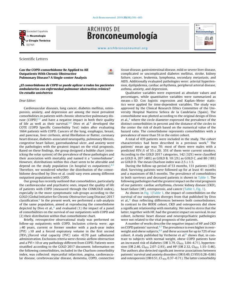
Arch Bronconeumol. 2019;55(11):591–603 www.archbronconeumol.org Scientific Letters Can the COPD-comorbidome Be Applied to All Outpatients With Chronic Obstructive Pulmonary Disease? A Single-center Analysis ¿El comorbidoma de COPD se puede aplicar a todos los pacientes ambulatorios con enfermedad pulmonar obstructiva crónica? Un estudio unicéntrico Dear Editor: Cardiovascular diseases, lung cancer, diabetes mellitus, osteoporosis, anxiety, and depression are among the most prevalent comorbidities in patients with chronic obstructive pulmonary disease (COPD)1,2 and have a negative impact in both their quality of life as well as their survival.3,4 Divo et al.4 developed the COTE (COPD Specific Comorbidity Test) index after evaluating 1664 patients with COPD. Cancers of the lung, esophagus, breast, and pancreas, liver cirrhosis, atrial fibrillation or flutter, coronary heart disease, diabetes-associated neuropathy, pulmonary fibrosis, congestive heart failure, gastroduodenal ulcer, and anxiety were the pathologies with the greatest impact on the vital prognosis. Based on these findings, the group designed a bubble chart (mimicking the solar system) of the prevalence of these pathologies and their association with mortality and named it a “comorbidome”. However, distributions within this chart seem to be alterable and depend on the study population (in hospital vs outpatients).4,5 Therefore, we wondered whether the distribution of the comorbidome described by Divo et al. could vary even among different outpatient populations with COPD. Our group has recently outlined that comorbidities, particularly the cardiovascular and psychiatric ones, impact the quality of life of patients with COPD (measured through the COMCOLD index), especially in the more symptomatic sub-groups according to the GOLD (Global Initiative for Chronic Obstructive Lung Disease) 2017 classification.6 In the present work, we performed a sub-analysis of the same population, aimed at reproducing the comorbidome depicted by Divo et al.,4 and evaluated (1) the impact of a panel of comorbidities on the survival of our outpatients with COPD and (2) their distribution within that comorbidome chart. Briefly, retrospective observational study was performed on follow-up outpatients with COPD. Inclusion criteria were: age ≥40 years, current or former smoker with a pack-year index (PYI) ≥10 and a forced expiratory volume in the first second (FEV1 )/forced vital capacity (FVC) ratio <0.70 past salbutamol administration. Exclusion criteria were chronic airflow obstruction and a PYI < 10 or any pathology different from COPD. Patients were stratified according to the GOLD 2017 document. Information on the following comorbidities, included in the Charlson comorbidity index, was collected: myocardial infarction, angina, cardiovascular disease, cerebrovascular disease, dementia, COPD, connective tissue disease, gastrointestinal disease, mild or severe liver disease, complicated or uncomplicated diabetes mellitus, stroke, kidney failure, cancer, leukemia, lymphoma, secondary metastasis, and AIDS. Additionally evaluated pathologies were: arterial hypertension, dyslipidemia, cardiac arrhythmia, peripheral arterial disease, asthma, anxiety, and depression. Qualitative variables were expressed as absolute values and percentages, while quantitative variables were summarized as means ± SD. Cox logistic regression and Kaplan–Meier statistics were applied for time-dependent variables. The study was approved by the Clinical Research Ethics Committee of the University Hospital Nuestra Señora de la Candelaria (Spain). The comorbidome was plotted according to the original design of Divo et al.,4 where the circle diameter expressed the prevalence of the distinct comorbidities in percent and the distance of the circles to the center the risk of death based on the numerical value of the hazard ratio. The comorbidome represents comorbidities with a prevalence of more than 5% in the entire cohort. A total of 439 patients were included in the study. The cohort characteristics had been described in a previous work.6 The patients’ mean age was 70; most of them were males with a predicted FEV1 % of 55 ± 20; 35% of them were current smokers. According to the GOLD 2017 categories, 142 (32%) were classified as GOLD A, 207 (48%) as GOLD B, 10 (2%) as GOLD C, and 80 (18%) as GOLD D. The mean Charlson index was 2.5 ± 1.5. During the follow-up period of 53 months, 131 patients (30%) died. Surviving patients were followed up for at least 41 months and a maximum of 88.5 months. The prevalence of comorbidities in both survivors and deceased patients is shown in Table 1. The following pathologies had the greatest impact on the vital prognosis of our patients: cardiac arrhythmia, chronic kidney disease (CKD), heart failure (HF), osteoporosis, and cancer (Table 1, Fig. 1). As shown in Fig. 1/Table 1, the impact of comorbidities on the survival of our outpatients diverges from that published by Divo et al.,4 thus reflecting differences between both comorbidomes. In contrast to the BODE cohort, CKD and osteoporosis did show a significant relationship with mortality. We need to stress that the latter, together with HF, had the greatest impact on survival. In our cohort, ischemic heart disease and neuropsychiatric pathologies were not related to the vital prognosis of the patients. A number of works describe the negative impact of HF and CKD on COPD patients’ survival.4,7 The prevalence is even higher in overweight and obese subjects,8,9 and these account for up to 72% of our cohort. A study published by Verberne et al.8 shows that, in contrast to subjects with normal weight, obese COPD patients have an increased risk of diabetes (OR 3.79, CI95% 3.04–4.71), hypertension (OR 2.46, CI95% 2.07–2.93), and HF (OR 2.32, CI95% 1.55–3.46). The authors also detected significant inverse associations between patients’ survival and anxiety disorders (OR 0.49, CI 95% 0.28–0.86) and osteoporosis (OR 0.51, CI 95% 0.37–0.71). The latter comorbidity 592 Scientific Letters / Arch Bronconeumol. 2019;55(11):591–603 Table 1 Comorbidities of COPD and their association with risk of death. Comorbidities Prevalence in non-deceased patients (n = 308) Prevalence in deceased patients (n = 131) Hazard ratioa (95% confidence interval) P Osteoporosis, n (%) HF, n (%) CKD, n (%) Neoplasia, n (%) Arrhythmia, n (%) Asthma, n (%) Peripheral arterial disease, n (%) Anxiety, n (%) CVA, n (%) IHD, n (%) Diabetes mellitus, n (%) SA, n (%) Liver disease, n (%) Obesity, n (%) Depression, n (%) Arterial hypertension, n (%) Dyslipidemia, n (%) 7 (2.3) 30 (9.7) 28 (9.1) 24 (7.8) 48 (15.6) 66 (21.4) 31 (10.1) 24 (7.8) 21 (6.8) 45 (14.6) 93 (30.2) 72 (23.4) 27 (8.8) 97 (31.5) 42 (13.6) 110 (35.7) 99 (32.1) 9 (6.9) 33 (25.2) 28 (21.5) 24 (18.3) 41 (31.3) 33 (25.2) 18 (13.7) 9 (6.9) 11 (8.4) 26 (19.8) 50 (38.2) 33 (25.2) 9 (6.9) 41 (31.3) 13 (9.9) 39 (29.8) 45 (34.4) 2.00 1.84 1.66 1.57 1.51 1.43 1.12 1.11 1.10 1.07 0.98 0.95 0.91 0.82 0.77 0.76 0.74 .045 .003 .020 .049 .033 .078 .650 .763 .757 .757 .926 .798 .792 .285 .372 .170 .112 1.02–3.95 1.23–2.75 1.08–2.56 1.00–2.44 1.03–2.21 0.96–2.13 0.68–1.85 0.56–2.19 0.59–2.05 0.70–1.65 0.69–1.41 0.64–1.41 0.46–1.80 0.56–1.11 0.43–1.37 0.51–1.12 0.52–1.07 Abbreviations: HF: heart failure. CKD: chronic kidney disease. IHD: ischemic heart disease. CVA: cerebrovascular accidents. SA: sleep apnea. a All the HR are estimated adjusted by age of patients. Prevalence 15% PADs 25% Asthma Arrhythmia Neoplasia Anxiety CKD 50% HF CVA Osteoporosis IHD DM DLP Sleep Apnea Arterial hypertension Liver disease Depression obesity Fig. 1. The “comorbidome” is a graphic expression of comorbities with more than 5% prevalence in the entire cohort. The area of the circles relates to the prevalence of the disease. The proximity to the center (mortality) express the strength of the association between the disease and the risk of death. This was scaled from the inverse of the HR (1/HR). The dotted line represents HR = 1. Beyond the line, HR are less than 1. The red bubble represents statistical significance association (HR > 1; P < .05). Abbreviations: HF: heart failure. CKD: chronic kidney disease. IHD: ischemic heart disease. CVA: cerebrovascular accidents. DM: diabetes mellitus. DLP: dyslipidemia. PADs: peripheral arterial disease. is classically described for patients with an emphysema phenotype, usually characterized by low weight, considerable air trapping, and a worse vital prognosis than the other phenotypes.10 We may not ignore the limitations of our study, i.e., the sample size, methodology—this is an observational study—and the fact that some of the comorbidities in the article by Divo et al.4 were not analyzed in our cohort. In contrast to the BODE cohort, patients with relevant comorbidities that prevented them from performing the 6-minute walk test were not excluded from our study. In addition, minimum follow-up was slightly longer in our study. In this sense, our work may better represent the patients with COPD that we find in healthcare practice. This prompted us to reflect on a general applicability of the COTE index (COPD specific comorbidity test) based on the comorbidome of Divo et al.,4 thinking that it should be substantiated in different patient populations. In summary, our study suggests that distributions within the comorbidome may vary according to the analyzed outpatient population. References 1. Mapel DW, Hurley JS, Frost FJ, Petersen HV, Picchi MA, Coultas DB. Health care utilization in chronic obstructive pulmonary disease. A case-control study in a health maintenance organization. Arch Intern Med. 2000;160:2653–8. 2. Decramer M, Janssens W. Chronic obstructive pulmonary disease and comorbidities. Lancet Respir Med. 2013;1:73–83. 3. Almagro P, Cabrera FJ, Diez J, Boixeda R, Alonso Ortiz MB, Murio C, et al. Comorbidities and short-term prognosis in patients hospitalized for acute exacerbation of COPD. The ESMI study. Chest. 2012;142:1126–33. 4. Divo M, Cote C, de Torres JP, Casanova C, Marin JM, Pinto-Plata V, et al. Comorbidities and risk of mortality in patients with chronic obstructive pulmonary disease. Am J Respir Crit Care Med. 2012;186:155–61. Scientific Letters / Arch Bronconeumol. 2019;55(11):591–603 5. Almagro P, Cabrera FJ, Diez-Manglano J, Boixeda R, Recio J, Mercade J, et al. Comorbidome and short-term prognosis in hospitalised COPD patients: the ESMI study. Eur Respir J. 2015;46:850–3. 6. Figueira Gonçalves JM, Martín Martínez MD, Pérez Méndez LI, García Bello MA, Garcia-Talavera I, Hernández SG, et al. Health status in patients with COPD according to GOLD 2017 classification: use of the COMCOLD score in routine clinical practice. COPD. 2018:1–8. 7. Fedeli U, De Giorgi A, Gennaro N, Ferroni E, Gallerani M, Mikhailidis DP, et al. Lung and kidney: a dangerous liaison? A population-based cohort study in COPD patients in Italy. Int J Chron Obstruct Pulmon Dis. 2017;12:443–50. 8. Verberne LDM, Leemrijse CJ, Swinkels ICS, van Dijk CE, de Bakker DH, Nielen MMJ. Overweight in patients with chronic obstructive pulmonary disease needs more attention: a cross-sectional study in general practice. NPJ Prim Care Respir Med. 2017;27:63. 9. Ting SM, Nair H, Ching I, Taheri S, Dasgupta I. Overweight, obesity and chronic kidney disease. Nephron Clin Pract. 2009;112:c121–7. 10. Burgel PR, Paillasseur JL, Peene B, Dusser D, Roche N, Coolen J, et al. Two distinct chronic obstructive pulmonary disease (COPD) phenotypes are associated with high risk of mortality. PLoS One. 2012;7:e51048. Juan Marco Figueira Gonçalves a,∗ , Miguel Ángel García Bello b , María Dolores Martín Martínez c , Ignacio García-Talavera a , Rafael Golpe d Primary Pulmonary Hemangiopericytoma夽 Hemangiopericitoma pulmonar primario Dear Editor: The high prevalence of epithelial lung tumors means that mesenchymal lung tumors are rarities seen very occasionally in routine clinical practice. We report the case of a patient referred to the respiratory medicine clinic for the study of recurrent self-limiting episodes of bloody expectoration. This was a female patient, 56 years of age, originally from Venezuela, who presented due to repeated self-limiting episodes of expectoration of blood while coughing, not affecting hemodynamic parameters, blood gases, or blood counts. Four years previously in her home country, she had been diagnosed on CT with a prevertebral solid tumor, determined on thoracotomy to be thoracic hemangioma, that was treated by radiation therapy (total dose unknown). Other surgical history included left inguinal herniorrhaphy, hysterectomy, and double adnexectomy. Physical examination was unremarkable, and both complete blood count and biochemistry results were within normal ranges. Chest X-ray showed the presence of a round subcarinal lesion. CT confirmed the presence of a mass enhanced by administration of contrast medium, measuring about 8.5×4.5 cm in diameter, located immediately behind the right pulmonary artery and accompanied by paraaortic and retroperitoneal lymphadenopathies. Fiberoptic bronchoscopy (Fig. 1) showed a purplish mass with a polylobulated surface located in the main carina, almost entirely occluding the entrance to the left main bronchus, but that did not prevent passage of the bronchoscope. The appearance of the endobronchial lesion and the patient’s history of an intrathoracic vascular lesion made the collection of endoscopic biopsies inadvisable. Some days later, rigid bronchoscopy was performed under general anesthesia, and biopsies were collected, complicated by severe bleeding controlled with diathermy, followed by arteriography with embolization of the branches feeding the right bronchial artery. Biopsy samples showed the presence of numerous endothelized vessels with fibrous walls and a dense proliferation of cells with elongated nuclei arranged in bundles with variable spatial 夽 Please cite this article as: Hernández Pérez JM, Pérez Negrín L, López Charry CV. Hemangiopericitoma pulmonar primario. Arch Bronconeumol. 2019;55:593–594. 593 a Pneumology and Thoracic Surgery Service, University Hospital Nuestra Señora de la Candelaria (HUNSC), Santa Cruz de Tenerife, Spain b Division of Clinical Epidemiology and Biostatistics, Research Unit, University Hospital Nuestra Señora de la Candelaria (HUNSC), and Primary Care Management, Santa Cruz de Tenerife, Spain c Clinical Analysis Service, University Hospital Nuestra Señora de la Candelaria (HUNSC), Santa Cruz de Tenerife, Spain d Respiratory Medicine Service, University Hospital Lucus Augusti, Lugo, Spain ∗ Corresponding author. E-mail address: juanmarcofi[email protected] (J.M. Figueira Gonçalves). https://doi.org/10.1016/j.arbres.2019.03.016 0300-2896/ © 2019 SEPAR. Published by Elsevier España, S.L.U. All rights reserved. arrangement with no necrosis or mitosis, that were positive for markers CD34 and CD31, Ki-67 in less than 5%, and negative for epithelial and muscle markers; these findings were consistent with pulmonary hemangiopericytoma. Between 2% and 6% of all primary mediastinal tumors are of mesenchymal origin. Of these, hemangiopericytoma is a rarity: it originates in the Zimmermann pericytes, which form part of the outer layer surrounding the endothelium of the capillaries and is now classified as perivascular tumor. It represents less than 2% of all soft tissue sarcomas,1 and approximately 1% of all tumors of vascular origin.2 The main sites tend to be the muscle tissue of the limbs, subcutaneous tissue, and the retroperitoneum. The chest (usually mediastinal) as the primary site of hemangiopericytoma is extremely rare, as attested by a review of literature which retrieved very few cases.3 Published cases of pulmonary involvement mostly began as solitary pulmonary nodules, and to our knowledge, this is the first case with endobronchial expression evidenced by bronchoscopy. This type of tumor usually begins with a wide variety of symptoms, which most notably include hemoptysis.4 Image testing, especially CT with and without contrast and magnetic reso- Fig. 1. Image obtained during bronchoscopy highlighting the presence of a polylobulated tumor at the entrance to the left main bronchus.
