Mouse anti-Progesterone Receptor, Clone 1A6
Anuncio
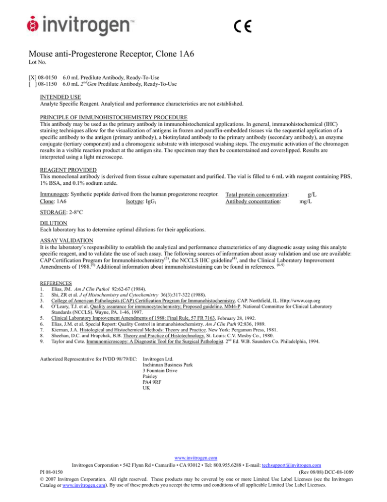
Mouse anti-Progesterone Receptor, Clone 1A6 Lot No. [X] 08-0150 [ ] 08-1150 6.0 mL Predilute Antibody, Ready-To-Use 6.0 mL 2ndGen Predilute Antibody, Ready-To-Use INTENDED USE Analyte Specific Reagent. Analytical and performance characteristics are not established. PRINCIPLE OF IMMUNOHISTOCHEMISTRY PROCEDURE This antibody may be used as the primary antibody in immunohistochemical applications. In general, immunohistochemical (IHC) staining techniques allow for the visualization of antigens in frozen and paraffin-embedded tissues via the sequential application of a specific antibody to the antigen (primary antibody), a biotinylated antibody to the primary antibody (secondary antibody), an enzyme conjugate (tertiary component) and a chromogenic substrate with interposed washing steps. The enzymatic activation of the chromogen results in a visible reaction product at the antigen site. The specimen may then be counterstained and coverslipped. Results are interpreted using a light microscope. REAGENT PROVIDED This monoclonal antibody is derived from tissue culture supernatant and purified. The vial is filled to 6 mL with reagent containing PBS, 1% BSA, and 0.1% sodium azide. Immunogen: Synthetic peptide derived from the human progesterone receptor. Clone: 1A6 Isotype: IgG1 Total protein concentration: Antibody concentration: g/L mg/L STORAGE: 2-8°C DILUTION Each laboratory has to determine optimal dilutions for their applications. ASSAY VALIDATION It is the laboratory’s responsibility to establish the analytical and performance characteristics of any diagnostic assay using this analyte specific reagent, and to validate the use of such assay. The following sources of information about assay validation and use are available: CAP Certification Program for Immunohistochemistry(3), the NCCLS IHC guideline(4), and the Clinical Laboratory Improvement Amendments of 1988.(5) Additional information about immunohistostaining can be found in references. (6-9) REFERENCES 1. Elias, JM. Am J Clin Pathol 92:62-67 (1984). 2. Shi, ZR et al. J of Histochemistry and Cytochemistry 36(3):317-322 (1988). 3. College of American Pathologists (CAP) Certification Program for Immunohistochemistry. CAP. Northfield, IL. Http://www.cap.org 4. O’Leary, T.J. et al. Quality assurance for immunocytochemistry; Proposed guideline. MM4-P. National Committee for Clinical Laboratory Standards (NCCLS). Wayne, PA. 1-46, 1997. 5. Clinical Laboratory Improvement Amendments of 1988: Final Rule, 57 FR 7163, February 28, 1992. 6. Elias, J.M. et al. Special Report: Quality Control in immunohistochemistry. Am J Clin Path 92:836, 1989. 7. Kiernan, J.A. Histological and Histochemical Methods: Theory and Practice. New York: Pergamon Press, 1981. 8. Sheehan, D.C. and Hrapchak, B.B. Theory and Practice of Histotechnology. St. Louis: C.V. Mosby Co., 1980. 9. Taylor and Cote. Immunomicroscopy: A Diagnostic Tool for the Surgical Pathologist. 2nd Ed. W.B. Saunders Co. Philadelphia, 1994. Authorized Representative for IVDD 98/79/EC: Invitrogen Ltd. Inchinnan Business Park 3 Fountain Drive Paisley PA4 9RF UK www.invitrogen.com Invitrogen Corporation • 542 Flynn Rd • Camarillo • CA 93012 • Tel: 800.955.6288 • E-mail: [email protected] PI 08-0150 (Rev 08/08) DCC-08-1089 © 2007 Invitrogen Corporation. All right reserved. These products may be covered by one or more Limited Use Label Licenses (see the Invitrogen Catalog or www.invitrogen.com). By use of these products you accept the terms and conditions of all applicable Limited Use Label Licenses. Anticuerpo Receptor de la Progesterona de ratón, Clon 1A6 Reactivo específico para electrolito [X] 08-0150 [ ] 08-1150 6,0 mL de anticuerpo prediluido, listo para usar 6,0 mL de anticuerpo prediluido de segunda generación, listo para usar PROPÓSITO DE USO Reactivo específico para electrolito Las características analíticas y de rendimiento no han sido determinadas. PRINCIPIO DEL PROCEDIMIENTO DE INMUNOHISTOQUÍMICA Este anticuerpo puede ser utilizado como el anticuerpo primario con el método de detección marcado con estreptavidina-biotina (LAB-SA)(1,2) o un método equivalente. En general, las técnicas de tinción inmunohistoquímicas (IHQ) permiten la visualización de antígenos en cortes de tejido congelados y infiltrados en parafina a través de la aplicación secuencial de un anticuerpo específico al antígeno (anticuerpo primario), un anticuerpo biotinilado al anticuerpo primario (anticuerpo secundario), un conjugado enzimático (componente terciario) y un sustrato cromogénico con pasos intermedios de lavado. La activación enzimática del cromógeno resulta en un producto de reacción visible en el sitio del antígeno. La muestra puede ser contra coloreada y cubierta con un cubreobjeto. Los resultados se interpretan con un microscopio de luz. REACTIVO SUMINISTRADO El anticuerpo Receptor de la Progesterona de ratón se deriva del sobrenadante del cultivo tisular y se diluye en tampón fosfato (PBS), Ph 7,4, y seroalbúmina bovina al 1% (BSA) con azida de sodio al 0,1% (NaN3) como conservante. Inmunogén: Péptido sintético derivado del Receptor de la Progesterona humano Isotipo: IgG1 Clon: 1A6 ALMACENAMIENTO: 2-8°C DILUCIÓN Cada laboratorio debe determinar las diluciones óptimas para su aplicación. VALIDACIÓN DEL ANÁLISIS El laboratorio es responsable por la determinación de las características analíticas y de rendimiento de cualquier análisis diagnóstico mediante este reactivo específico para electrolitos y por la validación del uso de dicho análisis. Se dispone de las siguientes fuentes de información sobre la validación y uso del análisis: Programa de certificación CAP para inmunohistoquímica(3), las recomendaciones sobre IHQ del NCCLS(4), y las modificaciones para mejoría del laboratorio clínico 1988.(5) En las referencias puede encontrarse información adicional sobre inmunotinción histológica.(6-9) 1. 2. 3. 4. 5. 6. 7. 8. 9. Elias, JM. Am J Clin Pathol 92:62-67 (1984). Shi, ZR et al. J of Histochemistry and Cytochemistry 36(3):317-322 (1988). College of American Pathologists (CAP) Certification Program for Immunohistochemistry. CAP. Northfield, IL. Http://www.cap.org O’Leary, T.J. et al. Quality assurance for immunocytochemistry; Proposed guideline. MM4-P. National Committee for Clinical Laboratory Standards (NCCLS). Wayne, PA. 1-46, 1997. Clinical Laboratory Improvement Amendments of 1988: Final Rule, 57 FR 7163, February 28, 1992. Elias, J.M. et al. Special Report: Quality Control in immunohistochemistry. Am J Clin Path 92:836, 1989. Kiernan, J.A. Histological and Histochemical Methods: Theory and Practice. New York: Pergamon Press, 1981. Sheehan, D.C. and Hrapchak, B.B. Theory and Practice of Histotechnology. St. Louis: C.V. Mosby Co., 1980. Taylor and Cote. Immunomicroscopy: A Diagnostic Tool for the Surgical Pathologist. 2nd Ed. W.B. Saunders Co. Philadelphia, 1994. MARCAS COMERCIALES Cap-Plus™, Clearmount™, Digest-All™, HistoGrip™, Histomount™, y Histostain®, NBA™, PicTure™ y Zymed® son marcas comerciales de Zymed Laboratories, Inc. Para obtener más información vea la versión inglesa de la ficha técnica o vaya a www.invitrogen.com. TIENE ALGUNA PREGUNTA SOBRE ESTE PRODUCTO, DIRÍJASE A SU DISTRIBUIDOR LOCAL. Representante autorizado de IVDD 98/79/EC: Invitrogen Ltd., Inchinnan Business Park, 3 Fountain Drive, Paisley, PA4 9RF, UK www.invitrogen.com Invitrogen Corporation • 542 Flynn Rd • Camarillo • CA 93012 • Tel: 800.955.6288 • E-mail: [email protected] PI 08-0150 (Rev 08/08) DCC-08-1089 © 2007 Invitrogen Corporation. All right reserved. These products may be covered by one or more Limited Use Label Licenses (see the Invitrogen Catalog or www.invitrogen.com). By use of these products you accept the terms and conditions of all applicable Limited Use Label Licenses. Souris anti-Recepteur de Progesterone, Clone 1A6 Réactif spécifique à l’analyte. [X] 08-0150 [ ] 08-1150 6,0 mL Anticorps Prédilué, Prêt à l'Utilisation 6,0 mL 2ndGén Anticorps Prédilué, Prêt à l'Utilisation UTILISATION VOULUE Réactif spécifique à l’analyte. Les caractéristiques analytiques et de performance ne sont pas établies. PRINCIPE DE LA PROCÉDURE D’IMMUNOHISTOCHIMIE Cet anticorps peut être utilisé comme anticorps primaire avec la méthode de détection étiquetée streptavidine-biotine (LAB-SA)(1,2) ou une méthode équivalente. En général, les techniques de coloration immunohistochimiques (IHC) permettent de visualiser des antigènes dans des tissus congelés et incorporés dans de la paraffine par l’intermédiaire de l’application séquentielle d’un anticorps spécifique à l’antigène (anticorps primaire), un anticorps biotinylé à l’anticorps primaire (anticorps secondaire), un conjugué enzymatique (composant tertiaire) et un substrat chromogénique avec des étapes de lavage interposées. L’activation enzymatique du chromogène produit une réaction visible du produit au site de l’antigène. Le spécimen peut alors être contre-coloré et recouvert d’une lamelle couvreobjet. Les résultats sont interprétés à l’aide d’un microscope optique. REACTIF FOURNI Souris anti-Recepteur de Progesterone est dérivé d’un surnageant d’une culture de tissu et dilué dans du Phosphate Tamponné Salin (PBS), pH 7.4, et 1% d'albumine de sérum de bovin (BSA) avec 0.1% azide de sodium(NaN3) comme préservatif. Immunogène: Peptide synthétique dérivé de Recepteur de Progesterone humain Isotype: IgG1 Clone: 1A6 STOCKAGE : à 2-8°C DILUTION Chaque laboratoire doit déterminer quelles dilutions sont optimales pour ses applications. VALIDATION DE L’ESSAI Il incombe au laboratoire d’établir les caractéristiques analytiques et de performance de tout essai de diagnostic utilisant ce réactif spécifique à l’analyte, et de valider l’utilisation de cet essai. Les sources d’information suivantes concernant la validation et l’utilisation de l’essai sont disponibles : programme de certification CAP pour l’immunohistochimie(3), la directive concernant l’IHC de NCCLS(4), et les modifications d’amélioration du laboratoire clinique de 1988.(5) Vous trouverez des données supplémentaires concernant l’immunohistocoloration dans les références.(6-9) 1. 2. 3. 4. 5. 6. 7. 8. 9. Elias, JM. Am J Clin Pathol 92:62-67 (1984). Shi, ZR et al. J of Histochemistry and Cytochemistry 36(3):317-322 (1988). College of American Pathologists (CAP) Certification Program for Immunohistochemistry. CAP. Northfield, IL. Http://www.cap.org O’Leary, T.J. et al. Quality assurance for immunocytochemistry; Proposed guideline. MM4-P. National Committee for Clinical Laboratory Standards (NCCLS). Wayne, PA. 1-46, 1997. Clinical Laboratory Improvement Amendments of 1988: Final Rule, 57 FR 7163, February 28, 1992. Elias, J.M. et al. Special Report: Quality Control in immunohistochemistry. Am J Clin Path 92:836, 1989. Kiernan, J.A. Histological and Histochemical Methods: Theory and Practice. New York: Pergamon Press, 1981. Sheehan, D.C. and Hrapchak, B.B. Theory and Practice of Histotechnology. St. Louis: C.V. Mosby Co., 1980. Taylor and Cote. Immunomicroscopy: A Diagnostic Tool for the Surgical Pathologist. 2nd Ed. W.B. Saunders Co. Philadelphia, 1994. MARQUE DE FABRIQUE Cap-Plus™, Clearmount™, Digest-All™, HistoGrip™, Histomount™, et Histostain®, NBA™, PicTure™, et Zymed® sont des marques des Zymed Laboratories, Inc. Pour des informations plus détaillées, veuillez vous référer aux feuilles de données en version anglaise ou aller sur www.invitrogen.com. SI VOUS AVEZ DES QUESTIONS SUR CE PRODUIT CONTACTEZ VOTRE DISTRIBUTEUR LOCAL. Représentant Autorisé pour IVDD 98/79/EC: Invitrogen Ltd., Inchinnan Business Park, 3 Fountain Drive, Paisley, PA4 9RF, UK www.invitrogen.com Invitrogen Corporation • 542 Flynn Rd • Camarillo • CA 93012 • Tel: 800.955.6288 • E-mail: [email protected] PI 08-0150 (Rev 08/08) DCC-08-1089 © 2007 Invitrogen Corporation. All right reserved. These products may be covered by one or more Limited Use Label Licenses (see the Invitrogen Catalog or www.invitrogen.com). By use of these products you accept the terms and conditions of all applicable Limited Use Label Licenses. Topo anti-Recettore Progesterone, Clone 1A6 Reagente analita-specifico [X] 08-0150 [ ] 08-1150 6,0 mL di anticorpo prediluito, pronto all’uso 6,0 mL di 2ndGen Anticorpo Prediluito, Pronto all’uso SCOPO D’UTILIZZO Reagente analita-specifico. Le caratteristiche analitiche e della performance non sono state stabilite. PRINCIPIO DI FUNZIONAMENTO DELLA PROCEDURA IMMUNOISTOCHIMICA Questo anticorpo può essere usato quale anticorpo primario nel metodo di rilevazione marcato con streptavidina-biotina (LAB-SA)(1,2) o metodi analoghi. In termini generali, le tecniche di colorazione immunoistochimica (IIC) consentono la visualizzazione di antigeni in tessuti congelati ed inclusi in paraffina tramite l’applicazione sequenziale di un anticorpo specifico sull’antigene (anticorpo primario), di un anticorpo biotilinato sull’anticorpo primario (anticorpo secondario), un coniugato enzimatico (componente terziario) ed un substrato cromogenico con fasi di lavaggio frapposte. L’attivazione enzimatica del cromogeno induce un prodotto di reazione visibile presso il sito dell’antigene. Il provino può quindi essere controcolorato ed inserito in un coprivetrino. I risultati possono essere interpretati usando un microscopio ottico.. REAGENTI FORNITI Topo anti-Recettore Progesterone è derivato da supernatante di coltura di tessuto e diluito in soluzione salina tamponata al fosfato (PBS), pH 7,4, e 1% di albumina di siero bovino (BSA) con 0,1% sodio azide (NaN3) come conservante. Immunogeno: Peptide sintetico derivato dal Recettore Progesterone umano Isotipo: IgG1 Clone: 1A6 CONSERVAZIONE: 2-8 °C DILUIZIONE Ciascun laboratorio dovrà determinare quali siano le diluizioni ottimali per le proprie applicazioni. CONVALIDA DELLE PROVE Ciascun singolo laboratorio dovrà determinare le caratteristiche analitiche e della performance delle prove diagnostiche eseguite utilizzando il presente reagente analita-specifico e quindi convalidare l’uso di dette prove. Sono disponibili le seguenti fonti di informazioni sulla convalida delle prove ed il relativo utilizzo: Programma di certificazione per l’immunoistochimica CAP(3), Linea guida sull’IIC del NCCLS(4) e Emendamenti di perfezionamento delle procedure cliniche di laboratorio del 1988.(5) Nel materiale di consultazione sono rinvenibili ulteriori informazioni sulla colorazione immunoistochimica.(6-9) 1. 2. 3. 4. 5. 6. 7. 8. 9. Elias, JM. Am J Clin Pathol 92:62-67 (1984). Shi, ZR et al. J of Histochemistry and Cytochemistry 36(3):317-322 (1988). College of American Pathologists (CAP) Certification Program for Immunohistochemistry. CAP. Northfield, IL. Http://www.cap.org O’Leary, T.J. et al. Quality assurance for immunocytochemistry; Proposed guideline. MM4-P. National Committee for Clinical Laboratory Standards (NCCLS). Wayne, PA. 1-46, 1997. Clinical Laboratory Improvement Amendments of 1988: Final Rule, 57 FR 7163, February 28, 1992. Elias, J.M. et al. Special Report: Quality Control in immunohistochemistry. Am J Clin Path 92:836, 1989. Kiernan, J.A. Histological and Histochemical Methods: Theory and Practice. New York: Pergamon Press, 1981. Sheehan, D.C. and Hrapchak, B.B. Theory and Practice of Histotechnology. St. Louis: C.V. Mosby Co., 1980. Taylor and Cote. Immunomicroscopy: A Diagnostic Tool for the Surgical Pathologist. 2nd Ed. W.B. Saunders Co. Philadelphia, 1994. MARCHI REGISTRATI Cap-Plus™, Clearmount™, Digest-All™, HistoGrip™, Histomount™, e Histostain®, NBA™, PicTure™, e Zymed® sono marchi registrati di Zymed Laboratories, Inc. Per informazioni più dettagliate vi preghiamo di consultare la versione inglese del catalogo oppure visitate il sito www.invitrogen.com. SE AVETE DOMANDE SUL PRODOTTO, CONTATTATE IL VOSTRO DISTRIBUTORE PIÙ`VICINO. Rappresentante Autorizzato per IVDD 98/79/EC: Invitrogen Ltd., Inchinnan Business Park, 3 Fountain Drive, Paisley PA4 9RF, UK www.invitrogen.com Invitrogen Corporation • 542 Flynn Rd • Camarillo • CA 93012 • Tel: 800.955.6288 • E-mail: [email protected] PI 08-0150 (Rev 08/08) DCC-08-1089 © 2007 Invitrogen Corporation. All right reserved. These products may be covered by one or more Limited Use Label Licenses (see the Invitrogen Catalog or www.invitrogen.com). By use of these products you accept the terms and conditions of all applicable Limited Use Label Licenses. Maus Anti-Progesterone Rezeptor, Klon 1A6 Analytspezifisches Reagenz [X] 08-0150 [ ] 08-1150 6.0 mL Vorverdünnter Antikörper, gebrauchsfertig 6.0 mL 2ndGen Vorverdünnter Sie Antikörper, Gebrauchsfertig BEABSICHTIGTER NUTZEN Analytspezifisches Reagenz. Analytische und Leistungsmerkmale wurden nicht festgelegt. IMMUNHISTOCHEMISCHES VERFAHRENSPRINZIP Dieser Antikörper kann als primärer Antikörper mit der gekennzeichneten Streptavidin-Biotin (LAB-SA)(1,2) Detektion oder einer gleichwertigen Methode verwendet werden. Generell ermöglichen immunhistochemische (IHC) Färbungsmethoden die Visualisierung der Antigene in gefrorenem und in Paraffin eingebetteten Geweben durch die sequenzielle Anwendung eines spezifischen Antikörpers gegen das Antigen (primärer Antikörper), eines biotinylierten Antikörpers gegen den primären Antikörper (sekundären Antikörper), eines Enzymkonjugats (tertiäre Komponente) und eines chromogenen Substrats mit zwischengeschalteten Waschschritten. Die enzymatische Aktivierung des Chromogens führt zu einer sichtbaren Reaktion an der Antigenstelle. Die Probe kann anschließend gegengefärbt und mit einem Coverslip versehen werden. Die Ergebnisse werden unter Verwendung eines Lichtmikroskops interpretiert. REAGENS GELIEFERT Maus Anti-Progesterone Rezeptor ist wird aus Gewebekultur-Supernatant abgeleitet und verdünnt in Phosphatgepufferter Salzlösung (PBS), pH 7.4, und 1%igem Rinder Serum Albumin (BSA) mit 0.1% Sodium Azide (NaN3) als ein Konservierungsmittel. Immunogen: Aus humanem Progesterone Rezeptor abgeleitetes synthetisches Peptid Isotyp: IgG1 Klon: 1A6 LAGERUNG: 2-8°C VERDÜNNUNG Jedes Labor muss die optimale Verdünnung für die jeweilige Anwendung selbst bestimmen. ASSAY-VALIDIERUNG Das Labor ist dafür verantwortlich, die analytischen und Leistungsmerkmale jedes Diagnose-Assays unter Verwendung dieses analytspezifischen Reagenz zu bestimmen und die Verwendung des Assay zu validieren. Folgende Informationsquellen über die Validierung und Verwendung des Assay stehen zur Verfügung: CAP-Zertifizierungsprogramm für Immunhistochemie(3), die NCCLS IHC Richtlinie(4) und die Clinical Laboratory Improvement Amendments von 1988.(5) Weitere Informationen über Immunhistochemische Färbung sind dem entsprechenden Referenzmaterial zu entnehmen.(6-9) 1. 2. 3. 4. 5. 6. 7. 8. 9. Elias, JM. Am J Clin Pathol 92:62-67 (1984). Shi, ZR et al. J of Histochemistry and Cytochemistry 36(3):317-322 (1988). College of American Pathologists (CAP) Certification Program for Immunohistochemistry. CAP. Northfield, IL. Http://www.cap.org O’Leary, T.J. et al. Quality assurance for immunocytochemistry; Proposed guideline. MM4-P. National Committee for Clinical Laboratory Standards (NCCLS). Wayne, PA. 1-46, 1997. Clinical Laboratory Improvement Amendments of 1988: Final Rule, 57 FR 7163, February 28, 1992. Elias, J.M. et al. Special Report: Quality Control in immunohistochemistry. Am J Clin Path 92:836, 1989. Kiernan, J.A. Histological and Histochemical Methods: Theory and Practice. New York: Pergamon Press, 1981. Sheehan, D.C. and Hrapchak, B.B. Theory and Practice of Histotechnology. St. Louis: C.V. Mosby Co., 1980. Taylor and Cote. Immunomicroscopy: A Diagnostic Tool for the Surgical Pathologist. 2nd Ed. W.B. Saunders Co. Philadelphia, 1994. WARENZEICHEN Cap-Plus™, Clearmount™, Digest-All™, HistoGrip™, Histomount™, und Histostain®, NBA™, PicTure™, und Zymed® sind Warenzeichen der Zymed Laboratories, Inc. Für genauere Information beziehen Sie sich bitte entweder auf die englische Version auf der Datenplatte oder auf www.invitrogen.com. SOLLTEN SIE IRGENDWELCHE FRAGEN ÜBER DIESES PRODUKT HABEN, WENDEN SIE SICH BITTE AN IHREN ÖRTLICHEN VERTEILER. Bevollmächtigter Repräsentant für IVDD 98/79/EC: Invitrogen Ltd., Inchinnan Business Park, 3 Fountain Drive, Paisley PA4 9RF,UK www.invitrogen.com Invitrogen Corporation • 542 Flynn Rd • Camarillo • CA 93012 • Tel: 800.955.6288 • E-mail: [email protected] PI 08-0150 (Rev 08/08) DCC-08-1089 © 2007 Invitrogen Corporation. All right reserved. These products may be covered by one or more Limited Use Label Licenses (see the Invitrogen Catalog or www.invitrogen.com). By use of these products you accept the terms and conditions of all applicable Limited Use Label Licenses.
