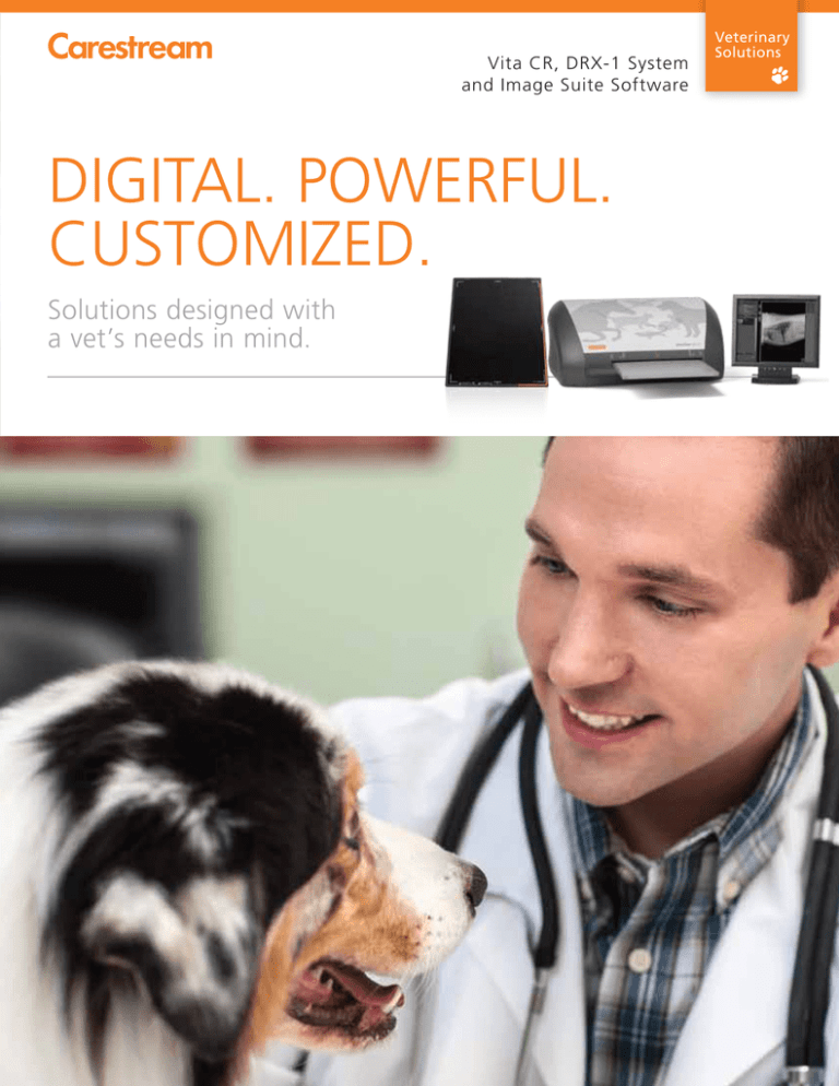
digital. powerful. customized.
Anuncio

Vita CR, DRX-1 System and Image Suite Software DIGITAL. POWERFUL. CUSTOMIZED. Solutions designed with a vet’s needs in mind. Veterinary Solutions NOW IS THE TIME TO GO DIGITAL. Meet the powerful digital-imaging solutions designed specifically for veterinary practices. CARESTREAM Image Suite software, combined with the CARESTREAM Vita CR System or DRX-1 System, allows you to upgrade your veterinary practice to digital, easily and affordably. These systems are optimized with precisely the features your veterinary practice needs – including vet-specific order entry and exam views, customized animal-image processing and a full suite of veterinary measurement tools. Now, you can go digital with confidence. RADIOGRAPHY 101: MAKING IT CLEAR. Jargon-heavy explanations about digital X-ray technology can be hard to follow: “CR, DR, CsI, Bucky, DQE, amorphous silicon, PACS, DICOM, quantum noise” – these terms could confuse anyone. But you don’t care about the acronyms, or the fine details of how the technology works – you just want to know how digital X-ray can benefit your practice. So let’s keep it simple. Let’s first briefly profile the three common approaches to radiography. Then, the rest of the brochure will give you a look at their advantages, features and benefits – to help you select the solution that’s best for you. The Three Common Approaches to Radiography: Film-based Radiology The process familiar to most people. It’s been around for more than a century. This method exposes a sheet of film with a beam of radiation to create an image. Film gets the job done, and can provide quality diagnostic images. But it has drawbacks as well: it’s time-consuming, requires ongoing film purchases, uses harsh chemicals and consumes valuable office space to store. 2 DRX FO 2530C E R V T Computed Radiography (CR) Digital Radiography (DR) This technology replaces the sheet of film with a cassette containing a phosphor screen. It’s exposed the same way film is, to create a latent image on the screen. The cassette is then inserted into a scanner that captures the analog image data, converts it to digital, and displays the new, electronic image on your screen. The cassette's screen is erased and can be used over and over again – reducing your consumable costs. The CR process is much faster than film, and delivers excellent image quality. It’s a very affordable way to go digital – and a very effective “bridge” for going from film to full DR imaging. This method is also referred to as Direct Digital Radiography, because it eliminates CR’s intermediate scanning step from the process. When performing the exposure, the image captured is already fully digital – and is sent instantly to your viewing station. DR delivers superb images, while boosting productivity and workflow to save you time and money. 3 MAKING THE TRANSITION FROM FILM TO DIGITAL. Tired of Working with Film and Chemicals? Choosing the Right Road Forward Film is by all means a time-proven and effective capture medium. As you know, however, it does have some disadvantages: Carestream has several ways to help convert your office to digital imaging. We’ll work closely with you to help you determine which fits your office the best. • X-ray film is a consumable, requiring frequent purchases. • Chemical fumes and odors can be strong and unpleasant. • Chemical disposal is highly regulated, not environmentally friendly and costly. • Storing years-worth of films takes up valuable space. • You must maintain a dedicated dark room for the developer. Upgrading to digital eliminates every one of these problem – and offers a whole set of brand new benefits. Making the Transition Going digital can seem like a big step for a smaller practice. You might be wondering, “Will the change be disruptive?” or “Will it take forever for me and staff to master the new technology?” “Will a digital system include all of the vet-specific features I need?” We can answer all your questions and support you through a smooth and easy transition. (And, just for the record, the answers to those questions are No, No, and Yes!) 4 Current Film Users: • If you’re currently imaging with film, we can help you step up to the practice-altering productivity benefits of our affordable CR solution – which in turn can serve as the bridge to a state-of-the-art Carestream DR system. • Or, you can go directly from film to our advanced, powerful DR offerings. Current CR Users: • Now may be the right time for you to move up to DR, as well. If your CR system has helped your practice grow and succeed, DR is the natural next step in that evolution. And, with CR or DR, you can use your existing generator and X-ray positioning equipment – no need to buy a new room. IMAGE SUITE 4: THE POWER BEHIND OUR CR AND DR SOLUTIONS. Engineered for Veterinary Applications Faster Results CR or DR? You win whichever you choose – because both systems are based on the same remarkable software platform. It’s called Image Suite, and we’ve just come out with our latest update: Version 4. When an animal is sick or injured, neither you, the owner nor the patient wants to endure a long wait for diagnostic images. So we designed these solutions to be fast. They deliver the high-quality images you need in minutes or less – so diagnoses can be made far more quickly. Many digital-imaging systems marketed to vets come with a generic, human-based clinical interface that you need to adapt. By contrast, Image Suite software offers an interface designed for the way vets work. And, the entire system is loaded with custom veterinary features you’re looking for. • Our order-entry screens have the fields you want and need, for selection of breed, species and more. • You can input unique identifiers like tattoos and chip IDs. • Veterinary-specific reference images are provided to enable accurate positioning, while sample veterinary X-rays provide guides for enhanced image quality. • Our customized animal-image processing database fine-tunes processing parameters for specific species, breeds, body parts and views. • In addition to common tools like ruler, angle and zoom, we also provide a suite of specialized veterinary measurement tools that helps you arrive at precise diagnoses faster than ever before. These tools include: • Hip Dysplasia Measurement Tool • VD Thorax Measurement Tool • Vertebral Scale Measurement Tool • Tibial Tuberoses Advancement Measurement Tool • Tibial Plateau Leveling Osteotomy Measurement Tool • Laminitis Measurement Tool Plus, reviewing diagnostic images on-screen with animal owners makes it far easier to explain the animal’s condition with clarity and speed – and show owners why your recommended treatment is the best option. Of course, rapid image access also means expedited workflow and enhanced productivity in your practice. A Quick and Easy Transition Our simple installation process means you’ll be up and running quickly – usually in less than two hours. Both the Vita CR with Image Suite 4 or the DRX-1 System with Image Suite 4 are exceptionally simple to learn – training time is minimal, and the workflow is designed to match what you do with film today. Ease of Use The Vita CR Image Suite 4 and DRX-1 System with Image Suite 4 utilize the identical user interface. Its design is highly intuitive, so you and your staff will be using it like experts in no time. And, should you decide to step up from CR to the DRX-1 System, there’s no new interface to learn. Imaging You Can Depend On In addition to speed, you’ll receive excellent image quality. These systems provide sharp, clear images that support more accurate review and diagnosis. They have been fine-tuned for the body parts and views of the animals you treat. So don’t settle for processing designed for a human ankle – when what you really need is processing optimized for a canine hock. 5 IF YOU CHOOSE TO GO CR, YOU’LL NEED: • Your existing generator and X-ray positioning equipment – no new purchase needed CARESTREAM D IRECT V IEW CR Screens and Cassettes • Image Suite 4 Software as described above Ideal for use with the DIRECTVIEW Vita CR System, our line of screens and cassettes offers a wide selection of sizes: • A Vita CR Scanner • 8 x 10 in. • 14 x 17 in. • And the screens and cassettes of your choice • 10 x 12 in. • 14 x 33 in. • 11 x 14 in. • 24 x 30 cm • 14 x 14 in. • 15 x 30 cm The CARESTREAM Vita CR Scanner. Quality Image Scanning Made Simple. After the image has been taken, the Vita CR scanner “reads” your exposed phosphor screen, and completes the process of creating your digital image. This affordable scanner is a compact, lightweight tabletop unit with a small footprint. This makes it an ideal fit for smaller veterinary practices, and it can also be taken into the field. With three options for throughput speed, Vita CR systems offer in-house, high-quality digital imaging to fit your workflow. CARESTREAM D RY V IEW 5700 Laser Imager Ideal for veterinary practices, the DRYVIEW 5700 Laser Imager offers speed, simplicity and true affordability when a hardcopy print is needed. IF YOU CHOOSE TO GO DR, YOU’LL NEED: • Your existing generator and X-ray positioning equipment – no new purchase needed DRX Detectors The right option for every application. • Image Suite 4 Software as described above • DRX-1 Detector for general exams • A DRX-1 System • DRX-1C Detector for dose-sensitive applications • And the DRX-Detector(s) of your choice • DRX 2530C Detector with a smaller-format design for easy positioning – ideal for equine patients • TDR Detector for high performance at an affordable cost The CARESTREAM DRX-1 System The CARESTREAM DRX-1 System introduced the power of the world’s first wireless, cassette-sized detector. It’s a single detector that slides right into any of your existing imaging equipment, transforming each into a full-digital capture device. There’s no need to modify your generator or Bucky, discard your wall stand or table, or radically alter your procedures. You're using familiar equipment, so the learning curve is very short. We've had thousands of generators safety tested and qualified to work seamlessly with DR, with no changes required. Let us help you transform your X-ray system into a Digital Radiography system today! 6 COMPUTED RADIOGRAPHY: THE BRIDGE FROM FILM TO DIRECT DIGITAL IMAGING. If you’re ready to move away from film in your practice, but aren’t yet ready to invest in a Carestream DR solution, CR can be the ideal intermediate step. First, your new CR system will give you tremendously improved productivity, convenience and time-savings. And when you are ready to go DR, it couldn’t be easier or more cost-effective. You’ll leverage your investment in your CR system’s software, console and user interface – just purchase a DRX-1 System and you’ll be ready to harness the full benefits of digital. Plus, you and your staff will be using the same user interface, so there’s little-to-no training involved and no disruption of productivity. 7 BOOST YOUR PRODUCTIVITY WITH THE DIGITAL SOLUTION VETS HAVE BEEN WAITING FOR. A Community of Service and Support For dependable service, look to our Customer Success Network. We work continuously to improve your imaging performance, help you to innovate as needs change and make the most of your budget and resources. Carestream’s Customer Success Network surrounds you with a dynamic team of experts, with a Single Point of Entry for easy, customized access to the right people in every situation. You and your patients will benefit from the expertise and best practices only Carestream can deliver – based on thousands of customer engagements worldwide and our 100-year heritage in medical-imaging innovation. © Carestream Health, Inc., 2013. CARESTREAM, DIRECTVIEW and DRYVIEW are trademarks of Carestream Health. CAT 200 0031 11/13 Veterinary Image Suite v3.0 Descripción Técnica Veterinary Image Suite V3.0 Descripción Técnica Página 1 de 10 Veterinary Image Suite v3.0 Descripción Técnica SISTEMA CARESTREAM IMAGE SUITE v3.0 Una Solución completa de adquisición y gestión de imágenes digitales Conozca CARESTREAM Image Suite v3.0 - un paquete de software de CR accesible, creado específicamente para consultorios privados y clínicas pequeñas. Es ideal para optimizar su flujo de trabajo hoy, y flexible para satisfacer sus necesidades crecientes en el mañana. Proporciona imágenes de alta calidad al tiempo que racionaliza su flujo de trabajo. Incluyendo la captura, la visualización, el procesamiento, la impresión y el almacenamiento de sus imágenes. Todo ello le permite incrementar la productividad y mejorar la atención a los pacientes. SISTEMA CR VITA VETERINARIO El sistema CR Vita tiene un diseño duradero y ligero, que puede resistir condiciones extremas. Es utilizable con ocho tamaños de cassette, que incluyen: 8 x 10, 10 x 12, 11 x 14, 14 x 14, 14 x 17 pulgadas, 15 x 30 cm, 24 x 30 cm y 14 x 33 pulgadas (para exámenes de espinografía). Además ofrece archivo a corto plazo, transmisión DICOM a PACS y a impresión, con una variedad de dispositivos de salida sin costo adicional. Con un rendimiento de más de 40 chasis por hora (14 x 17 pulgadas), el sistema también ofrece la capacidad de consultar listas de trabajo y la posibilidad de grabación en CD/DVD. Image Suite permite: • Registro de paciente (Generación de Lista de Trabajo – Worklist) • Adquisición de Imagen • Procesamiento de Imagen • Visualización / Anotaciones / Edición Página 2 de 10 Veterinary Image Suite v3.0 Descripción Técnica • Mini-PACS (Opcional) • Reporte (Opcional) • Impresión • Almacenamiento (Opcional) • Web Viewers: Hasta 8 licencias de visualización remota (Opcional) Flujo de trabajo simple Agendamiento Adquisición Visualización/Edición/ Diagnóstico Impresión/Archivo Agendamiento Identificación Específica del Animal (Color, Especie, Tatuajes, ID chip, etc.): Aumenta la capacidad de identificar fácilmente los pacientes y por lo tanto la reducción de errores potenciales. Registre pacientes de manera remota, incrementando su productividad y disminuyendo el tiempo de espera del paciente. Incluya múltiples imágenes en una sola orden de manera sencilla. Selección de gráficos de figura, que permiten una rápida e intuitiva selección de parte del cuerpo y vista. El software veterinario incluye diferentes animales y regiones anatómicas predeterminadas. Todas las entradas de partes del cuerpo están vinculadas a algoritmos de procesamiento específicos (algoritmo de optimización multifrecuencia/eliminación de ruido, etc.) que proporcionan siempre resultados de diagnóstico óptimos en las radiografías. Página 3 de 10 Veterinary Image Suite v3.0 Descripción Técnica Habilidad para añadir y editar partes del cuerpo existentes. El software básico incluye las siguientes características: Módulo de Adquisición o Procesamiento de Imagen DirectView o Black Surround Masking o Grid Detection and Suppression o Low Exposure Optimization o Guías de Exposición Grabado de CD/DVD Servicios DICOM: o DICOM Store SCU o DICOM Print o DICOM Modality Worklist Software de Agendamiento Web Módulo de Adquisición Image Suite proporciona imágenes de alta calidad que se procesan automáticamente con gran precisión. También puede establecer los parámetros de procesamiento de imágenes que prefiera como valores por defecto durante el flujo de trabajo normal y todavía podrá ajustar los datos de imagen sin procesar en la estación de trabajo central si es necesario. Las imágenes generadas cumplen con el estándar DICOM 3.0. El módulo de adquisición de Image Suite solamente almacenará localmente hasta 2000 imágenes. Cuando el límite es superado, el software dejará de adquirir imágenes, a menos que se adquiera el mini-PACS opcional ó se coloque mayor capacidad de disco. Página 4 de 10 Veterinary Image Suite v3.0 Descripción Técnica La versión del software Image Suite que permite el procesamiento de imágenes en el equipo, posee software de mejoramiento de imágenes: Enhanced Visualization Automatic (EVA): Procesamiento de imagen multi-frecuencia. Visualización de alto y bajo contraste de tejido óseo y blando. Procesamiento consistente por parte del cuerpo y mejorado de partes del cuerpo desconocidas. Permite incrementar latitud conservando detalle de contraste en zonas sub y sobre expuestas de la imagen. Black Surround/Masking Software: La función de marco negro mejorada aumenta automáticamente la resolución de la imagen, ya que elimina el flare para una mejor visualización. Identifica automáticamente el área de interés y esconde el espacio circundante. Se puede seleccionar manualmente el área que se desea para obtener una colimación correcta. Grid Detection and Suppression Software: Detección y Supresión de grilla automático. El usuario puede indicar que la imagen se procese con activación de Grilla o sin ella. Low Exposure Optimization Software: Software de disminución automática de ruidos en el procesamiento de la imagen. Él mismo reduce el ruido en áreas de baja exposición para preservar el detalle. DICOM STORE: Permite que desde el CR se puedan enviar imágenes en formato DICOM tanto a la Workstation como a cualquier Terminal conectada a la red de imágenes de su institución. Las imágenes generadas cumplen con el estándar DICOM 3.0. DICOM PRINT: Permite enviar desde el CR una ó varias imágenes a cualquier impresora DICOM. DICOM MODALITY WORKLIST: Permite obtener información del paciente directamente de HIS/RIS, eliminando así la necesidad de que el técnico introduzca esta información en el sistema CR, ganando tiempo y evitando la posibilidad de errores en el ingreso de los datos. Grabado de CD/DVD: Brinda la capacidad de grabar en CD o DVD las imágenes de uno o varios pacientes, en formato DICOM y JPEG, para posterior visualización de las mismas en cualquier PC. Dentro del CD/DVD también se graba un visualizador de imágenes DICOM autoejecutable. Guía de Posicionamiento: Ejemplos intuitivos y simples para asegurar la posición apropiada para imágenes consistentes de alta calidad. Las imágenes anteriores y la lista de trabajo están siempre visibles a la derecha. Página 5 de 10 Veterinary Image Suite v3.0 Descripción Técnica La imagen del CR será procesada automáticamente utilizando el EV-A. El técnico o médico podrá entonces visualizar / editar / diagnosticar la imagen. La imagen con profundidad de bit total se encuentra en la Workstation para poder ajustar brillo, latitud, contraste, ruido, etc. Herramientas de Procesamiento de Imágenes “Ventaneo” inverso (negativo). Presentación de una imágen o grupo de imágenes. Zoom y Pan interactivos. Rotación y flip. Cine display mode. Lente de magnificación. Exportar imágenes de diferentes tipos (BMP, JPEG) Pantalla de previsualización de imágenes clave. Window/Level de imágenes. Ajustes de Brillo, Latitud, Contraste, Nitidez, Nivel de ruido, etc. Conectividad y archivo Soporte completo del DICOM standard. DICOM storage SCU y SCP (Opcional). DICOM print SCU. Tipos de modalidades DICOM soportadas incluyen: CT, MR, CR, US. (Opcional) Posibilidad de almacenamiento en CD/DVD (Single Media Archive) o Disco Rígido Externo USB (HDD) (Opcional). Exportación de cualquier tipo de imágenes al formato DICOMDIR estándar. Anotaciones y Mediciones Anotaciones de texto y flechas. Medición de pelvis. Herramientas de medicion de distancias y angulos. Esconder titulos de la imágen. Marcar y previsualizar imágenes clave. Fotografiado y previsualización de film. Impresión Personalizada Impresión matricial o no matricial. El inicio y el pie de página pueden ser personalizados para la institución. Impresión en tamaño real. Re-dimensionamiento de las imagenes seleccionadas en el pre-visualizador de impresión con el objetivo de visualizar el resultado de la impresión. Página 6 de 10 Veterinary Image Suite v3.0 Descripción Técnica Imprime una escala en tamaño real en cada imagen. Las imágenes y los reportes pueden ser enviados desde la Host Workstation hacia una variedad de dispositivos de salida incluyendo impresoras, CD/DVD y otros sistemas PACS. CDs/DVD pueden ser utilizados para archivo de todos los estudios (Shelf Archiving) o para CDs individuales de pacientes. Cuando se quema un CD de paciente, se incluye un visor DICOM para asegurar que las imágenes pueden ser visualizadas en cualquier lugar. Image Suite tiene una capacidad robusta de impresión multi-formato para una variedad de formatos de salida, eliminando la necesidad de programas adicionales de impresión. La interfase de usuario es muy sencilla y de fácil manejo. Compatible con monitores touch screen. Permite la rotación, señalización, previsualización y zoom entre otras herramientas. Y a su vez contempla la visualización y envío a impresión en tamaño real. Posibilidad de incluir escáner de código de barras para automatizar la asignación de los cassettes a cada estudio, mejorando así el flujo de trabajo. Página 7 de 10 Veterinary Image Suite v3.0 Descripción Técnica Herramientas de medición avanzada: Mayor facilidad de uso para un mejor diagnóstico. • HD: Displasia de cadera (hip dysplasia) • VD Thorax: Evaluación del tamaño cardíaco • VHS: Vertebral Scale System para medir el tamaño del corazón en perros • TTA: medición de avance de tuberosidad tibial (tibial tuberoses advancement measurement) • TPLO: medición de nivelación de meseta tibial (tibial plateau leveling osteotomy measurement) • Laminitis: Medición radiológica de la pata de caballos Hip Dysplasia Tibial Tuberosity Advancement VD Thorax Tibial Plateau Leveling Osteotomy Vertebral Heart Score Laminitis Página 8 de 10 Veterinary Image Suite v3.0 Descripción Técnica Licencias Opcionales: Mini-PACS Archiving Software: Provee la capacidad de almacenar imágenes o PACS/Archive Module. o 4 Visualizadores Web (Expandible a 8 usuarios concurrentes). o Capacidad de hacer backup programado ó a demanda de imágenes. o Almacenamiento de imágenes en modo “Para Procesamiento” y modo “Para Presentación”. o Software de Importación de Imágenes. o Hacer backups a CDs y/o USBs extraíbles. PACS/Archive Module Workstation PACS Archive List Workstation PACS Viewer Workstation PACS Print Web Archive List Web Viewer Report Página 9 de 10 Veterinary Image Suite v3.0 Descripción Técnica Clinical Report Software* o Provee la habilidad de crear/editar/visualizar reportes adjuntados a los estudios, tanto desde la Host Workstation, como de los visualizadores Web. Licencias Web Viewer* o Posibilidad de agregar hasta un máximo de 8 visualizadores web. ACCES del MÉDICO* ON-SITE O REMOTO IMAGE SUITE HOST WORKSTATION Web Viewing / Editing / Reporting * Licencia DICOM Store Service Class Provider (SCP)* o Permite el envío de imágenes desde otras modalidades (CT, MR, US) DICOM STORE SCP * CR, DR, MR, CT, US IMAGE SUITE HOST WORKSTATION Página 10 de 10


